


Microbiology is a foundational pillar of modern nursing practice. Understanding the microscopic world of bacteria, viruses, fungi, and other microorganisms is crucial for effective patient care, infection control, and clinical diagnostics. Special emphasis is placed on the relevance of these topics to nursing practice, ensuring that readers can directly apply this knowledge in healthcare settings.
Introduction
Microorganisms are omnipresent – in the air, water, soil, and even within the human body. While many microbes are harmless or beneficial, others can cause diseases and complicate patient recovery. For nurses, a sound understanding of microbiology is indispensable for several reasons:
- Implementing effective infection prevention and control measures
- Understanding the basis of various infectious diseases
- Interpreting laboratory reports and microscopy findings
- Educating patients and communities about hygiene and disease prevention
- Collaborating efficiently with other healthcare professionals
History of Microbiology
Early Discoveries and Milestones
The history of microbiology is a testament to human curiosity and scientific progress. The discipline originated from the quest to understand the causes of diseases, spoilage of food, and the processes of fermentation and decay.
- Ancient Times: Although the existence of invisible “germs” was speculated in ancient India, Greece, and Rome, there was no direct evidence of microorganisms. The “germ theory” was only a philosophical idea.
- 17th Century: The invention of the microscope marked a turning point. For the first time, scientists could observe tiny living entities, challenging the prevailing theories of spontaneous generation.
- 19th Century: The “Golden Age of Microbiology” saw rapid advancements, including the identification of specific microbes causing diseases, the development of vaccines, and sterilisation techniques.
- 20th Century and Beyond: The discovery of antibiotics, the structure of DNA, and the rise of molecular biology transformed microbiology into a cornerstone of medicine and public health.
Impact on Healthcare
Microbiology has revolutionised healthcare by enabling:
- Identification and control of infectious diseases (e.g., tuberculosis, cholera, influenza)
- Development of vaccines and antimicrobial therapies
- Implementation of aseptic techniques in surgery and patient care
- Understanding of normal human flora and its role in health and disease
- Advances in biotechnology and diagnostics
Fathers of Microbiology
Several pioneering scientists are celebrated as the “fathers” of microbiology. Their discoveries and inventions laid the foundation for modern medical and clinical microbiology.
1. Antonie van Leeuwenhoek (1632–1723)
Often called the “Father of Microbiology,” van Leeuwenhoek was a Dutch tradesman and scientist. Using handcrafted microscopes, he was the first to observe and describe single-celled organisms, which he called “animalcules.” His meticulous observations of bacteria, protozoa, sperm cells, and blood cells opened up a new world, demonstrating that life existed beyond what the naked eye could see.
2. Louis Pasteur (1822–1895)
A French chemist and microbiologist, Pasteur’s work disproved the theory of spontaneous generation and established the germ theory of disease. He developed the process of pasteurisation to prevent spoilage in food and beverages, created vaccines for rabies and anthrax, and introduced aseptic techniques in laboratories and hospitals. Pasteur’s contributions underpin modern infection control and immunisation programmes.
3. Robert Koch (1843–1910)
A German physician, Koch is renowned for identifying the causative agents of tuberculosis, cholera, and anthrax. He formulated “Koch’s postulates,” a set of criteria to establish a causal relationship between a microbe and a disease. Koch’s methods for isolating, culturing, and staining bacteria are fundamental to clinical microbiology and diagnostics.
4. Other Notable Figures
- Joseph Lister (1827–1912): Introduced antiseptic surgery, dramatically reducing post-operative infections.
- Edward Jenner (1749–1823): Pioneered vaccination with his work on smallpox.
- Alexander Fleming (1881–1955): Discovered penicillin, the first antibiotic, revolutionising treatment of bacterial infections.
- Selman Waksman (1888–1973): Discovered streptomycin, the first antibiotic effective against tuberculosis.
Introduction to Microscopy
Microscopy is the technique of using microscopes to view objects and areas of objects that cannot be seen with the naked eye. It is indispensable in microbiology, enabling the study of microorganisms, cell structures, and pathological processes.
Definition and Importance in Microbiology
A microscope magnifies tiny specimens, allowing scientists and healthcare professionals to examine their shape, size, structure, and behaviour. For nurses, microscopy is vital for:
- Identifying pathogens in clinical specimens (e.g., blood, urine, sputum)
- Monitoring wound infections and hospital-acquired infections
- Assessing effectiveness of sterilisation and disinfection protocols
- Understanding cellular changes in various diseases
Basic Principles of Microscopy
- Magnification: Enlarging the image of a specimen to view its details.
- Resolution: The ability to distinguish two close objects as separate; higher resolution reveals finer details.
- Contrast: Enhancing differences between the specimen and its background to improve visibility.
Different types of microscopes use varying principles – light, fluorescence, or electrons – to achieve these objectives.
Brightfield Microscope
Structure and Components
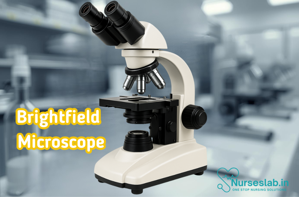
The brightfield microscope, also known as the compound light microscope, is the most commonly used in clinical and educational settings. Its main components include:
- Eyepiece (Ocular lens): Where the observer looks through; typically provides 10x magnification.
- Objective lenses: Usually 4x, 10x, 40x, and 100x (oil immersion) magnifications.
- Stage: The platform that holds the specimen slide.
- Light source: Provides illumination, usually from below the stage.
- Condenser: Focuses light onto the specimen.
- Coarse and fine focus knobs: Adjust the focus of the image.
- Arm and base: Provide structural support.
Working Principle
In brightfield microscopy, visible light passes through the specimen and into the objective lens. The specimen appears darker against a bright, illuminated background. Staining techniques (such as Gram staining) are often used to enhance contrast and highlight specific structures.
Applications in Nursing
- Examining blood smears for malaria, anaemia, or leukaemia
- Identifying bacteria in wound swabs or urine samples
- Studying tissue biopsies for pathological changes
- Monitoring infection control measures in clinical settings
Advantages
- Simple to use and widely available
- Cost-effective compared to advanced microscopes
- Suitable for routine examination of stained specimens
Limitations
- Limited resolution (up to 0.2 micrometres)
- Cannot visualise very small viruses or detailed internal structures
- Live, unstained specimens are difficult to observe clearly
Fluorescent Microscope
Principle of Fluorescence
Fluorescence microscopy utilises the property of certain substances to emit light of a specific wavelength when excited by light of a shorter wavelength. In this technique, fluorescent dyes (fluorochromes) are used to stain the specimen. When exposed to ultraviolet or blue light, these dyes emit visible light, making the labelled structures glow brightly against a dark background.
Components of a Fluorescent Microscope
- Light source: High-intensity mercury or xenon lamp (or LEDs) for excitation.
- Excitation filter: Allows only specific wavelengths to reach the specimen.
- Dichroic mirror: Reflects excitation light towards the specimen and transmits emitted fluorescence to the eyepiece.
- Emission filter: Permits only the emitted fluorescent light to reach the observer’s eye.
- Objective lenses: Specially designed to transmit ultraviolet and visible light efficiently.
- Eyepiece and camera: For direct observation and documentation.
Uses in Clinical Microbiology
- Rapid identification of Mycobacterium tuberculosis in sputum using auramine-rhodamine stain
- Detection of malarial parasites, fungi, and other pathogens in clinical samples
- Visualisation of antibodies or antigens using immunofluorescence techniques
- Assessment of cell viability and apoptosis in research and diagnostics
Advantages
- High specificity and sensitivity for detecting labelled targets
- Enables observation of living cells and dynamic processes
- Allows multiplexing – simultaneous detection of multiple targets
Limitations
- Requires expensive equipment and special fluorescent dyes
- Specimens may bleach (lose fluorescence) over time
- Background fluorescence can interfere with interpretation
Electron Microscope
Electron microscopy represents the pinnacle of resolution and magnification in imaging technology. Instead of light, it uses a beam of electrons to visualise specimens, enabling observation of subcellular structures and viruses.
Types of Electron Microscopes
- Transmission Electron Microscope (TEM): Electrons pass through ultra-thin sections of the specimen, revealing internal structures in exquisite detail. TEM is used to study organelles, viruses, and molecular complexes.
- Scanning Electron Microscope (SEM): Electrons scan the surface of a specimen, producing detailed three-dimensional images of its external features. SEM is ideal for studying surface morphology and biofilms.
Working Mechanism
In both TEM and SEM, electrons are generated by an electron gun and focused onto the specimen using electromagnetic lenses. In TEM, transmitted electrons are detected to form an image, while in SEM, secondary electrons emitted from the surface are collected to create a topographical image. Specimens must be specially prepared, fixed, and often coated with metal for electron microscopy.
Applications in Microbiology
- Identification of viruses (e.g., influenza, herpesvirus, SARS-CoV-2)
- Detailed visualisation of bacterial cell walls, flagella, and spores
- Study of ultrastructural changes in tissues and cells during infection
- Research into biofilms, nanomedicine, and antimicrobial mechanisms
Comparison with Light Microscopes
| Feature | Light Microscope (Brightfield/Fluorescent) | Electron Microscope (TEM/SEM) |
| Source of Illumination | Visible or UV light | Electron beam |
| Resolution | ~0.2 micrometres | Up to 0.2 nanometres (TEM) |
| Specimen Preparation | Simple; living or fixed | Complex; fixed, dehydrated, often metal-coated |
| Image | Colour (natural or stained) | Black and white, high contrast |
| Applications | Routine clinical work, teaching | Research, virology, ultrastructural studies |
| Cost | Affordable | Very expensive |
Relevance to Nursing Practice
A thorough understanding of microbiology and microscopy empowers nurses to excel in patient care and infection control. Here’s how this knowledge directly benefits nursing practice:
1. Infection Prevention and Control
- Recognising sources and modes of transmission of infectious agents
- Implementing hand hygiene, sterilisation, and disinfection protocols
- Monitoring hospital-acquired infections and outbreaks
2. Diagnostics and Specimen Collection
- Proper collection, labelling, and transport of clinical specimens for microbiological analysis
- Interpreting laboratory and microscopy reports to inform patient care decisions
- Assisting in point-of-care testing and rapid diagnostics
3. Patient and Community Education
- Educating patients about communicable diseases and preventive measures
- Promoting vaccination and adherence to treatment regimens
- Raising awareness about antibiotic resistance and rational use of medicines
4. Professional Collaboration
- Communicating effectively with microbiologists, physicians, and infection control teams
- Participating in quality improvement and research initiatives
5. Lifelong Learning
Microbiology is a rapidly evolving field. Nurses must remain updated with emerging pathogens, new diagnostic techniques, and changing guidelines to ensure optimal patient outcomes.
Conclusion
Microbiology and microscopy are integral to nursing education and practice. From the historical breakthroughs of van Leeuwenhoek, Pasteur, and Koch to the advanced imaging techniques of today, these disciplines have transformed the way diseases are understood, prevented, and treated. For nurses, mastery of applied microbiology is not just an academic requirement but a practical necessity for safeguarding patient health, preventing infections, and contributing to the broader goals of public health. As science advances, nurses will continue to play a pivotal role at the intersection of microbiology and patient care.
REFERENCES
- Apurba S Sastry, Essential Applied Microbiology for Nurses including Infection Control and Safety, First Edition 2022, Jaypee Publishers, ISBN: 978-9354659386
- Joanne Willey, Prescott’s Microbiology, 11th Edition, 2019, Innox Publishers, ASIN- B0FM8CVYL4.
- Anju Dhir, Textbook of Applied Microbiology including Infection Control and Safety, 2nd Edition, December 2022, CBS Publishers and Distributors, ISBN: 978-9390619450
- Gerard J. Tortora, Microbiology: An Introduction 13th Edition, 2019, Published by Pearson, ISBN: 978-0134688640
- Durrant RJ, Doig AK, Buxton RL, Fenn JP. Microbiology Education in Nursing Practice. J Microbiol Biol Educ. 2017 Sep 1;18(2):18.2.43. https://pmc.ncbi.nlm.nih.gov/articles/PMC5577971/
Stories are the threads that bind us; through them, we understand each other, grow, and heal.
JOHN NOORD
Connect with “Nurses Lab Editorial Team”
I hope you found this information helpful. Do you have any questions or comments? Kindly write in comments section. Subscribe the Blog with your email so you can stay updated on upcoming events and the latest articles.


