


Last Updated on February 13, 2026 by Nurseslab.in Editorial Team
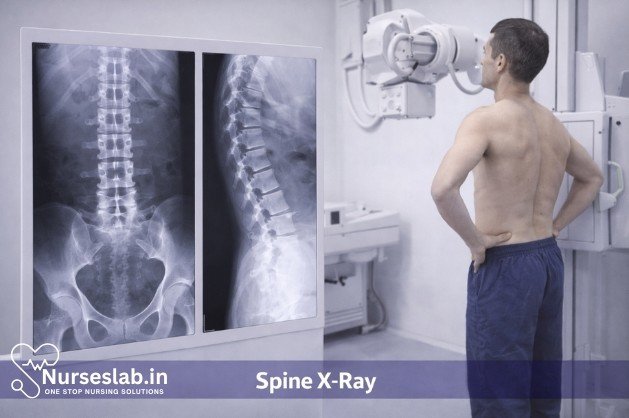
A spine X‑ray is a quick, non‑invasive imaging test used to evaluate vertebral alignment, fractures, degenerative changes, and structural abnormalities. It supports diagnosis and treatment planning in orthopedics, neurology, and primary care.
Introduction
Spine X-rays have long been an essential diagnostic tool in clinical practice, playing a pivotal role in the evaluation of spinal pathology. They offer a non-invasive and relatively quick method of visualising the bony structures of the vertebral column, aiding clinicians in the diagnosis of a variety of conditions. With the increasing prevalence of musculoskeletal disorders and the growing need for accurate assessment, spine X-ray diagnostics remain a cornerstone in both emergency and elective medical settings.
Purpose of Spine X-Ray Diagnostics
Clinical Scenarios
The primary purpose of spine X-ray diagnostics is to evaluate and investigate a wide spectrum of spinal conditions. Common clinical indications include:
- Acute trauma, such as suspected vertebral fractures following accidents or falls
- Chronic back pain where underlying structural abnormalities are suspected
- Assessment of spinal alignment in conditions like scoliosis or kyphosis
- Detection of degenerative changes associated with ageing or arthritis
- Evaluation of congenital malformations and developmental anomalies
- Monitoring postoperative changes or hardware placement
- Screening for infections, tumours, or metastatic disease
Conditions Detected
Spine X-rays are particularly useful for identifying:
- Fractures and dislocations
- Degenerative disc disease and osteophytes
- Abnormal curvatures, such as scoliosis and lordosis
- Congenital vertebral anomalies
- Bone infections (osteomyelitis)
- Neoplastic lesions, both benign and malignant
While X-rays are limited in their ability to visualise soft tissues, they provide critical information about bone integrity, alignment, and structural changes.
Patient Preparation
Pre-procedure Instructions
Proper patient preparation is vital to ensure the accuracy and safety of spine X-ray diagnostics. Key steps include:
- Explaining the procedure to the patient, addressing any concerns or anxieties
- Confirming the patient’s identity and verifying the indication for the X-ray
- Checking for pregnancy in female patients of childbearing age, as radiation exposure should be avoided
- Requesting the removal of jewellery, metallic objects, and clothing that may interfere with image quality
- Ensuring the patient’s comfort and privacy throughout the process
Safety Considerations
Radiation safety is paramount in spine X-ray diagnostics. Measures include:
- Utilising protective lead aprons to shield non-targeted body parts
- Minimising exposure by using the lowest effective radiation dose
- Adhering to the ALARA (As Low As Reasonably Achievable) principle
- Ensuring proper collimation to restrict the X-ray beam to the area of interest
- Maintaining a safe distance for staff during exposure
X-Ray Techniques for the Spine
Types of Views
Spine X-ray examinations typically involve several standard views to provide comprehensive visualisation:
| Region | Standard Views | Purpose |
| Cervical Spine | AP (anteroposterior), lateral, odontoid (open mouth) | Assess vertebral alignment, fractures, and C1/C2 articulation |
| Thoracic Spine | AP, lateral | Visualise vertebral bodies, disc spaces, and kyphosis |
| Lumbar Spine | AP, lateral, oblique | Evaluate vertebral body integrity, spondylolisthesis, and disc degeneration |
| Sacrum & Coccyx | AP, lateral | Assess trauma, congenital anomalies, and degenerative changes |
Equipment Used
Modern spine X-ray diagnostics employ advanced radiographic equipment, including:
- Digital radiography systems for high-resolution imaging
- Conventional X-ray machines for standard film-based procedures
- Portable X-ray units for bedside or emergency use
- Fluoroscopy devices for dynamic studies (e.g., flexion-extension views)
Patient Positioning
Accurate positioning is crucial for optimal image quality and diagnostic value. General principles include:
- Ensuring the patient is relaxed and stationary during exposure
- Aligning the anatomical area of interest with the X-ray beam
- Using positioning aids such as sponges or supports for comfort and stability
- Adhering to standard protocols for each spinal region
For example, in a lateral lumbar spine view, the patient lies on their side with knees flexed, arms positioned away from the spine, and the X-ray beam centred over the lumbar vertebrae.
Procedure Steps
Step-by-Step Process
- Patient Identification and Consent: Confirm patient details and obtain informed consent, explaining the procedure and risks.
- Preparation: Instruct the patient to remove any interfering objects and change into a hospital gown if necessary.
- Positioning: Assist the patient into the required position based on the region and view needed.
- Equipment Setup: Adjust the X-ray machine settings, ensuring proper collimation and exposure parameters.
- Exposure: The radiographer steps behind a protective barrier and activates the X-ray, capturing the required images.
- Image Review: Verify image quality and repeat exposures if artefacts or inadequate visualisation occur.
- Completion: The patient is assisted off the table, and post-procedure instructions are provided.
Patient Experience
Spine X-ray procedures are generally brief, lasting between 10 to 20 minutes depending on the complexity. Patients may experience mild discomfort from maintaining certain positions but are otherwise unharmed. Exposure to radiation is minimal and strictly regulated.
Interpretation of Spine X-Rays
Reading Images
Interpretation of spine X-rays requires systematic analysis by trained radiologists or clinicians. The process typically follows these steps:
- Assessment of image quality, including exposure, positioning, and clarity
- Identification of anatomical landmarks and alignment of vertebral bodies
- Evaluation for fractures, dislocations, or abnormal curvatures
- Inspection for degenerative changes such as disc space narrowing, osteophyte formation, or sclerosis
- Detection of abnormal soft tissue shadows, calcifications, or foreign bodies
- Comparison with previous imaging studies for progression or resolution of pathology
Common Findings
Frequent radiographic findings in spine X-rays include:
- Compression fractures, often seen in osteoporosis
- Spondylolisthesis, characterised by vertebral slippage
- Disc space narrowing and endplate changes in degenerative disc disease
- Abnormal curvature patterns in scoliosis or kyphosis
- Evidence of post-surgical hardware or fusion
Reporting
Formal reporting involves structured documentation of findings, typically including:
- Patient demographics and clinical indication
- Technical details of the examination
- Summary of radiographic findings
- Impression or diagnosis, with recommendations for further evaluation if necessary
Clear and concise reporting is essential for effective clinical communication and patient management.
Risks and Limitations
Radiation Exposure
One of the principal risks associated with spine X-ray diagnostics is exposure to ionising radiation. Although the doses are generally low, cumulative exposure over time can increase the risk of adverse effects, particularly in vulnerable populations such as children and pregnant women. Adherence to safety protocols and judicious use of imaging are key to minimising risks.
Diagnostic Limitations
Spine X-rays, while invaluable for bony pathology, have notable limitations:
- Poor visualisation of soft tissues, such as intervertebral discs, ligaments, and nerves
- Limited sensitivity for early or subtle bone changes
- Potential for missed injuries or pathologies that require advanced imaging (e.g., MRI, CT scans)
- Artefacts from patient movement or external objects can obscure findings
Therefore, spine X-rays are often complemented by other modalities for comprehensive assessment.
Recent Advancements in Spine X-Ray Diagnostics
Digital Imaging
The transition from conventional film-based X-rays to digital radiography has revolutionised spinal imaging. Advantages of digital systems include:
- Enhanced image quality with superior resolution and contrast
- Reduced radiation doses due to improved detector sensitivity
- Immediate image availability for rapid review and remote consultation
- Ease of image storage, retrieval, and sharing via electronic medical records
- Advanced post-processing capabilities for better visualisation and analysis
AI-Assisted Interpretation
Artificial Intelligence (AI) is increasingly being integrated into spine X-ray diagnostics, offering several benefits:
- Automated detection of vertebral fractures, alignment abnormalities, and degenerative changes
- Reduction in reporting times and increased diagnostic accuracy
- Decision support for clinicians, particularly in resource-limited settings
- Continuous learning algorithms that improve performance with larger datasets
AI tools are not a substitute for clinical expertise but serve as valuable adjuncts, enhancing workflow efficiency and patient outcomes.
Other Technological Innovations
Additional advancements include:
- Low-dose imaging protocols for paediatric and high-risk populations
- Integration with Picture Archiving and Communication Systems (PACS) for seamless data management
- Development of mobile and handheld X-ray devices for field and emergency use
- 3D reconstruction techniques for improved anatomical assessment
Nursing Care for Patients Undergoing Spine X-Ray Diagnostic Procedures
Spine X-ray diagnostic procedures are essential tools in evaluating patients with suspected spinal pathology, such as trauma, degenerative disease, infection, or malignancy. Nurses play a pivotal role in the holistic care of patients undergoing these imaging studies, ensuring patient safety, comfort, and accurate diagnostic results.
Pre-Procedural Nursing Care
Patient Assessment
The nurse begins by performing a thorough assessment, which includes:
- Medical History: Identify indications for the X-ray, previous spinal conditions, and history of allergies (especially to contrast agents if used).
- Medication Review: Note any medications that may impact the procedure, such as anticoagulants, pain medications, or sedatives.
- Physical Assessment: Evaluate the patient’s mobility, pain levels, ability to cooperate, and need for assistance with positioning.
Patient Preparation and Education
Educating the patient is crucial to reduce anxiety and enhance cooperation. Key points include:
- Procedure Explanation: Describe the purpose, steps, and expected duration of the X-ray. Clarify that the test is painless but may require remaining still in specific positions.
- Radiation Safety: Reassure the patient about the minimal risks associated with diagnostic radiation exposure, and discuss the use of protective lead shields.
- Instructions: Advise the patient to remove jewelry, metal objects, or clothing that may interfere with imaging. Ensure the patient has fasted if contrast studies are anticipated.
- Consent: Confirm that informed consent has been obtained as per hospital policy.
Psychological Support
Many patients experience anxiety regarding diagnostic procedures. Nurses should:
- Provide emotional support and reassurance.
- Address fears about radiation, pain, or diagnosis.
- Encourage questions and provide clear, honest answers.
Physical Preparation
On the day of the procedure, nurses should:
- Verify patient identity and procedure details.
- Assist with changing into a gown and removing interfering items.
- Assess for pregnancy in women of reproductive age and notify radiology staff if applicable.
- Ensure the patient’s comfort and safety prior to transfer to the radiology department.
Intra-Procedural Nursing Care
Patient Positioning and Assistance
Proper positioning is critical for accurate imaging. Nurses may assist the radiographer in:
- Helping the patient onto the X-ray table, especially those with limited mobility.
- Providing pillows or supports to maintain required positions (e.g., supine, lateral, or oblique).
- Ensuring patient comfort, particularly for those with pain or neurological impairment.
- Monitoring for signs of discomfort or distress and intervening as necessary.
Safety Precautions
Nurses have a responsibility to minimize radiation exposure:
- Ensure lead aprons/shields are used for non-imaged body parts.
- Verify that all staff and accompanying individuals are appropriately protected or outside the exposure area.
- Confirm that the procedure room is prepared and equipment is functioning.
Monitoring and Support
During the procedure, nurses should:
- Monitor the patient’s vital signs if indicated, especially in trauma or unstable patients.
- Observe the patient’s response, looking for anxiety, pain, or changes in condition.
- Provide verbal support and instructions, encouraging the patient to remain still to avoid retakes.
- Assist with repositioning between different views as requested by the radiographer.
Post-Procedural Nursing Care
Immediate Post-Procedure Assessment
After the X-ray, nurses should:
- Assist the patient in getting off the X-ray table and back into a comfortable position.
- Assess for any immediate adverse effects, such as increased pain, dizziness, or anxiety.
- Provide assistance as needed for mobility or transport back to the ward.
Patient Education and Follow-Up
Nurses are responsible for ongoing patient education, including:
- Informing the patient about the next steps, such as when results will be available and who will discuss them.
- Explaining any activity restrictions or precautions if indicated (e.g., after trauma or contrast studies).
- Encouraging the patient to report any new symptoms, such as persistent pain, numbness, or weakness.
Documentation
Accurate and comprehensive documentation is essential:
- Record the procedure date, time, type of X-ray, and patient response.
- Note any complications, interventions, or special precautions taken.
- Document patient education and instructions provided.
Special Considerations
Pediatric Patients
Children may require additional reassurance, distraction techniques, and parental involvement. Nurses should use age-appropriate language and may need to immobilize the child gently to obtain quality images.
Patients with Disabilities or Limited Mobility
These patients may need extra assistance with positioning, transfers, and communication. Nurses should advocate for their comfort and dignity throughout the procedure.
Pregnant Patients
X-rays are generally avoided in pregnant women unless absolutely necessary. Nurses should confirm pregnancy status before imaging and ensure appropriate fetal shielding if the procedure must proceed.
Contrast Studies
Occasionally, contrast agents are used to enhance imaging. Nurses must assess for allergies, monitor for adverse reactions, and provide post-procedural hydration instructions.
Complications and Nursing Interventions
While spine X-rays are low-risk, nurses should be alert for possible complications:
- Radiation Exposure: Minimize exposure and educate the patient about safety.
- Pain or Discomfort: Provide analgesia or repositioning as needed.
- Anxiety: Offer reassurance and support.
- Adverse Reactions (Contrast): Monitor for allergic reactions and intervene promptly.
Role of Communication and Collaboration
Effective nursing care relies on communication and teamwork:
- Coordinate with radiology staff to ensure smooth workflow.
- Communicate patient needs and concerns clearly.
- Advocate for the patient’s comfort and safety at every stage.
REFERENCES
- United States Food and Drug Administration. Medical X-ray imaging http://www.fda.gov/Radiation-EmittingProducts/RadiationEmittingProductsandprocedures/medicalimaging/medicalx-rays/default.htm . Last updated 2/21/2023.
- National Cancer Institute (U.S.). Radiation https://www.cancer.gov/about-cancer/causes-prevention/risk/radiation . Last updated 3/7/2019.
- National Institute of Biomedical Imaging and Bioengineering (U.S.). X-rays https://www.nibib.nih.gov/science-education/science-topics/x-rays. Updated 6/2022.
- Van den Wyngaert, T. (2023). Normal Spine: X-ray and CT Anatomy. In: Van den Wyngaert, T., Gnanasegaran, G., Strobel, K. (eds) Clinical Atlas of Bone SPECT/CT. Springer, Cham. https://doi.org/10.1007/978-3-031-26449-8_76
- National Library of Medicine (U.S.). Thoracic spine x-ray http://www.nlm.nih.gov/medlineplus/ency/article/003806.htm. Review date 7/28/2021.
- Radiopaedia. Spine radiography https://radiopaedia.org/articles/spine-radiography. Last revised 3/23/2023.
Stories are the threads that bind us; through them, we understand each other, grow, and heal.
JOHN NOORD
Connect with “Nurses Lab Editorial Team”
I hope you found this information helpful. Do you have any questions or comments? Kindly write in comments section. Subscribe the Blog with your email so you can stay updated on upcoming events and the latest articles.


