


Retinoscopy is an essential objective method for assessing refractive errors by observing the reflex of light from the retina. It helps determine myopia, hyperopia, and astigmatism, making it vital in optometry, ophthalmology, and clinical eye‑care training.
Introduction
Retinoscopy is a cornerstone diagnostic procedure in the field of ophthalmology and optometry, widely used for the objective assessment of refractive errors in the human eye. Since its inception in the late 19th century, retinoscopy has evolved into an indispensable tool for eye care professionals, especially in settings where subjective responses from patients are unreliable or unavailable. The technique, though seemingly simple, requires a thorough understanding of optical principles, careful technique, and precise interpretation of findings.
Definition and Purpose of Retinoscopy
What is Retinoscopy?
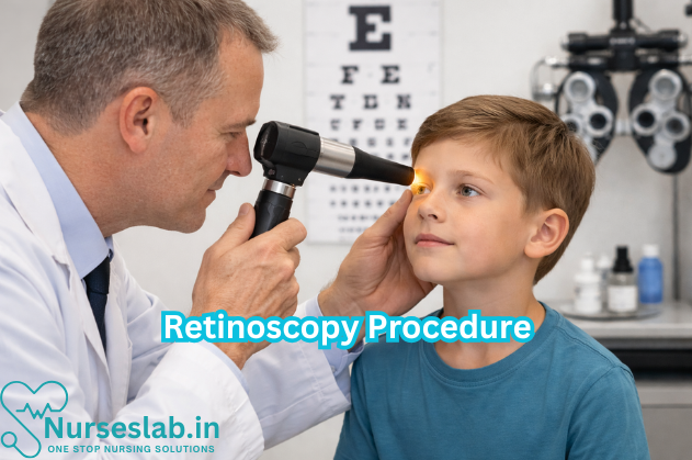
Retinoscopy, also known as skiascopy, is an objective method for determining the refractive status of the eye. The procedure involves projecting a beam of light into the eye using a retinoscope and observing the movement of the reflected light (retinal reflex) from the patient’s retina. By introducing various lenses in front of the eye, the examiner seeks to neutralise the reflex, thereby determining whether the eye is emmetropic (normal), myopic (short-sighted), or hypermetropic (long-sighted), and quantifying the refractive error.
Purpose of Retinoscopy
The primary purpose of retinoscopy is to objectively assess the refractive state of the eye, particularly in individuals who are unable to provide reliable subjective responses, such as young children, non-verbal patients, or those with communication difficulties. It serves as a fundamental procedure for prescribing corrective lenses and for the early detection of conditions that can impair visual development, such as amblyopia and strabismus.
Indications and Contraindications
Indications
Retinoscopy is indicated in a variety of clinical scenarios, including:
- Assessment of refractive errors (myopia, hypermetropia, astigmatism)
- Evaluation of visual complaints in non-verbal or uncooperative patients
- Screening children for amblyogenic factors
- Preoperative evaluation before ocular surgeries
- Assessment of refractive status in patients with intellectual disabilities
- Baseline examination in paediatric and community eye care programmes
Contraindications
While retinoscopy is generally a safe and non-invasive procedure, certain situations may limit its effectiveness or feasibility:
- Significant media opacities (e.g., dense cataract, corneal opacity, vitreous haemorrhage) that prevent visualisation of the retinal reflex
- Severe photophobia or poor cooperation despite attempts at reassurance
- Acute ocular surface disorders (e.g., severe conjunctivitis, keratitis) where patient comfort is compromised
Equipment and Preparation
Required Equipment
- Retinoscope: The primary instrument, available in two main types:
- Spot retinoscope: Projects a circular beam of light
- Streak retinoscope: Projects a linear beam, preferred for astigmatism assessment
- Trial lenses and trial frame: A set of spherical and cylindrical lenses of varying powers used to neutralise the reflex
- Lens rack or skiascopic ruler: Alternative to trial lens set, especially useful for quick estimation
- Occluder: For monocular assessment, to cover the non-tested eye
- Measuring tape or ruler: To ensure accurate working distance
- Dimly lit room: Essential for optimal visualisation of the reflex
Preparation of Patient and Examiner
- Explain the procedure to the patient (or guardian, in case of children) to ensure cooperation and minimise anxiety
- Seat the patient comfortably at eye level with the examiner, typically at a working distance of 50 cm or 66 cm (corresponding to lens powers of +2.00 D or +1.50 D respectively)
- Remove the patient’s spectacles if worn
- Ensure the patient’s head is straight and eyes are open wide, fixating on a distant non-accommodative target to relax accommodation
- The examiner should ensure their own refractive error is corrected, and maintain a steady hand and consistent working distance throughout the procedure
Step-by-Step Retinoscopy Procedure
Types of Retinoscopy
- Static Retinoscopy: Performed while the patient fixates on a distant target, assessing the refractive error when accommodation is at rest
- Dynamic Retinoscopy: Performed while the patient fixates on a near target, evaluating the accommodative response
Static Retinoscopy: Detailed Steps
Positioning:
- Seat the patient comfortably in a dimly lit room
- Examiner sits at a fixed distance (commonly 66 cm or 50 cm) directly in front of the patient
Initial Observation:
- Ask the patient to look at a distant fixation target to minimise accommodation
- Hold the retinoscope at the chosen working distance, aligned with the patient’s visual axis
Projecting the Light:
- Switch on the retinoscope and direct the light beam into the patient’s pupil
- Move the retinoscope horizontally and vertically, observing the movement of the red reflex
Interpretation of Reflex:
- With movement: The reflex moves in the same direction as the retinoscope; suggests hypermetropia or low myopia
- Against movement: The reflex moves in the opposite direction; suggests myopia greater than the inverse of the working distance
- Neutral reflex: No movement of the reflex; indicates emmetropia or the refractive error is neutralised by the lens in place
Neutralisation:
- Introduce trial lenses (starting with low plus or minus powers) in front of the patient’s eye
- Continue observing the reflex while changing lenses, aiming to reach the point where the reflex shows no movement (neutralisation)
Working Distance Correction:
- After neutralisation, subtract the dioptric equivalent of the working distance (e.g., +1.50 D for 66 cm, +2.00 D for 50 cm) from the lens power at neutralisation to determine the actual refractive error
Repeat for the Other Eye:
- Cover the first eye and repeat the process on the contralateral eye
Recording Results:
- Document the findings, including sphere, cylinder, and axis (if astigmatism is present)
Dynamic Retinoscopy
Dynamic retinoscopy assesses the accommodative function by performing the procedure while the patient fixates on a near target (usually at 40 cm). The examiner observes the reflex and determines the accommodative response, useful in diagnosing accommodative disorders, especially in children.
Interpretation of Findings
Understanding Retinal Reflexes
The essential part of retinoscopy is interpreting the movement of the retinal reflex:
- With movement: Indicates hypermetropia or lesser degrees of myopia (less than the working distance dioptric value)
- Against movement: Indicates myopia greater than the working distance dioptric value
- No movement (neutral): Indicates that the refractive error is balanced by the lens in place
Neutralisation and Calculation
Upon achieving neutrality, the examiner must account for the working distance by subtracting the dioptric equivalent from the neutralising lens power. For example, if neutrality is achieved with a +3.00 D lens at a working distance of 66 cm (+1.50 D), the actual refractive error is +1.50 D (+3.00 D – +1.50 D).
Recording Results
The refractive status is documented in terms of sphere, cylinder, and axis. In cases of astigmatism, different powers are required to neutralise the reflex in different meridians, and the axis is determined by rotating the streak retinoscope to align with the principal meridians.
Advantages of Retinoscopy
- Objectivity: Does not rely on subjective feedback from patients, making it ideal for children and non-verbal individuals
- Accuracy: Provides a reliable estimation of refractive error, forming the basis for spectacle prescription
- Versatility: Useful across various age groups and clinical scenarios
- Early Detection: Facilitates early diagnosis of amblyogenic factors and refractive errors
- Cost-effective: Requires minimal equipment and can be performed in resource-limited settings
Limitations of Retinoscopy
- Operator Dependent: Requires considerable skill and experience for accurate results
- Influence of Accommodation: Inadequate control of accommodation, especially in young patients, can lead to inaccurate findings
- Media Opacities: Dense cataracts or corneal opacities can obscure the retinal reflex
- Limited Precision in High Astigmatism: Determining exact cylinder axis and power can be challenging in irregular astigmatism
- Patient Cooperation: Poor cooperation or excessive movement may compromise accuracy
Clinical Significance
Retinoscopy holds immense clinical value in both routine practice and special situations:
- Refractive Error Assessment: Forms the foundation for prescribing corrective lenses, especially in paediatric and special needs populations
- Paediatric Ophthalmology: Essential for detecting refractive errors that may lead to amblyopia or strabismus if left uncorrected
- Screening Programmes: Widely used in school screening and community outreach initiatives for early detection of visual impairment
- Preoperative Work-up: Provides baseline refractive status prior to ocular surgeries
- Special Populations: Beneficial in patients with intellectual disabilities, developmental delays, or speech impairments where subjective refraction is not feasible
Nursing Care of Patients Undergoing Retinoscopy Procedure
While retinoscopy is considered a safe and non-invasive procedure, the role of nursing care is paramount in ensuring patient comfort, accurate results, and the prevention of complications.
Pre-Procedure Nursing Care
Patient Assessment
- Obtain a thorough ophthalmic and medical history, including prior eye conditions, surgeries, medications, and allergies.
- Assess the patient’s level of understanding and ability to cooperate with the procedure, particularly in children or individuals with special needs.
- Evaluate baseline visual acuity and document findings.
Preparation of the Environment and Equipment
- Ensure the examination room is well-lit and organized, with all necessary equipment (retinoscope, trial lenses, lens rack, eye drops, cotton swabs, gloves, etc.) readily available.
- Maintain infection control standards by cleaning and disinfecting reusable instruments before and after use.
- Prepare cycloplegic or mydriatic eye drops as prescribed, and verify correct dosage and patient identity prior to administration.
Patient Preparation and Education
- Explain the purpose, steps, and sensations associated with retinoscopy in language appropriate to the patient’s age and comprehension level.
- Address any anxieties or misconceptions, emphasizing the safety and painless nature of the procedure.
- Instruct patients (or caregivers) on the importance of remaining still and focusing on a designated target during the examination.
- For pediatric patients, use age-appropriate language, visual aids, or play therapy techniques to foster cooperation.
- Obtain informed consent, ensuring the patient or legal guardian understands the procedure and potential risks.
Administration of Eye Drops
- Explain the purpose of cycloplegic/mydriatic drops (to dilate the pupils and temporarily paralyze accommodation for accurate measurement).
- Demonstrate proper hand hygiene and don gloves before administration.
- Ask the patient to tilt their head back and look up; gently retract the lower eyelid and instill the prescribed number of drops without touching the eye or lashes.
- Instruct the patient to keep their eyes closed gently for a few moments to allow absorption and minimize systemic absorption by applying gentle pressure to the inner canthus (punctal occlusion), especially in children or elderly patients.
- Monitor for adverse reactions, such as stinging, redness, or allergic response.
Intra-Procedure Nursing Care
Patient Positioning and Support
- Assist the patient into a comfortable and stable seated position, with chin resting on the appropriate support if using a slit lamp or similar apparatus.
- Ensure children are securely seated on a caregiver’s lap or in a pediatric examination chair to minimize movement.
- Provide reassurance and verbal guidance throughout the procedure.
Assisting the Ophthalmologist/Optometrist
- Hand over instruments and trial lenses as required.
- Monitor patient cooperation and intervene gently if the patient becomes restless or anxious.
- Encourage the patient to maintain steady fixation and avoid blinking excessively.
- For uncooperative pediatric patients, use distraction techniques or gentle restraint as per institutional policy and with parental consent.
Monitoring for Adverse Reactions
- Observe for signs of discomfort, excessive tearing, or photophobia due to pupil dilation.
- Remain vigilant for rare but serious reactions to mydriatic agents, such as increased intraocular pressure, allergic responses, or systemic effects (flushing, tachycardia, dry mouth).
- Be prepared to provide immediate assistance or notify the ophthalmologist if an adverse reaction occurs.
Post-Procedure Nursing Care
Patient Observation and Safety
- Monitor the patient for 15-30 minutes after the procedure, especially if mydriatic/cycloplegic agents were used.
- Assess for lingering discomfort, blurred vision, or signs of allergic reaction.
- Instruct the patient to avoid rubbing their eyes and to report any unusual symptoms immediately.
Patient Education and Discharge Instructions
- Inform patients that vision may remain blurred and sensitivity to light may persist for several hours following the use of eye drops.
- Advise the use of sunglasses or protective eyewear when outdoors until normal vision returns.
- Caution patients against driving, operating machinery, or engaging in activities requiring clear vision until the effects of the drops have worn off.
- For pediatric patients, instruct caregivers to supervise closely and prevent accidental injury due to impaired vision.
- Explain any prescribed follow-up care or appointments, and provide written instructions if necessary.
- Discuss the importance of adhering to any new corrective lens prescriptions or further ophthalmologic evaluations as recommended.
Documentation
- Record all aspects of care, including patient assessment, administration of eye drops (type, dose, time), patient’s response, and any adverse reactions.
- Document patient education provided and the patient’s understanding and acceptance of instructions.
- Note any challenges encountered during the procedure and interventions implemented.
Special Considerations
Pediatric Patients
- Utilize child-friendly explanations and involve caregivers in the process.
- Be patient and flexible, allowing time for the child to become comfortable with the environment and equipment.
- Use distraction techniques such as toys, videos, or storytelling.
- Monitor closely for systemic absorption of eye drops and associated side effects, as children are more susceptible.
Geriatric Patients
- Assess for comorbidities such as glaucoma, which may contraindicate certain mydriatic agents.
- Ensure clear communication and accommodate any sensory or cognitive impairments.
- Assist with mobility and provide support to prevent falls due to temporary visual impairment.
Patients with Disabilities or Nonverbal Patients
- Engage caregivers or family members to facilitate communication and cooperation.
- Use nonverbal cues, gestures, or communication boards as appropriate.
- Allow extra time and adapt the environment to suit the patient’s needs.
Complications and Nursing Interventions
Potential Complications
- Allergic reaction to eye drops (redness, itching, swelling, rash).
- Systemic side effects (tachycardia, dry mouth, headache) from mydriatic/cycloplegic agents.
- Increased intraocular pressure, especially in predisposed individuals.
- Photophobia and transient blurred vision.
Nursing Interventions
- Monitor vital signs and ocular status closely post-procedure.
- Provide prompt intervention for allergic or systemic reactions, including notifying the medical team and administering emergency medications as prescribed.
- Educate patients and caregivers on recognizing warning signs that require immediate medical attention.
- Ensure a safe environment until normal vision is restored.
REFERENCES
- Bagga, D.K., Woodhouse, J.M. (2024). Retinoscopy. In: Das, T., Satgunam, P. (eds) Ophthalmic Diagnostics. Springer, Singapore. https://link.springer.com/chapter/10.1007/978-981-97-0138-4_3#citeas
- Elliott DB. Refraction and prescribing. In: Elliott DB, ed. Clinical Procedures in Primary Eye Care. 5th ed. Elsevier. 2021:65-108.
- Enaholo ES, Musa MJ, Zeppieri M. Objective Refraction Technique: Retinoscopy. 2023 Oct 28. In: StatPearls [Internet]. Treasure Island (FL): StatPearls Publishing; 2025 Jan. https://pubmed.ncbi.nlm.nih.gov/37983333/
- Evans BJW. Detecting Binocular Vision Anomalies in Primary Eyecare Practice. In: Evans BJW, ed. Pickwell’s Binocular Vision Anomalies. 6th ed. Elsevier. 2022:11-44.
- Gaiser H, Reilly J, Gulmiri A. Refraction. In: Reilly J, Gaiser H, Young B, eds. Clinical Procedures for Ocular Examination. 5th ed. McGraw Hill; 2024.
- Kaur K, Gurnani B. Subjective Refraction Techniques (https://www.ncbi.nlm.nih.gov/books/NBK580482/). 2023 Jun 11. In: StatPearls [Internet]. Treasure Island (FL): StatPearls Publishing; 2025 Jan.
Stories are the threads that bind us; through them, we understand each other, grow, and heal.
JOHN NOORD
Connect with “Nurses Lab Editorial Team”
I hope you found this information helpful. Do you have any questions or comments? Kindly write in comments section. Subscribe the Blog with your email so you can stay updated on upcoming events and the latest articles.


