


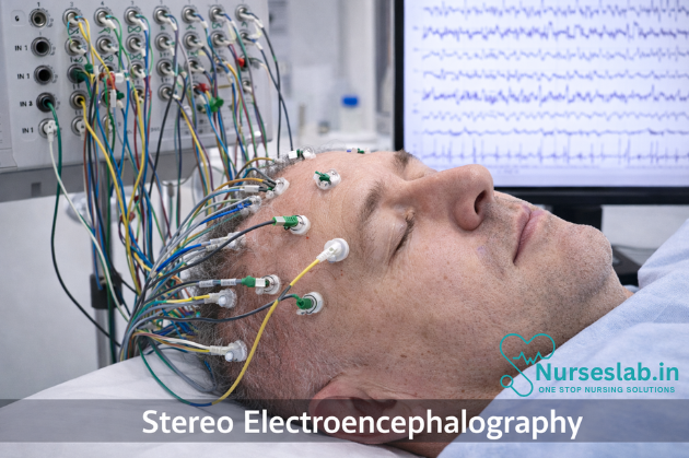
Stereo electroencephalography (SEEG) is a minimally invasive technique that uses implanted depth electrodes to record electrical activity from deep brain structures. It helps identify seizure onset zones in complex or drug‑resistant epilepsy and guides neurosurgical planning.
Introduction
Stereo Electroencephalography (SEEG) has emerged as a cornerstone in the presurgical evaluation of patients with drug-resistant epilepsy. Since its inception in the late 1960s by Jean Talairach and Jean Bancaud in France, SEEG has revolutionised the approach to localising epileptogenic zones deep within the brain. Unlike traditional surface EEG and subdural grid techniques, SEEG enables three-dimensional mapping of cortical and subcortical structures, offering unparalleled precision in the diagnosis and treatment planning for complex epilepsy cases.
Definition and Purpose of SEEG
SEEG is an invasive neurophysiological technique that involves the stereotactic placement of multiple depth electrodes into specific brain regions to record electrical activity from both cortical and subcortical structures. The primary purpose of SEEG is to precisely localise the epileptogenic zone—the area of the brain responsible for generating seizures—particularly when non-invasive methods such as scalp EEG, MRI, and PET fail to provide conclusive information. SEEG differs from other EEG techniques in its ability to sample deep and bilateral brain structures simultaneously, allowing for a comprehensive assessment of seizure networks rather than isolated cortical regions.
The clinical objectives of SEEG include:
- Identifying the epileptogenic zone for possible surgical resection or ablation
- Delineating the functional anatomy of eloquent cortex to minimise post-surgical deficits
- Understanding complex seizure networks, especially in patients with non-lesional or multifocal epilepsy
Indications:
SEEG is recommended in specific clinical scenarios where non-invasive diagnostic modalities are insufficient or ambiguous. The most common indications include:
- Drug-resistant focal epilepsy: Patients who have failed to achieve seizure control with at least two appropriate anti-epileptic drugs.
- Discordant or inconclusive non-invasive findings: When scalp EEG, MRI, PET, or SPECT results are inconsistent or fail to localise the seizure onset zone.
- Multiple or bilateral potential epileptogenic zones: Particularly in cases of multifocal epilepsy, tuberous sclerosis, or bilateral temporal lobe epilepsy.
- Proximity to eloquent cortex: When the presumed epileptogenic zone is near regions responsible for language, motor, or sensory functions, requiring detailed functional mapping.
- Prior unsuccessful epilepsy surgery: For patients with persistent seizures after previous surgical intervention.
SEEG is primarily utilised in adults and children with complex epilepsy who are candidates for surgical treatment but require further localisation to optimise outcomes and minimise risks.
Pre-Procedure Preparation
Patient Selection
Careful patient selection is crucial to maximise the diagnostic yield and minimise risks associated with SEEG. Multidisciplinary epilepsy teams—including epileptologists, neurosurgeons, neuroradiologists, neuropsychologists, and specialised nursing staff—collaborate to evaluate each candidate. Factors such as seizure semiology, imaging findings, prior EEG data, neuropsychological assessment, and overall health status are considered.
Imaging and Planning
High-resolution structural MRI is essential to identify lesions and anatomical landmarks. Functional imaging, such as PET, SPECT, and magnetoencephalography (MEG), may provide additional information regarding metabolic abnormalities or network connectivity. These data are integrated into advanced planning software, which assists in generating a three-dimensional representation of the patient’s brain for accurate electrode trajectory planning.
Consent and Preoperative Assessment
Informed consent is obtained after a thorough discussion of the procedure, potential risks, alternatives, and expected outcomes. Preoperative evaluation includes routine blood tests, coagulation profile, and anaesthetic assessment. Antiepileptic medication regimens are reviewed and may be modified prior to the procedure to facilitate seizure recording.
Technical Aspects:
SEEG utilises specialised equipment designed for precision and safety:
- Stereotactic frames or frameless navigation systems: These provide a reference for accurate electrode placement based on preoperative imaging.
- Depth electrodes: Multi-contact, thin electrodes (typically 0.8–1.2 mm in diameter) constructed of biocompatible materials such as platinum-iridium or stainless steel. Each electrode contains multiple recording contacts spaced at regular intervals (e.g., 2–5 mm apart).
- Planning software: Advanced neurosurgical navigation platforms integrate MRI, CT, and functional imaging to create patient-specific 3D brain models, allowing precise targeting while avoiding vascular structures.
Procedural Steps:
The SEEG procedure is performed in a highly controlled environment, typically under general anaesthesia, although some centres may utilise awake protocols depending on the clinical context.
- Preoperative Imaging and Planning
- High-resolution MRI and CT scans are obtained to visualise brain anatomy and cranial landmarks.
- Imaging data are imported into navigation software to plan electrode trajectories, ensuring avoidance of blood vessels and critical structures.
- Patient Positioning and Preparation
- The patient is positioned supine or as required, and the head is stabilised using a stereotactic frame or a frameless fixation device.
- The scalp is prepped and draped in a sterile fashion.
- Stereotactic Registration
- The navigation system is registered to the patient’s anatomy using fiducial markers or anatomical landmarks.
- Burr Hole Creation
- Small burr holes (typically 2–3 mm in diameter) are drilled in the skull at pre-planned entry points.
- Electrode Insertion
- Sterile depth electrodes are inserted through the burr holes along the planned trajectories, guided by the navigation system.
- Each electrode is secured to the skull using fixation bolts or plates to prevent displacement.
- Post-Implantation Imaging
- A post-operative CT scan is performed to verify electrode positions and rule out complications such as haemorrhage.
- Monitoring and Data Acquisition
- The patient is transferred to an epilepsy monitoring unit (EMU), where continuous video-EEG monitoring is conducted for several days to weeks.
- Antiepileptic medications may be tapered to increase the likelihood of recording spontaneous seizures.
- Electrode Removal
- After sufficient data are collected, electrodes are removed at the bedside or in the operating theatre, usually under local anaesthesia.
Throughout the monitoring period, patients are closely observed for neurological status and potential complications.
Risks and Complications
While SEEG is considered relatively safe compared to other invasive techniques, it is not without risks. The most common complications include:
- Haemorrhage: Intracerebral or subdural bleeding is the most serious complication, with reported rates of 1–2%. Meticulous planning and avoidance of vascular structures mitigate this risk.
- Infection: Superficial or deep infections, including meningitis or cerebral abscess, occur in approximately 1–3% of cases. Strict aseptic technique and perioperative antibiotics are essential preventive measures.
- Electrode displacement: Rarely, electrodes may shift, compromising data quality or causing tissue injury. Secure fixation techniques are employed to prevent this.
- Seizure induction: The procedure may provoke seizures, particularly during monitoring, but this is often therapeutically desired to localise the seizure onset zone.
- Other complications: These include transient neurological deficits, scalp haematoma, cerebrospinal fluid leak, and, rarely, venous infarction.
Risk mitigation strategies involve rigorous patient selection, advanced imaging and planning, experienced surgical teams, and close postoperative monitoring.
Benefits and Outcomes
The diagnostic yield of SEEG is high, with studies reporting successful localisation of the epileptogenic zone in 70–90% of appropriately selected patients. The key benefits include:
- Accurate localisation: Enables precise mapping of seizure onset and propagation, even in deep or bilateral brain regions.
- Minimally invasive approach: Compared to subdural grid implantation, SEEG requires fewer and smaller cranial openings, reducing morbidity.
- Tailored surgical intervention: Facilitates targeted resection or ablation, improving the likelihood of seizure freedom while preserving critical functions.
- Comprehensive network analysis: Allows for investigation of complex seizure networks, guiding surgical and non-surgical interventions.
- Favourable safety profile: Lower rates of infection and neurological deficits compared to other invasive EEG methods.
Patient outcomes following SEEG-guided surgery are favourable, with many centres reporting long-term seizure freedom rates of 60–70% in selected populations.
Interpretation of SEEG Findings
SEEG interpretation requires specialised expertise in neurophysiology and epileptology. The process involves:
- Visual and quantitative analysis: Reviewing ictal (seizure) and interictal (between seizures) EEG patterns across all electrode contacts.
- Seizure onset zone identification: Determining the earliest site of abnormal electrical activity, its propagation pathways, and its relationship to functional brain regions.
- Functional mapping: Electrical stimulation through SEEG electrodes is used to identify eloquent cortex (e.g., motor, language, sensory areas), guiding surgical planning.
- Integration with imaging: Correlating SEEG data with MRI and other imaging modalities to refine localisation and surgical strategy.
The ultimate goal is to delineate the epileptogenic zone while preserving vital functions, thereby maximising post-surgical seizure freedom and quality of life.
Nursing Care for Patients Undergoing Stereo Electroencephalography
Stereo electroencephalography (SEEG) is a minimally invasive diagnostic procedure used to localize epileptogenic zones in patients with drug-resistant epilepsy. By implanting depth electrodes into specific brain regions, SEEG enables clinicians to record electrical activity, thereby facilitating precise mapping of seizure origins.
Pre-Procedure Nursing Care
1.Patient Assessment
Comprehensive patient assessment is the foundation of effective SEEG care. Nurses should obtain a detailed medical history, focusing on seizure characteristics, prior treatments, comorbidities, allergies, and psychosocial factors. Baseline neurological and physical examinations are essential to identify pre-existing deficits and inform post-procedure comparisons. Assessment of laboratory results, including coagulation profiles and infection markers, helps determine procedural readiness and risk stratification.
2.Informed Consent
Nurses are instrumental in facilitating the informed consent process. They must ensure that patients (and families) understand the rationale for SEEG, the procedural steps, potential risks, benefits, and alternatives. Nurses should verify that consent is voluntary and that all questions are addressed, collaborating with physicians to clarify complex information as needed. Documentation of consent is a key responsibility, reflecting ethical and legal standards.
3.Patient Education
Education is integral to reducing anxiety and promoting cooperation. Nurses should provide clear, jargon-free explanations of SEEG, including electrode placement, expected sensations, duration, and the importance of remaining still during the procedure. Written materials, visual aids, and one-on-one discussions can support comprehension. Addressing concerns about pain, appearance, and recovery fosters trust and engagement.
4.Psychological Support
SEEG can be emotionally taxing for patients and families. Nurses should assess psychological readiness, offer reassurance, and connect patients with counseling services if needed. Empathetic communication and active listening help alleviate fears and encourage open dialogue, contributing to a supportive environment.
5.Preparation Protocols
Pre-procedure preparation includes ensuring fasting status, administering prophylactic antibiotics as ordered, and confirming the availability of blood products for patients at risk of bleeding. Nurses must coordinate preoperative imaging (e.g., MRI, CT) and mark electrode entry sites. Removal of jewelry, securing intravenous access, and verifying patient identification are standard protocols. Skin preparation and hair shaving at electrode sites may be required to minimize infection risk.
Intra-Procedure Nursing Responsibilities
1.Monitoring Vital Signs
Continuous monitoring of vital signs is crucial during SEEG electrode implantation, which is typically performed under general anesthesia or conscious sedation. Nurses should track heart rate, blood pressure, respiratory rate, oxygen saturation, and temperature, promptly reporting deviations to the surgical team. Maintenance of normothermia and hemodynamic stability is vital for procedural success.
2.Assisting the Surgical Team
Nurses function as key members of the intraoperative team, assisting with patient positioning, equipment setup, and sterile field maintenance. They help manage anesthesia, pass instruments, and document procedural milestones. Effective communication and anticipation of surgeon needs facilitate workflow and reduce procedural time.
3.Infection Control
Strict adherence to aseptic technique is essential to prevent surgical site infections. Nurses must ensure proper hand hygiene, sterile draping, and use of personal protective equipment. Monitoring for breaches in sterility and immediate correction of lapses safeguard patient safety.
4.Patient Safety
Patient safety encompasses fall prevention, pressure injury avoidance, and protection from inadvertent injury during electrode placement. Nurses should ensure appropriate padding, secure immobilization, and regular assessment of skin integrity. Documentation of intraoperative events supports continuity of care.
Post-Procedure Nursing Care
1.Immediate Recovery
Following SEEG implantation, patients are transferred to a monitored setting for recovery. Nurses must assess airway patency, consciousness level, and vital signs, watching for signs of distress. Early detection of complications such as airway obstruction or hemodynamic instability is critical.
2.Neurological Assessments
Frequent neurological assessments are required to detect new deficits, changes in mental status, or seizure activity. Nurses should use standardized scales (e.g., Glasgow Coma Scale) and compare findings to pre-procedure baselines. Any deterioration warrants immediate notification of the medical team.
3.Wound Care
Electrode entry sites require meticulous care to prevent infection and promote healing. Nurses should inspect sites for redness, swelling, discharge, or bleeding, and change dressings according to protocol. Documentation of wound status and interventions is necessary for ongoing management.
4.Pain Management
Pain is a common concern after SEEG. Nurses should assess pain using validated scales, administer prescribed analgesics, and monitor for side effects. Non-pharmacological interventions such as cold packs, relaxation techniques, and distraction may also be beneficial.
5.Infection Surveillance
Post-procedure infection risk is heightened due to invasive electrode placement. Nurses must monitor for fever, elevated white blood cell counts, and local signs of infection. Early reporting and initiation of empiric antibiotics can prevent serious complications. Adherence to hand hygiene and environmental cleaning protocols further reduces risk.
Patient and Family Education
1.Explaining the Procedure
Ongoing education is vital for empowering patients and families. Nurses should review the purpose of SEEG, expected hospital stay duration, and potential findings. Visual aids and models can enhance understanding.
2.Expected Outcomes
Patients should be informed about possible outcomes, including identification of seizure foci, surgical candidacy, or need for further evaluation. Nurses should clarify the implications of results and facilitate discussions with the medical team.
3.Post-Procedure Instructions
Education should include wound care, activity restrictions, signs of complications, and medication adherence. Nurses should provide written instructions and confirm comprehension through teach-back methods. Scheduling follow-up appointments and providing contact information for concerns ensure continuity of care.
4.Addressing Concerns
Patients and families often have questions about recovery, prognosis, and lifestyle impact. Nurses should encourage open communication, provide evidence-based answers, and refer to specialists as needed. Emotional support and reassurance help mitigate anxiety and foster resilience.
Complication Management
1.Identifying Potential Complications
Complications of SEEG include infection, hemorrhage, neurological deficits, electrode displacement, and cerebrospinal fluid (CSF) leakage. Nurses must maintain high vigilance for subtle signs of deterioration.
2.Managing Infection
Prompt recognition and management of infection are paramount. Nurses should initiate cultures, administer antibiotics as ordered, and collaborate with infectious disease specialists. Removal of infected electrodes may be necessary in severe cases.
3.Managing Hemorrhage
Signs of bleeding include sudden neurological changes, headache, decreased consciousness, or swelling at electrode sites. Nurses should perform rapid assessments, notify the medical team, and prepare for possible imaging or surgical intervention.
4.Managing Neurological Deficits
New deficits such as weakness, aphasia, or visual changes require immediate evaluation. Nurses should document findings, provide supportive care, and assist with further diagnostic testing. Rehabilitation referrals may be indicated for persistent deficits.
5.Other Complications
Electrode displacement or CSF leakage may present as headache, swelling, or fluid drainage. Nurses should report these findings, maintain strict bedrest if ordered, and assist with corrective procedures.
Interdisciplinary Collaboration
1.Team-Based Care
Optimal SEEG care relies on collaboration between nurses, neurologists, neurosurgeons, anesthesiologists, and allied health professionals. Regular team meetings, shared documentation, and clear communication channels enhance patient safety and outcomes.
2.Role of Nurses in the Team
Nurses act as patient advocates, care coordinators, and educators, bridging gaps between disciplines. They facilitate information exchange, clarify care plans, and ensure that patient preferences are respected. Their observations and assessments are critical for timely intervention and decision-making.
3.Collaboration with Allied Health
Physical therapists, occupational therapists, speech-language pathologists, and social workers may be involved in post-SEEG rehabilitation and support. Nurses should coordinate referrals, monitor progress, and communicate findings to the broader team.
REFERENCES
- Alomar S, Jones J, Maldonado A, Gonzalez-Martinez J. The Stereo-Electroencephalography Methodology. Neurosurg Clin N Am. 2016 Jan;27(1):83-95. doi: 10.1016/j.nec.2015.08.003. PMID: 26615111.
- Centers for Disease Control & Prevention (U.S.) Types of Seizures https://www.cdc.gov/epilepsy/about/types-of-seizures.htm. Last reviewed 9/30/2020.
- Delgado-Garcia G, Frauscher B. Future of Neurology & Technology: Stereoelectroencelphalography in Presurgical Epilepsy Evaluation (https://pubmed.ncbi.nlm.nih.gov/34799457/). Neurology. 2022;98:e437-e440.
- Epilepsy Foundation. LITT Thermal Ablation https://www.epilepsy.com/treatment/surgery/types/litt-thermal-ablation). Last reviewed 2/11/2020.
- Bratu IF, Bénar CG, Villalon SM, Ciavatti L, Mazel F, Bartolomei F, Trébuchon A. Mapping Postictal Aphasia through Signal Complexity: A Stereo-Electroencephalography Study. Ann Neurol. 2025 Nov;98(5):1080-1095. doi: 10.1002/ana.27307. Epub 2025 Jul 11. PMID: 40646712; PMCID: PMC12577677.
- Iida K, Otsubo H. Stereoelectroencephalography: Indication and Efficacy (https://www.ncbi.nlm.nih.gov/pmc/articles/PMC5566696/). Neurol Med Chir (Tokyo). 2017;57(8):375-385.
- Kim LH, Parker JJ, Ho AL, et al. Postoperative outcomes following pediatric intracranial electrode monitoring: A case for stereoelectroencephalography (SEEG) (https://pubmed.ncbi.nlm.nih.gov/32028127/). Epilepsy Behav. 2020;104(Pt A):106905.
- Wang L, Jin W, Zhang Y, et al. Stereoelectroencephalography-guided radiofrequency thermocoagulation in drug-resistant focal epilepsy (https://www.ncbi.nlm.nih.gov/pmc/articles/PMC8908190/).
- Lu, R., Wang, M., Wang, Y., Zhao, R., Li, H., Zhang, Y., & Zhou, Y. (2024). Safety, Accuracy, and Efficacy of Robot-Assisted Stereo Electroencephalography in Children of Different Ages. Neurosurgery, 95(1), 137–145. https://doi.org/10.1227/neu.0000000000002853
Stories are the threads that bind us; through them, we understand each other, grow, and heal.
JOHN NOORD
Connect with “Nurses Lab Editorial Team”
I hope you found this information helpful. Do you have any questions or comments? Kindly write in comments section. Subscribe the Blog with your email so you can stay updated on upcoming events and the latest articles.


