


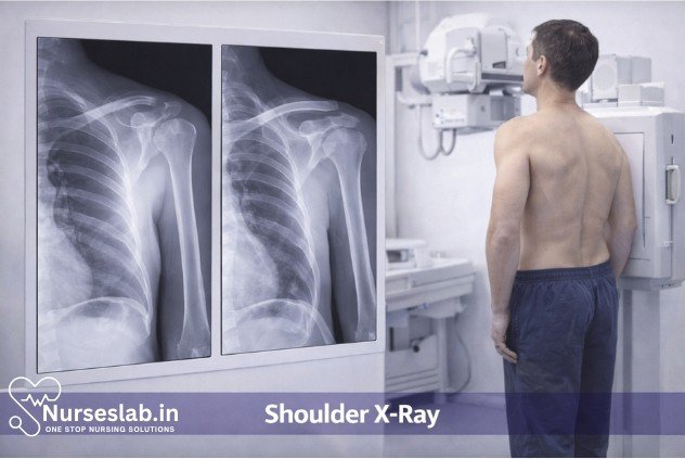
A shoulder X‑ray is a quick, non‑invasive imaging test used to evaluate the bones and joint spaces of the shoulder. It helps diagnose fractures, dislocations, arthritis, rotator cuff–related changes, and other structural abnormalities essential for orthopedic assessment.
Introduction
The shoulder joint, known for its remarkable range of motion and complex anatomical structure, is susceptible to a variety of injuries and pathological conditions. Radiographic evaluation through shoulder X-rays remains a cornerstone in the diagnostic assessment of shoulder complaints.
Purpose and Significance of Shoulder X-Rays
Shoulder X-rays are a non-invasive imaging technique used to visualise the bony structures of the shoulder, including the humeral head, glenoid cavity, scapula, clavicle, and surrounding soft tissues. The primary purpose of this diagnostic tool is to detect and assess fractures, dislocations, degenerative changes, infections, tumours, and congenital anomalies.
Indications
Shoulder X-rays are indicated in a wide array of clinical scenarios, including but not limited to:
- Acute trauma: Suspected fractures, dislocations, or subluxations resulting from falls, sports injuries, or road traffic accidents.
- Chronic pain: Persistent shoulder pain unresponsive to conservative treatment, raising suspicion of degenerative joint disease or rotator cuff pathology.
- Limited range of motion: Stiffness or mechanical restriction suggesting adhesive capsulitis, osteoarthritis, or joint ankylosis.
- Swelling or deformity: Visible abnormalities or swelling warranting exclusion of bony lesions or soft tissue calcifications.
- Suspected infection: Clinical features of septic arthritis or osteomyelitis, such as fever, redness, and localised tenderness.
- Neoplastic processes: Evaluation for primary bone tumours or metastatic lesions.
- Congenital or developmental disorders: Assessment of anatomical variations or malformations in paediatric populations.
- Pre- and post-operative assessment: Planning for surgical interventions and monitoring post-surgical outcomes.
It is crucial to select appropriate imaging based on the patient’s clinical presentation and history, ensuring optimal diagnostic yield while minimising unnecessary radiation exposure.
Preparation: Patient Preparation and Equipment Setup
Proper preparation is essential to obtain high-quality shoulder X-ray images and to ensure patient safety. The following aspects must be considered:
Patient Preparation
- Explanation of the procedure: The patient should be informed about the purpose, steps, and expected duration of the examination. Addressing concerns and answering questions helps alleviate anxiety and improves cooperation.
- Consent: While formal written consent is not always required for routine radiographs, verbal consent and confirmation of understanding are essential.
- Clothing and accessories: The patient should remove clothing, jewellery, or metallic objects from the upper body as these can obscure anatomical details or produce artefacts.
- Pregnancy status: In female patients of reproductive age, it is important to rule out pregnancy due to the potential teratogenic effects of ionising radiation. If pregnancy cannot be excluded, alternative imaging or shielding should be considered.
- Positioning and comfort: The patient should be instructed to maintain the required position while ensuring comfort, as involuntary movement can degrade image quality.
Equipment Setup
- X-ray machine: Ensure the radiographic unit is calibrated and functioning correctly, with appropriate exposure settings for the patient’s body habitus.
- Image receptor: Use a digital detector or film cassette positioned to capture the region of interest.
- Lead shielding: Gonadal and thyroid shields should be used as appropriate to reduce radiation exposure to non-target organs.
- Markers: Place right (R) or left (L) markers on the image receptor to indicate the side being imaged.
- Room preparation: Ensure the imaging suite is clean, well-lit, and free of unnecessary obstructions to facilitate safe movement of the patient and staff.
Procedure: Step-by-Step Guide
The shoulder X-ray typically comprises several standard views, each tailored to reveal specific anatomical details. The most commonly performed projections include the anteroposterior (AP), scapular Y, and axillary views. The following step-by-step guide outlines the general workflow:
Patient Identification and Verification:
- Confirm the patient’s identity using at least two identifiers (e.g., name and date of birth).
- Verify the imaging request and clinical indication to ensure appropriateness.
Patient Positioning:
- Assist the patient to stand, sit, or lie down as per the required projection.
- Ensure the affected shoulder is adequately exposed and positioned.
Selection of Projections:
- Anteroposterior (AP) View: The patient faces the X-ray detector with the back against the cassette. The arm is slightly abducted and externally rotated.
- Scapular Y View: The patient is turned so the scapula forms a Y-shape on the image. This view is crucial for assessing dislocations.
- Axillary (Inferosuperior) View: The arm is abducted to 90°, and the X-ray beam is directed through the axilla. This view demonstrates the relationship of the humeral head to the glenoid.
- Other Special Views: Additional projections such as the Grashey (true AP), Stryker notch, or West Point views may be performed based on clinical need.
Exposure Settings:
- Select appropriate kilovoltage (kV) and milliampere-seconds (mAs) based on patient size and the view required.
- Collimate the X-ray beam to the area of interest to minimise radiation dose.
Image Acquisition:
- Instruct the patient to remain still and hold their breath during exposure to prevent motion artefacts.
- Activate the X-ray exposure and verify immediate image capture on the digital workstation or film.
- Image Review and Repeat Imaging (if necessary):
- Review the images for quality, coverage, and diagnostic adequacy.
- Repeat the projection if the initial image is suboptimal due to positioning or movement.
Post-Procedure Communication:
- Inform the patient that the procedure is complete and provide relevant aftercare instructions.
- Document the procedure and findings as per institutional protocols.
Image Acquisition: Technical Considerations and Positioning
High-quality shoulder X-rays depend on meticulous attention to technical parameters and patient positioning. The following considerations are essential:
Technical Parameters
- Exposure Factors: Use low-to-moderate kV (60–70 kV) and appropriate mAs to balance image contrast and minimise noise.
- Focus-to-Detector Distance (FDD): Typically set at 100–120 cm to reduce magnification and improve image sharpness.
- Collimation: Limit the field to include the entire shoulder girdle and proximal humerus while excluding unnecessary regions.
- Grid Use: Employ an anti-scatter grid for larger patients to enhance image quality by reducing scatter radiation.
Positioning Techniques
- AP View: The patient stands or sits upright. The scapula is flat against the detector, and the arm is externally rotated to visualise the greater tuberosity.
- Scapular Y View: The patient is rotated 45–60° towards the affected side. The arm hangs naturally to prevent distortion.
- Axillary View: The patient abducts the arm while leaning laterally, and the X-ray beam passes through the axilla to the detector placed above the shoulder.
- Immobilisation: Use supports or sandbags as necessary to maintain the required position, especially in uncooperative or paediatric patients.
Interpretation:
Accurate interpretation of shoulder X-rays requires a systematic approach, sound anatomical knowledge, and awareness of common pathologies. The following framework can guide interpretation:
- Image Quality Assessment:
- Check for correct patient identification, side markers, and projection labels.
- Assess exposure, contrast, sharpness, and absence of artefacts.
- Anatomical Review:
- Examine the humeral head, glenoid cavity, acromion, coracoid process, and clavicle for alignment and integrity.
- Evaluate joint spaces for uniformity and signs of effusion or narrowing.
- Pathological Findings:
- Fractures: Look for cortical disruptions, step-offs, or abnormal angulation.
- Dislocations: Assess the position of the humeral head relative to the glenoid. Anterior dislocations are most common.
- Arthritis: Identify joint space narrowing, osteophyte formation, and subchondral sclerosis.
- Calcifications: Detect soft tissue calcifications, which may indicate chronic tendinopathy or previous trauma.
- Tumours: Observe for lytic or sclerotic lesions, periosteal reaction, or abnormal bone architecture.
- Comparison and Correlation:
- Compare with previous imaging, if available, to assess progression or healing.
- Correlate radiographic findings with clinical symptoms and examination results.
- Reporting:
- Document findings in a structured radiology report, highlighting significant abnormalities, incidental findings, and recommendations for further imaging if needed.
Risks and Limitations:
While shoulder X-rays are generally safe and well-tolerated, certain risks and limitations must be acknowledged:
- Radiation Exposure: Although the radiation dose is relatively low, cumulative exposure can pose risks, especially in paediatric and pregnant patients. Adherence to the ALARA (As Low As Reasonably Achievable) principle is essential.
- Pain and Discomfort: Positioning may exacerbate pain in patients with acute injuries. Careful handling and effective communication can mitigate discomfort.
- Contraindications: Absolute contraindications are rare, but relative caution is warranted in pregnant women and those unable to cooperate with positioning.
- Limitations: X-rays primarily visualise bone and are less sensitive for soft tissue injuries such as rotator cuff tears, labral lesions, or early-stage infections. Advanced imaging (MRI or CT) may be required for comprehensive evaluation.
- False Negatives/Positives: Subtle fractures, occult lesions, or early pathological changes may be missed, underscoring the importance of clinical correlation and, if necessary, further imaging.
Aftercare: Post-Procedure Instructions and Follow-Up
After the completion of a shoulder X-ray, patients generally require minimal aftercare. However, certain instructions and follow-up actions are recommended:
- Return to Normal Activities: Patients can usually resume normal activities immediately unless otherwise advised due to the underlying clinical condition.
- Symptom Monitoring: Advise patients to report any new or worsening symptoms, such as increased pain, swelling, or restricted mobility.
- Radiation Precautions: Reassure patients that the radiation dose is minimal and unlikely to cause harm with a single exposure.
- Follow-Up Appointments: Instruct patients to follow up with their referring clinician to discuss the results, further management, or additional investigations if required.
- Documentation: Ensure that the radiographic images and reports are promptly uploaded to the patient’s electronic health record for review and continuity of care.
Nursing Care of Patients Undergoing Shoulder X-Ray Diagnostic Procedure
Nurses are integral to ensuring the safety, comfort, and effective management of patients undergoing shoulder X-ray procedures. This document provides an in-depth overview of the nursing responsibilities and care considerations for patients before, during, and after a shoulder X-ray, with an emphasis on patient safety, communication, and holistic care.
Pre-Procedure Nursing Care
1. Patient Assessment and Identification
- Verify Patient Identity: Ensure accurate identification using two patient identifiers (e.g., full name and date of birth), following hospital policy.
- Assess the Clinical Indication: Review the physician’s order for the X-ray, understand the reason for the test, and clarify any ambiguous instructions.
- Evaluate Patient History: Assess for allergies (especially to contrast if planned), recent shoulder surgeries, pregnancy (in females of childbearing age), and the presence of metallic implants or devices.
2. Patient Preparation
- Explain the Procedure: Clearly describe the purpose, steps, and duration of the procedure to the patient. Address any questions or concerns to reduce anxiety.
- Informed Consent: While formal consent is usually not required for standard X-rays, ensure the patient understands the procedure and agrees to continue.
- Remove Metallic Objects: Instruct the patient to remove jewelry, watches, hairpins, or clothing with metal fasteners from the neck and shoulder area, as these can interfere with image quality.
- Appropriate Attire: Provide a hospital gown if necessary to ensure the area is accessible and to maintain patient dignity.
- Assess Pain Level: Evaluate the patient’s pain and provide analgesia if required, especially in cases of trauma or suspected fracture.
- Pregnancy Precautions: Inquire about possible pregnancy in female patients of reproductive age. If pregnant, notify the radiology team and follow protocols to minimize fetal exposure.
3. Safety Considerations
- Radiation Protection: Ensure the use of lead aprons or shields to protect non-imaged body parts, particularly the thyroid gland and reproductive organs, when appropriate.
- Mobility and Fall Risk Assessment: Evaluate the patient’s ability to ambulate or transfer, especially after recent injury or surgery. Arrange for assistance or mobility aids as needed.
- Infection Control: Adhere to standard precautions, maintaining a clean environment and using gloves if there are open wounds or dressings on the shoulder.
Intra-Procedure Nursing Care
1. Patient Positioning and Comfort
- Assist with Positioning: Help the patient assume the correct position for each X-ray view (e.g., standing, sitting, or lying down; arm rotation as required).
- Support Injured Limbs: Use pillows, foam supports, or sandbags to stabilize an injured or painful shoulder and prevent further discomfort or movement during imaging.
- Monitor for Pain or Distress: Observe for signs of pain or anxiety. Pause the procedure if the patient is unable to tolerate a position, and communicate with the radiographer for adjustments.
2. Communication and Reassurance
- Provide Ongoing Explanation: Inform the patient about each step, when to remain still, and when images are being taken. This helps reduce anxiety and ensures cooperation for optimal image quality.
- Maintain Privacy and Dignity: Keep the patient covered as much as possible and limit exposure of the body to only what is necessary for imaging.
- Observe for Adverse Events: Be alert for any sudden changes in patient condition, such as dizziness or fainting, especially in trauma cases or patients with orthostatic hypotension.
3. Collaboration with Radiology Team
- Coordinate Care: Work closely with the radiographer to ensure the patient is positioned correctly and safely throughout the procedure.
- Assist with Immobilization: If necessary, gently help immobilize the shoulder during exposure to prevent blurred images.
- Lead Shielding: Confirm that lead shielding is in place for patient safety before exposures are made.
Post-Procedure Nursing Care
1. Immediate Post-Procedure Actions
- Assist with Repositioning: Help the patient return to a comfortable position or assist with transferring off the X-ray table if mobility is limited.
- Assess for Discomfort: Re-evaluate the patient’s pain level and provide analgesia if required.
- Monitor for Adverse Reactions: Observe for any new pain, dizziness, or distress following the procedure, especially if the patient was moved extensively during imaging.
2. Patient Education and Follow-Up
- Provide Information: Inform the patient that results will be interpreted by a radiologist and communicated to the referring physician, who will discuss findings and next steps.
- Advise on Activity: Instruct the patient to limit movement of the affected shoulder as appropriate, especially if a fracture or dislocation is suspected, until a definitive diagnosis and management plan are provided.
- Care of Dressings or Splints: If any immobilization devices or dressings are in place, review their care and when to seek medical attention for complications (e.g., increased pain, swelling, numbness).
- Address Concerns: Allow time for the patient to ask questions or express concerns about the procedure or their condition.
3. Documentation
- Record the Procedure: Document the date, time, type of X-ray, patient’s tolerance, any interventions (e.g., pain relief), and post-procedure observations.
- Report Abnormal Findings: Communicate any immediate concerns (e.g., new deformity, severe pain) to the healthcare team for prompt evaluation.
Special Considerations
1. Care for Vulnerable Populations
- Pediatric Patients: Use age-appropriate explanations, involve caregivers, and utilize distraction techniques. Ensure extra protection from radiation and minimal restraint.
- Elderly Patients: Be mindful of frailty, cognitive impairment, and increased risk of falls. Provide additional support and reassurance.
- Patients with Disabilities: Modify positioning and communication strategies to accommodate physical or sensory impairments.
2. Infection Prevention
- Wound Care: For patients with open wounds, ensure proper dressing is maintained and minimize manipulation of the affected area.
- Equipment Cleaning: Ensure X-ray equipment and positioning aids are cleaned between patients to prevent cross-contamination.
3. Handling Emergencies
- Acute Deterioration: Be prepared to respond to sudden changes in patient condition, such as shock or respiratory compromise, particularly in trauma cases.
- Incident Reporting: Document and report any adverse events or near-misses according to facility protocol.
Interprofessional Collaboration
Optimal nursing care during a shoulder X-ray involves close collaboration with radiographers, physicians, and other health professionals. Effective communication ensures that patient needs are met promptly and safely, and that all members of the care team are informed of relevant findings and concerns.
Ethical and Cultural Considerations
- Respect for Autonomy: Honour the patient’s right to refuse or question the procedure, and provide alternatives or additional explanations as needed.
- Cultural Sensitivity: Be aware of cultural beliefs that may affect the patient’s perception of the procedure, privacy, or exposure. Adapt explanations and care accordingly.
- Confidentiality: Maintain privacy regarding the patient’s diagnosis, procedure, and results at all times.
REFERENCES
- American Academy of Orthopaedic Surgeons (AAOS). X-Rays, CT scans and MRIs. https://orthoinfo.aaos.org/en/treatment/x-rays-ct-scans-and-mris/.
- Arthritis Foundation. Shoulder Anatomy https://www.arthritis.org/health-wellness/about-arthritis/where-it-hurts/shoulder-anatomy.
- Merck Manual (Consumer Version). Tests for Musculoskeletal Disorders https://www.merckmanuals.com/home/bone,-joint,-and-muscle-disorders/diagnosis-of-musculoskeletal-disorders/tests-for-musculoskeletal-disorders.
- Radiological Society of North America (RNSA) and American College of Radiology (ACR). Professions in Diagnostic Radiology. https://www.radiologyinfo.org/en/info/professions-diagnostic-radiology.
- U.S. Food & Drug Administration (FDA). We Want You to Know About X-rays: Get the Picture on Protection https://www.fda.gov/radiation-emitting-products/resources-you-radiation-emitting-products/we-want-you-know-about-x-rays-get-picture-protection.
- Sahoo, B., Agrawal, K. (2023). Normal X-ray and CT Anatomy of Shoulder and Elbow. In: Van den Wyngaert, T., Gnanasegaran, G., Strobel, K. (eds) Clinical Atlas of Bone SPECT/CT. Springer, Cham. https://doi.org/10.1007/978-3-031-26449-8_172
Stories are the threads that bind us; through them, we understand each other, grow, and heal.
JOHN NOORD
Connect with “Nurses Lab Editorial Team”
I hope you found this information helpful. Do you have any questions or comments? Kindly write in comments section. Subscribe the Blog with your email so you can stay updated on upcoming events and the latest articles.


