


Last Updated on May 28, 2026 by Nurseslab.in Editorial Team
Introduction
Understanding tooth anatomy is essential for nursing professionals, as oral health is a crucial component of overall well-being. Nurses are often the first point of contact for patients and play a significant role in early detection of dental problems, patient education, and facilitating oral care. A strong grasp of dental anatomy empowers nurses to recognise common oral issues, assist in clinical assessments, and provide holistic care.
Oral Health: An Overview
Oral health is intrinsically linked to systemic health. Poor oral hygiene can contribute to various medical conditions, including cardiovascular disease, diabetes, respiratory infections, and adverse pregnancy outcomes. As frontline healthcare providers, nurses must be equipped to assess oral health, identify abnormalities, and educate patients about preventive measures.
Basic Structure of Teeth
Each tooth is a complex organ with distinct parts and tissues, each serving unique functions. The main structural components of a tooth can be divided into the crown, neck, and root. These segments are made up of specialised tissues: enamel, dentin, pulp, and cementum. Supporting structures, such as the periodontal ligament and alveolar bone, anchor teeth within the jaw.
Crown, Neck, and Root
- Crown: The visible part of the tooth above the gumline. It is covered by a hard, protective layer called enamel.
- Neck: The region where the crown meets the root. It is located at the gumline and is also known as the cervical area.
- Root: The portion embedded in the jawbone, anchoring the tooth in place. The root is covered by cementum and contains the pulp canal.
Tooth Tissues Explained
- Enamel: The outermost, hardest tissue in the human body. It protects the underlying structures from mechanical and chemical damage.
- Dentin: A yellowish tissue beneath the enamel and cementum, making up the bulk of the tooth. It is less hard than enamel but provides flexibility and support.
- Pulp: The innermost soft tissue, consisting of nerves, blood vessels, and connective tissue. The pulp nourishes the tooth and responds to stimuli.
- Cementum: A calcified layer covering the tooth root, helping anchor the tooth to the jawbone via the periodontal ligament.
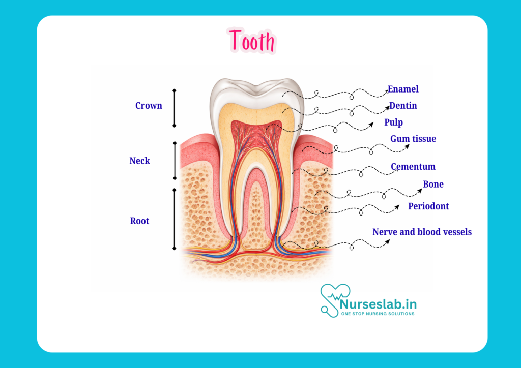
Supporting Structures
- Periodontal Ligament: A network of fibres that connects the tooth root to the alveolar bone, providing support and shock absorption.
- Alveolar Bone: The part of the jawbone that forms the sockets for tooth roots.
- Gingiva (Gums): The soft tissue surrounding the teeth and covering the alveolar bone, playing a key role in oral health.
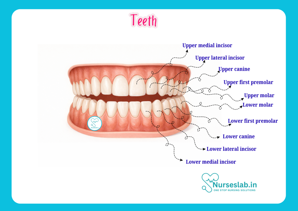
Types of Teeth: Functions and Features
Humans have four main types of teeth, each with specific roles in chewing, speech, and appearance. Understanding these differences is vital for clinical assessment and patient care.
Incisors
- Located at the front of the mouth (central and lateral incisors).
- Sharp, chisel-shaped edges for cutting food.
- Eight incisors in total: four upper and four lower.
Canines
- Located next to the incisors (also called cuspids).
- Pointed shape for tearing food.
- Four canines: two upper and two lower.
Premolars
- Situated behind the canines (also called bicuspids).
- Flat surfaces with ridges for crushing and grinding food.
- Eight premolars: four upper and four lower.
Molars
- Located at the back of the mouth.
- Broad, flat surfaces for grinding food into smaller particles.
- Usually twelve molars: six upper and six lower (including wisdom teeth).
Distinguishing Features
- Incisors and canines are primarily for cutting and tearing.
- Premolars and molars handle grinding and chewing.
- Each type has unique root and crown shapes, aiding in identification during examination.
Tooth Development and Eruption
Tooth development is a dynamic process starting before birth and continuing into adolescence. Understanding this process helps nurses anticipate issues related to teething, eruption, and dental anomalies.
Stages of Tooth Development
- Initiation (Bud Stage): Formation of tooth buds from the dental lamina in the embryonic jaw.
- Cap Stage: The tooth bud grows and forms a cap-like structure, with differentiation of enamel organ, dental papilla, and dental sac.
- Bell Stage: Further differentiation leads to the formation of enamel, dentin, and pulp tissues.
- Apposition and Maturation: Deposition and mineralisation of dental tissues.
Primary (Deciduous) vs Permanent Teeth
- Primary Teeth: Also known as milk or baby teeth, usually 20 in number. They begin to erupt around 6 months of age and are typically all present by 2½ to 3 years.
- Permanant Teeth: Replace primary teeth, with a full set of 32 teeth (including wisdom teeth). Eruption begins around age 6 and continues into late adolescence or early adulthood.
Eruption Timelines
| Type of Tooth | Primary Teeth Eruption (Months) | Permanent Teeth Eruption (Years) |
| Central Incisors | 6-10 | 6-8 |
| Lateral Incisors | 10-16 | 7-9 |
| Canines | 17-23 | 9-12 |
| First Premolars | Not present | 10-12 |
| Second Premolars | Not present | 11-13 |
| First Molars | 14-18 | 6-7 |
| Second Molars | 23-31 | 12-13 |
| Third Molars (Wisdom Teeth) | Not present | 17-21 |
Detailed Anatomy of Tooth Tissues
A closer look at the tissues comprising the tooth reveals their specialised functions and clinical significance.
Enamel
- Composed mainly of hydroxyapatite crystals (a form of calcium phosphate).
- Hardest substance in the human body, yet brittle and susceptible to acid erosion and decay.
- No nerve or blood supply, so damage is irreversible and painless until deeper layers are affected.
- Protects underlying dentin and pulp from physical and chemical insults.
Dentin
- Yellowish, less mineralised than enamel but more flexible.
- Contains microscopic tubules that communicate with the pulp, transmitting sensations of pain and temperature.
- Acts as a shock absorber and supports enamel integrity.
- Exposed dentin due to enamel loss can lead to sensitivity and discomfort.
Pulp Chamber and Nerves
- Central cavity of the tooth, housing nerves, blood vessels, and connective tissue.
- Vital for tooth nutrition, growth, and response to injury.
- Pulpitis (inflammation of the pulp) can cause severe pain and may require endodontic treatment (root canal).
- Pulp extends into root canals, which may be single or multiple depending on the tooth type.
Cementum
- Calcified tissue covering the root surface.
- Anchors the tooth to the periodontal ligament.
- Less hard than dentin and enamel; can be affected by gum disease and trauma.
- Continuous deposition throughout life helps maintain tooth stability.
Periodontal Ligament
- Composed of collagen fibres connecting the cementum to the alveolar bone.
- Acts as a shock absorber during chewing and biting.
- Contains blood vessels and nerves, contributing to tooth support and sensation.
- Inflammation (periodontitis) can compromise tooth stability and lead to tooth loss.
Functions of Teeth
Teeth serve several essential roles beyond mastication. Their health impacts nutrition, communication, self-esteem, and overall systemic health.
- Mastication: Chewing food into smaller, digestible pieces. Efficient mastication is vital for proper digestion and nutrient absorption.
- Speech: Teeth help form sounds and words, especially consonants. Missing teeth can affect clarity of speech.
- Aesthetics: Teeth contribute to facial appearance, smile, and self-confidence. Dental irregularities can impact psychological well-being.
- Overall Health Impact: Poor dental health is linked to systemic diseases such as diabetes, heart disease, and respiratory illness. Maintaining healthy teeth is thus essential for general health.
Oral Cavity and Supporting Structures
The oral cavity houses not just teeth but also several supporting tissues that play vital roles in maintaining oral health.
Gingiva (Gums)
- Firm, pink mucosal tissue surrounding the teeth.
- Protects underlying bone and supports teeth.
- Healthy gingiva is essential for tooth stability and prevention of periodontal disease.
Alveolar Bone
- Part of the jawbone that forms sockets for the tooth roots.
- Supports and retains teeth in position.
- Bone loss due to disease or trauma can lead to tooth mobility and loss.
Oral Mucosa
- Lining of the oral cavity, including the inner cheeks, lips, tongue, and floor of the mouth.
- Acts as a barrier against pathogens and mechanical injury.
- Maintains moisture and facilitates speech and swallowing.
Clinical Relevance for Nurses
Nurses play a critical role in promoting oral health, identifying dental issues, and supporting patients in both hospital and community settings.
Common Dental Conditions
- Dental Caries (Tooth Decay): Caused by bacterial acid production leading to enamel and dentin destruction. Early detection allows for timely intervention.
- Gingivitis: Inflammation of the gums, characterised by redness, swelling, and bleeding. Reversible with good oral hygiene.
- Periodontitis: Advanced gum disease involving bone loss and potential tooth loss. Requires professional management.
- Dental Trauma: Includes fractures, luxations, and avulsions. Immediate care can preserve tooth function and appearance.
- Oral Lesions: Ulcers, white patches, or growths may indicate infections or systemic conditions; prompt referral is vital.
Oral Assessment Techniques
- Inspect lips, gums, teeth, and oral mucosa for colour, integrity, and lesions.
- Assess for signs of infection, bleeding, swelling, or abnormal growths.
- Check for loose, fractured, or missing teeth.
- Evaluate patient-reported symptoms such as pain, sensitivity, or difficulty chewing.
Nursing Interventions and Patient Education
- Provide guidance on proper brushing and flossing techniques.
- Encourage regular dental check-ups and cleanings.
- Advise on dietary choices that support oral health (e.g., limiting sugary foods and drinks).
- Support patients with special needs, such as the elderly, children, or those with disabilities, in maintaining oral hygiene.
- Identify and manage oral complications associated with systemic illnesses or medical treatments (e.g., dry mouth in diabetics, mucositis in cancer patients).
REFERENCES
- Ross and Wilson, Anatomy and Physiology in Health and Illness, Fourteenth Edition, 1 July 2022, ISBN-13: 978-0323834612.
- Roger Watson, Anatomy and Physiology for Nurses, 14th Edition, 12-06-2018, ISBN: 9780702077418
- P.R Asha Latha, Text Book of Applied Anatomy & Physiology for Nurses, 7th Edition,3 January 2024, ISBN-13: 978-9356968622.
- Bryan H. Derikson, Tortora’s Principles of Anatomy and Physiology, 16th Edition, August 2023, ISBN: 978- 1119400066.
- Anatomy.co.uk, Reproductive System, Last updated on April 24, 2025, https://anatomy.co.uk/reproductive-system
Stories are the threads that bind us; through them, we understand each other, grow, and heal.
JOHN NOORD
Connect with “Nurses Lab Editorial Team”
I hope you found this information helpful. Do you have any questions or comments? Kindly write in comments section. Subscribe the Blog with your email so you can stay updated on upcoming events and the latest articles.


