


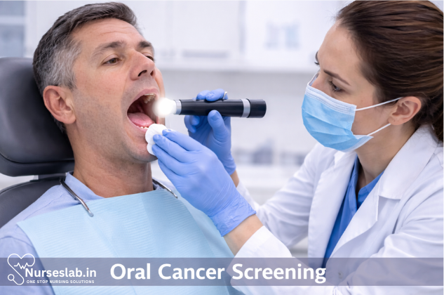
Oral cancer screening involves a visual and physical examination of the mouth, tongue, gums, and throat to detect early signs of cancer or precancerous changes. It helps identify lesions, lumps, and risk factors, supporting early diagnosis and improved outcomes.
Introduction
Oral cancer constitutes a significant public health concern worldwide, particularly in regions with high prevalence of tobacco and betel nut usage. In India, oral cancer accounts for a substantial proportion of all malignancies, with thousands of new cases diagnosed annually. Early detection remains pivotal in improving survival rates and reducing morbidity associated with this disease.
Importance of Oral Cancer Screening
Impact on Patient Outcomes
Screening for oral cancer allows for identification of pre-malignant lesions and early-stage malignancies, which are often amenable to less invasive therapies and have significantly better prognoses compared to advanced stages. The five-year survival rate for early-detected oral cancer can be as high as 80–85%, whereas late-stage detection drops survival rates below 50%. Routine screening in clinical practice can thus directly impact patient morbidity and mortality.
Risk Factors for Oral Cancer
- Tobacco Use: Smoking and smokeless tobacco are leading contributors to oral carcinogenesis.
- Alcohol Consumption: Excessive alcohol intake synergistically increases risk, especially when combined with tobacco.
- Betel Nut Chewing: Common in South Asia, areca nut compounds are carcinogenic.
- Human Papillomavirus (HPV): Increasingly recognised as a risk factor, particularly for oropharyngeal cancers.
- Chronic Trauma: Poorly fitting dentures, sharp teeth, and chronic mucosal irritation can predispose to malignant changes.
- Poor Oral Hygiene and Nutritional Deficiencies: Vitamin A and iron deficiencies have been implicated.
Understanding these risk factors is essential for targeted screening and patient education.
Diagnostic Methods in Oral Cancer Screening
Visual Examination
The cornerstone of oral cancer screening is a thorough visual inspection of the oral cavity. This includes assessment of the lips, buccal mucosa, tongue (dorsal, lateral, and ventral surfaces), floor of mouth, hard and soft palate, retromolar trigone, and oropharynx. Lesions suspicious for malignancy may appear as erythroplakia (red patches), leukoplakia (white patches), ulcerations, indurations, exophytic growths, or non-healing sores. Good illumination and use of mouth mirrors are essential for comprehensive evaluation.
Palpation
Palpation complements visual examination by detecting submucosal masses, indurations, and lymphadenopathy. Bimanual palpation of the floor of the mouth and tongue helps identify deep lesions, while examination of cervical lymph nodes is vital for staging and prognostication.
Adjunctive Techniques
- Toluidine Blue Staining: A vital dye that selectively stains dysplastic and neoplastic tissues, enhancing lesion visibility. The procedure involves rinsing the oral cavity, applying the dye, and assessing retention in suspicious areas.
- Brush Biopsy: A minimally invasive method using a cytology brush to collect transepithelial cellular samples from lesions for cytological analysis. Particularly useful for non-homogeneous or large lesions where scalpel biopsy may be impractical.
- Autofluorescence Devices: Instruments such as VELscope use tissue autofluorescence to identify abnormal mucosa, which appears as loss of fluorescence compared to healthy tissue.
- Chemiluminescence: Devices like Vizilite use acetic acid and chemiluminescent light to highlight abnormal epithelial changes.
Imaging Modalities
- Intraoral Radiography: Useful for evaluating bone involvement in advanced lesions.
- Ultrasound: Assists in assessing tumour depth and lymph node involvement.
- CT and MRI Scans: Provide detailed anatomical information, crucial for staging and surgical planning.
- PET Scans: Employed in metastatic work-up and monitoring treatment response.
Step-by-Step Screening Procedure
1. Patient History
- Obtain a comprehensive medical and dental history, focusing on risk factors such as tobacco, alcohol, betel nut use, prior oral lesions, family history of cancer, and symptoms like persistent ulcers, pain, dysphagia, or hoarseness.
- Enquire about duration and progression of symptoms.
2. Clinical Examination
- Inspect the face, neck, and lips for asymmetry, swelling, or lesions.
- Conduct a systematic intraoral examination using a mouth mirror and adequate lighting.
- Palpate all accessible areas for masses, induration, tenderness, or fixation.
- Examine cervical lymph nodes for enlargement, consistency, mobility, and tenderness.
3. Use of Diagnostic Aids
- Apply adjunctive techniques (toluidine blue, autofluorescence, chemiluminescence) to highlight suspicious mucosal changes.
- Employ brush biopsy for cytological sampling when appropriate.
4. Biopsy Process
Definitive diagnosis of oral cancer requires histopathological confirmation. The steps include:
- Selection of Biopsy Site: Choose the most representative area, avoiding necrotic or inflamed regions.
- Preparation: Ensure aseptic technique. For incisional biopsy, local anaesthesia is administered.
- Biopsy Procedure: Using a scalpel, excise a wedge of tissue including normal and abnormal mucosa. For small lesions, excisional biopsy (complete removal) may be performed.
- Handling and Labelling: Place specimen in 10% formalin, label appropriately, and send to the pathology laboratory.
Interpretation of Results
Criteria for Positive Findings
- Histopathology: Diagnosis is based on identification of dysplasia, carcinoma in situ, or invasive squamous cell carcinoma.
- Cytology: Brush biopsy or exfoliative cytology may reveal atypical cells, but histology remains the gold standard.
- Imaging: Evidence of bone erosion, soft tissue extension, or lymph node metastasis supports diagnosis and staging.
Differential Diagnosis
Several benign and malignant conditions mimic oral cancer, necessitating careful differential diagnosis:
- Chronic traumatic ulcers
- Lichen planus
- Leukoplakia (non-dysplastic)
- Infective lesions (e.g., tuberculosis, syphilis)
- Benign tumours (fibroma, papilloma)
Correlation of clinical, cytological, and histopathological findings is crucial for accurate diagnosis.
Follow-Up Protocols
- Patients with positive findings should be referred to oncology or oral surgery for further management, including staging, treatment planning, and multidisciplinary care.
- Negative or equivocal findings warrant periodic re-examination, especially in high-risk individuals.
- Documentation of lesion characteristics, photographs, and ongoing monitoring are recommended for all suspicious cases.
Recent Advancements in Oral Cancer Diagnostics
New Technologies
- Molecular Diagnostics: Techniques such as PCR, FISH, and next-generation sequencing identify genetic mutations and biomarkers associated with oral cancer, enabling early detection and personalised therapy.
- Salivary Biomarker Analysis: Non-invasive testing of saliva for proteins, DNA, and RNA markers shows promise for screening and monitoring.
- AI-Assisted Screening: Artificial intelligence algorithms are being developed to analyse clinical images and cytology slides, improving diagnostic accuracy and standardisation.
- Liquid Biopsy: Circulating tumour DNA and exosomes in blood or saliva can be detected using sensitive assays.
- Optical Coherence Tomography (OCT): This imaging modality provides high-resolution, real-time imaging of oral tissues, aiding in differentiation between benign and malignant changes.
Improvements in Adjunctive Techniques
- Enhanced autofluorescence devices with better specificity.
- Development of multiplex chemiluminescence for simultaneous detection of multiple biomarkers.
- Integration of telemedicine platforms for remote assessment and triage, especially in underserved areas.
Challenges and Limitations
Barriers to Effective Screening
- Lack of public awareness and education regarding oral cancer risk factors and symptoms.
- Insufficient training of primary healthcare providers in recognising early lesions.
- Resource constraints in rural and low-income settings, limiting access to advanced diagnostics.
- Social stigma associated with oral cancers, particularly in women and younger patients.
False Positives and Negatives
Adjunctive techniques, while useful, can sometimes yield false positive or false negative results, leading to unnecessary anxiety or missed diagnoses. Toluidine blue, for instance, may stain inflammatory lesions, while autofluorescence can be affected by pigmentation or vascularity. Histopathology remains essential for definitive diagnosis.
Limitations of Current Practices
- Reliance on clinical experience, which may vary among practitioners.
- Lack of universally accepted screening protocols.
- Limited availability of molecular diagnostic tests in routine practice.
Nursing Care of Patients Undergoing Oral Cancer Screening Procedure
Nurses are at the frontline of patient care during these screenings, providing education, emotional support, clinical assistance, and post-procedure guidance.
2. Role of the Nurse in Oral Cancer Screening
Nurses play a multifaceted role in oral cancer screening, acting as educators, advocates, care coordinators, and clinical assistants. Their responsibilities include patient assessment, education, procedural support, documentation, and psychological care. Effective nursing care ensures patient safety, comfort, and understanding throughout the screening process.
Pre-Procedure Nursing Care
2.1 Patient Assessment
- Medical and Dental History:
- Obtain a thorough medical and dental history, including risk factors such as tobacco use, alcohol consumption, family history of cancer, prior oral lesions, and exposure to human papillomavirus (HPV).
- Current Medications and Allergies:
- Document all current medications and known allergies to anticipate potential interactions or adverse reactions during the procedure.
- Baseline Vital Signs:
- Record baseline vital signs (blood pressure, pulse, temperature, respiration rate) to detect any changes during or after the procedure.
- Oral Assessment:
- Perform a preliminary oral examination to identify visible lesions, ulcers, or abnormalities and document findings for comparison post-procedure.
2.2 Patient Education and Preparation
- Explaining the Procedure:
- Provide a clear, jargon-free explanation of the screening process, its purpose, steps involved, and any expected sensations or discomforts. Use visual aids or models if available.
- Addressing Concerns and Anxiety:
- Encourage patients to express their fears or concerns. Offer reassurance and answer questions honestly to alleviate anxiety.
- Consent:
- Ensure informed consent is obtained, explaining risks, benefits, and alternatives. Verify patient understanding before proceeding.
- Pre-procedure Instructions:
- Advise patients on any necessary fasting, oral hygiene measures, or medication adjustments required prior to the screening.
2.3 Environmental and Equipment Preparation
- Privacy and Comfort:
- Prepare a private, well-lit, and clean environment. Ensure the patient is comfortably seated and draped appropriately.
- Equipment Check:
- Gather necessary equipment (gloves, tongue depressors, mirrors, lights, adjunctive diagnostic tools, biopsy kits if indicated) and verify functionality and sterility.
Intra-Procedure Nursing Care
3.1 Assisting the Clinician
- Instrument Handling:
- Assist the dentist or physician by passing instruments, preparing adjunctive devices, and ensuring a smooth workflow.
- Patient Support:
- Monitor the patient’s comfort and reassure them throughout the examination. Address complaints of discomfort promptl
- Observation and Documentation:
- Document findings meticulously, noting the location, size, color, and texture of any lesions. Record any interventions performed.
3.2 Monitoring for Complications
- Vasovagal Response:
- Be alert for signs of fainting or dizziness, especially in anxious patients. Take appropriate action if symptoms occur (e.g., lower the head, provide oxygen if needed).
- Allergic Reactions:
- Monitor for signs of allergic reactions, particularly if dyes or topical anesthetics are used. Be prepared to initiate emergency protocols if necessary.
- Bleeding:
- If a biopsy is performed, watch for excessive bleeding and manage it according to protocol (applying pressure, using hemostatic agents).
3.3 Infection Control
- Hand Hygiene:
- Adhere strictly to hand hygiene protocols before and after patient contact.
- Personal Protective Equipment (PPE):
- Use appropriate PPE (gloves, masks, protective eyewear) to prevent cross-contamination.
- Disinfection and Sterilization:
- Ensure all instruments are properly sterilized before use and that the environment is disinfected after the procedure.
Post-Procedure Nursing Care
4.1 Immediate Post-Procedure Assessment
- Vital Signs:
- Reassess and document vital signs, comparing them with baseline values.
- Observation:
- Monitor for delayed adverse reactions such as bleeding, swelling, or allergic responses, especially if a biopsy was performed.
4.2 Patient Education and Discharge Instructions
- Results Communication:
- Explain when and how the patient will receive screening results, and the significance of any findings. If a biopsy was performed, clarify the expected timeline for pathology results.
- Oral Care Instructions:
- Provide instructions on maintaining oral hygiene, avoiding irritants (e.g., tobacco, alcohol, spicy foods), and recognizing signs of infection or complications.
- Activity Restrictions:
- Advise on any necessary activity modifications, especially if tissue sampling was done (e.g., avoiding hard or hot foods until healing occurs).
- Follow-Up Appointments:
- Schedule and emphasize the importance of follow-up visits for result review, further diagnostic workup, or monitoring of lesions.
- Emergency Instructions:
- Instruct patients to seek immediate care if they experience severe bleeding, difficulty breathing, fever, or other concerning symptoms post-procedure.
4.3 Emotional and Psychological Support
- Counseling:
- Recognize that the possibility of cancer can cause significant anxiety. Offer supportive counseling and, if needed, referrals to mental health professionals or support groups.
- Family Involvement:
- Encourage family participation in care and education, as appropriate, to provide a support system for the patient.
Special Considerations
5.1 Culturally Sensitive Care
Respect cultural beliefs and practices that may influence the patient’s perception of cancer, health, and medical procedures. Use professional interpreters when language barriers exist and provide educational materials in the patient’s preferred language where possible.
5.2 Care for Vulnerable Populations
- Elderly Patients:
- Be mindful of comorbidities, cognitive impairment, and medication polypharmacy that may affect screening and follow-up care.
- Patients with Disabilities:
- Modify approaches to accommodate physical or cognitive limitations, ensuring accessibility and understanding.
5.3 Documentation and Legal Considerations
- Accurate Record-Keeping:
- Document all assessments, patient education, consent, procedural details, and post-procedure instructions meticulously.
- Confidentiality:
- Maintain strict confidentiality of patient information in line with legal and institutional policies.
REFERENCES
- Cancer Facts & Figures 2019. American Cancer Society. https://www.cancer.org/research/cancer-facts-statistics/all-cancer-facts-figures/cancer-facts-figures-2019.html.
- National Cancer Institute. Oral Cavity, Pharyngeal, and Laryngeal Cancer Screening (PDQ®)-Patient Version https://www.cancer.gov/types/head-and-neck/patient/oral-screening-pdq#section/_24.
- Lingen MW, et al. Evidence-based clinical practice guideline for the evaluation of potentially malignant disorders in the oral cavity. Journal of the American Dental Association. 2018; doi:10.1016/j.adaj.2017.07.032.
- The Oral Cancer Foundation. Cancer screening protocols https://oralcancerfoundation.org/discovery-diagnosis/cancer-screening-protocols/.
- Stefanac SJ, et al., eds. Patient evaluation and assessment. In: Diagnosis and Treatment Planning in Dentistry. 3rd ed. Elsevier; 2017. https://www.clinicalkey.com.
- The Oral Cancer Foundation. Oral Cancer Facts (https://oralcancerfoundation.org/facts/).
- Warnakulasuriya S, Kerr AR. Oral Cancer Screening: Past, Present, and Future (https://www.ncbi.nlm.nih.gov/pmc/articles/PMC8529297/). J Dent Res. 2021 Nov;100(12):1313-1320.
Stories are the threads that bind us; through them, we understand each other, grow, and heal.
JOHN NOORD
Connect with “Nurses Lab Editorial Team”
I hope you found this information helpful. Do you have any questions or comments? Kindly write in comments section. Subscribe the Blog with your email so you can stay updated on upcoming events and the latest articles.


