


Introduction
A liver biopsy is a medical procedure in which a small sample of liver tissue is removed for laboratory examination. This procedure plays a crucial role in diagnosing, assessing, and managing a variety of liver diseases, including hepatitis, fatty liver disease, cirrhosis, and liver cancer. As the liver performs essential metabolic, detoxification, and synthetic functions in the body, accurate diagnosis of its pathology is vital for effective treatment and prognosis.
Indications for Liver Biopsy
There are several reasons a healthcare provider might recommend a liver biopsy, including:
- Diagnosis of unexplained liver abnormalities: When blood tests, imaging, or clinical symptoms indicate liver dysfunction without a clear cause, biopsy provides definitive information.
- Assessment of disease severity: For chronic liver conditions like hepatitis B and C, nonalcoholic fatty liver disease (NAFLD), or autoimmune hepatitis, biopsy helps determine the extent of inflammation and scarring (fibrosis).
- Evaluation of masses or tumors: Biopsy can distinguish benign from malignant lesions and guide further management.
- Monitoring liver transplant health: In transplant recipients, biopsy may detect rejection, infection, or recurrence of original disease.
- Unexplained jaundice or hepatomegaly: When other investigations are inconclusive, a biopsy can clarify the diagnosis.
- Drug-induced liver injury: Suspected adverse reactions to medications can be confirmed and characterized by histological analysis.
Types of Liver Biopsy
There are several approaches to obtaining a liver tissue sample:
- Percutaneous (needle) biopsy: The most common technique, where a needle is inserted through the skin and into the liver to obtain tissue.
- Transjugular biopsy: The biopsy needle is introduced through a large vein in the neck (the jugular vein), a method used when patients have bleeding risks or ascites.
- Laparoscopic (surgical) biopsy: Performed during minimally invasive or open surgery, often when the liver is being directly visualized for another reason.
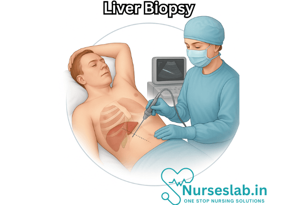
Percutaneous Liver Biopsy
This is the standard and most frequently performed method. The patient lies on their back or slightly on the left side. After identifying the biopsy site, usually in the right upper abdomen below the ribs, the area is cleaned and numbed with local anesthetic. Under ultrasound or CT guidance (for precision and safety), a special biopsy needle is inserted, and a small core of liver tissue is quickly removed.
Transjugular Liver Biopsy
This approach is reserved for patients with bleeding disorders, significant ascites, or those at high risk of complications. A catheter is inserted into the jugular vein in the neck, guided through the venous system into the hepatic veins. A biopsy needle is then passed through the catheter into the liver tissue. This technique avoids puncturing the liver capsule, reducing bleeding risk.
Laparoscopic Liver Biopsy
Laparoscopic biopsy is performed in the operating room under general anesthesia, usually when patients are already undergoing surgery for another reason (e.g., investigation of abdominal pain, suspected tumors). Small incisions and a camera allow direct visualization of the liver surface, from which a tissue sample is taken.
Pre-procedural Considerations and Preparation
Adequate preparation is essential to minimize risks and ensure the procedure’s success.
- Medical history and examination: The provider will review the patient’s history, medications, allergies, and prior bleeding tendencies.
- Blood tests: These check clotting parameters (INR, PT, aPTT, platelet count) to reduce bleeding risk.
- Imaging: Ultrasound or CT scans may be performed to map the liver’s size, position, and any abnormal areas.
- Medication adjustments: Blood thinners or antiplatelet agents may need to be stopped several days before the procedure.
- Fasting: Patients are generally asked to fast for 6–8 hours before the procedure.
The Biopsy Procedure
Step-by-Step Outline (Percutaneous Approach)
- Positioning: The patient lies supine with the right arm above the head to widen spaces between ribs.
- Site identification: The biopsy site is marked, often using ultrasound guidance for accuracy and safety.
- Sterilization and anesthesia: The skin is cleaned with antiseptic, and local anesthetic is injected to numb the area.
- Needle insertion: The physician makes a small incision and inserts the biopsy needle between the ribs into the liver.
- Sample collection: The needle is advanced into the liver, a core sample is taken, and the needle is withdrawn. Multiple samples may be collected if needed.
- Pressure and dressing: Gentle pressure is applied to the site to minimize bleeding, and a sterile bandage is placed.
- Recovery: The patient is monitored for a few hours (typically 2–4) for signs of bleeding or complications.
Post-procedure Care and Recovery
- Patients are monitored in a recovery area for several hours, with blood pressure, pulse, and oxygen levels checked regularly.
- Pain at the biopsy site or right shoulder is common and usually mild, managed with over-the-counter pain medicines.
- Avoid strenuous activity, heavy lifting, or vigorous exercise for at least 1–2 days.
- If there are signs of complications such as severe pain, shortness of breath, fainting, or persistent bleeding, immediate medical attention is required.
Risks and Complications
Although generally safe, a liver biopsy carries some risks:
- Pain: The most common side effect, usually brief and mild.
- Bleeding: Occurs in less than 1% of cases, but can be serious and may require intervention.
- Infection: Rare, but possible at the biopsy site or internally.
- Injury to organs: Accidental puncture of the lung, gallbladder, or other organs is uncommon, especially with image-guided techniques.
- Reaction to anesthesia: Allergic or adverse reactions, though extremely rare.
- Formation of arteriovenous fistula: Rarely, abnormal connections between blood vessels can develop.
Interpretation of Results
The liver tissue obtained is sent to a pathology laboratory, where it is processed, stained, and examined under a microscope. The pathologist evaluates the sample for:
- Degree and type of inflammation
- Fibrosis (scarring) stage and distribution
- Presence of fat, iron, or copper accumulation
- Tumor cells or abnormal growths
- Specific features of certain diseases (e.g., viral inclusions, granulomas)
Findings guide diagnosis, staging, and management, and in some cases, help determine prognosis.
Alternatives to Liver Biopsy
As technology has advanced, several non-invasive methods can provide information about liver health:
- Elastography: Ultrasound-based tests like FibroScan assess liver stiffness, correlating with fibrotic changes.
- Blood biomarker panels: Laboratory tests can estimate fibrosis or inflammation (e.g., FibroTest, APRI, FIB-4 Index).
- Imaging: MRI and CT scans can sometimes evaluate certain liver conditions, though they cannot match biopsy’s diagnostic precision for many diseases.
However, biopsy remains the gold standard when a definitive tissue diagnosis is necessary.
Nursing Care of Patients Undergoing Liver Biopsy Procedure
The procedure involves significant risks and requires meticulous nursing care before, during, and after the biopsy to ensure patient safety, comfort, and optimal outcomes.
Pre-Procedure Nursing Care
Nursing professionals play a critical role in preparing patients for liver biopsy, aiming to minimize risks and alleviate anxiety.
Patient Education and Consent
- Explain the Procedure: Clearly describe the steps of the liver biopsy, its purpose, and expected outcomes. Use simple language and visual aids if necessary.
- Discuss Risks and Benefits: Inform the patient about potential risks (bleeding, infection, pain, accidental injury to surrounding organs) and benefits of the procedure.
- Obtain Informed Consent: Ensure the patient (or legal guardian) signs the consent form after all questions have been addressed.
Pre-Procedure Assessment
- Review Medical History: Check for bleeding disorders, liver function, allergies, and medications, especially anticoagulants and antiplatelets.
- Laboratory Investigations: Verify recent results for complete blood count, coagulation profile (PT, INR, aPTT), liver function tests, and viral markers.
- Physical Examination: Assess for signs of ascites and check vital signs.
Patient Preparation
- Fasting Requirements: Instruct the patient to fast for at least 6 hours prior to the procedure to reduce the risk of aspiration.
- Medication Adjustment: Consult with the physician regarding temporary discontinuation of anticoagulants or antiplatelet agents.
- Intravenous Access: Ensure a patent IV line for possible administration of sedatives or emergency medications.
- Bladder Management: Advise the patient to empty their bladder before the procedure.
- Skin Preparation: Cleanse the biopsy site with an antiseptic solution and remove any jewelry or metallic objects.
Psychological Support
- Alleviating Anxiety: Offer reassurance and address the patient’s fears, encouraging them to voice concerns.
- Family Involvement: Allow family members to be present for support, if appropriate and permitted.
Intra-Procedure Nursing Care
Assisting the Physician
- Maintain Asepsis: Help set up a sterile field and ensure all equipment is sterile and functioning.
- Monitor Vital Signs: Continuously observe the patient’s heart rate, blood pressure, respiration, and oxygen saturation.
- Patient Positioning: Assist the patient into a supine or left lateral position, with the right arm above the head to widen the intercostal spaces for easy access.
- Support During Procedure: Offer verbal comfort and guidance to help the patient remain still.
- Immediate Response: Be ready to manage adverse reactions such as pain, vasovagal response, or sudden bleeding.
Specimen Handling
- Labeling and Transport: Properly label the specimen container and promptly deliver it to the laboratory for analysis.
- Documentation: Record the time, site, number of attempts, and type of needle used in the patient’s chart.
Post-Procedure Nursing Care
Monitoring and Observation
- Vital Signs: Monitor the patient’s blood pressure, pulse, respiration, and oxygen saturation every 15 minutes for the first 2 hours, then hourly for the next 4-6 hours as per protocol.
- Bleeding and Hemorrhage: Observe for signs of internal bleeding (tachycardia, hypotension, pallor, abdominal pain, distension, restlessness). Inspect the biopsy site for bleeding, hematoma, or swelling.
- Pain Assessment: Evaluate pain intensity and location, administer prescribed analgesics, and observe for referred pain to the right shoulder (indicating possible peritoneal irritation).
- Respiratory Status: Watch for signs of respiratory distress due to pneumothorax or pain.
Patient Positioning and Activity
- Bed Rest: Instruct the patient to remain on bed rest for 4-6 hours after the procedure, preferably lying on the right side to apply pressure and minimize bleeding.
- Gradual Activity: Advise against vigorous activity or heavy lifting for the next 24 hours.
Diet and Hydration
- Resume Fluids: Allow sips of water after the procedure, progressing to a normal diet if no complications arise.
- Nausea Management: Administer antiemetics if needed and monitor for vomiting, which could indicate complications.
Complication Surveillance
- Hemorrhage: Be alert for sudden abdominal pain, drop in blood pressure, or shock. Prepare to initiate emergency response if indicated.
- Infection: Observe for fever, chills, redness, or drainage at the biopsy site.
- Pneumothorax: Watch for sudden breathlessness, chest pain, or decreased breath sounds, and promptly report to the physician.
- Biliary Peritonitis: Assess for symptoms such as abdominal distension, jaundice, or severe pain, and report findings.
Discharge Planning and Patient Education
Instructions for Home Care
- Wound Care: Keep the biopsy site clean and dry. Report any excessive bleeding, drainage, or signs of infection.
- Activity Restrictions: Avoid strenuous exercise and heavy lifting for at least 24-48 hours.
- Medication: Take prescribed medications as directed, and do not resume anticoagulant therapy without consulting the healthcare provider.
- Symptoms to Report: Advise the patient to seek immediate medical attention for severe pain, breathlessness, persistent vomiting, or signs of shock.
Follow-Up Care
- Appointment Scheduling: Arrange for follow-up visits to review biopsy results and monitor recovery.
- Lab Tests: Inform about the need for repeat blood tests or imaging if complications are suspected.
Documentation
- Accurate Records: Document pre-procedure assessment, intra-procedure observations, post-procedure monitoring, patient education, and discharge instructions.
- Incident Reporting: Record any complications or adverse events promptly and thoroughly.
Special Considerations
Pediatric and Geriatric Patients
- Pediatric Care: Use age-appropriate communication, comfort measures, and parental involvement. Monitor closely for anxiety and complications.
- Geriatric Care: Be aware of increased risk of bleeding, comorbidities, and medication interactions. Monitor for confusion and falls.
Patients with Coagulopathies
- Correcting Coagulation Disorders: Administer fresh frozen plasma or vitamin K as prescribed to reduce bleeding risk.
- Postpone Procedure: If clotting parameters are not within safe limits, the procedure may be postponed.
REFERENCES
- Chowdhury AB, et al. Liver biopsy for assessment of chronic liver diseases: A synopsis. Clinical and Experimental Medicine. 2023; doi:10.1007/s10238-022-00799-z.
- National Institute of Diabetes and Digestive and Kidney Diseases (U.S.). Liver Biopsy. https://www.niddk.nih.gov/health-information/diagnostic-tests/liver-biopsy.
- Radiological Society of North America. Liver Biopsy. https://www.radiologyinfo.org/en/info/liver-biopsy.
- Liver biopsy. National Institute of Diabetes and Digestive and Kidney Diseases. https://www.niddk.nih.gov/health-information/diagnostic-tests/liver-biopsy.
- Pandey N, Hoilat GJ, John S. Liver Biopsy. [Updated 2023 Jul 24]. In: StatPearls [Internet]. Treasure Island (FL): StatPearls Publishing; 2025 Jan-. Available from: https://www.ncbi.nlm.nih.gov/books/NBK470567/
Stories are the threads that bind us; through them, we understand each other, grow, and heal.
JOHN NOORD
Connect with “Nurses Lab Editorial Team”
I hope you found this information helpful. Do you have any questions or comments? Kindly write in comments section. Subscribe the Blog with your email so you can stay updated on upcoming events and the latest articles.


