


A nuclear stress test evaluates blood flow to the heart at rest and during exercise using radioactive tracers and advanced imaging. It helps diagnose coronary artery disease, assess chest pain, and guide treatment decisions for cardiac patients.
Introduction
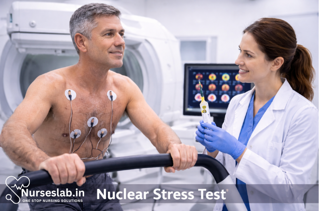
A nuclear stress test is a specialized diagnostic procedure used to evaluate the blood flow to the heart muscle both at rest and during physical exertion or pharmacologically-induced stress. It is an integral part of the non-invasive assessment of coronary artery disease (CAD) and other cardiovascular conditions.
Background and Rationale
Cardiovascular diseases, particularly coronary artery disease, remain the leading cause of morbidity and mortality worldwide. Early detection and accurate assessment of myocardial perfusion (the flow of blood to the heart muscle) are crucial for effective management and prevention of adverse cardiac events. Traditional stress tests, such as the exercise electrocardiogram (ECG), provide valuable information but may be limited in sensitivity and specificity. Nuclear stress testing enhances diagnostic accuracy by directly visualizing areas of reduced blood flow.
Historical Perspective
Nuclear cardiology emerged in the mid-20th century with the advent of radioactive tracers capable of highlighting physiological processes in the body. Over decades, the technology has evolved, with improvements in imaging agents, gamma cameras, and image processing techniques, making nuclear stress testing a cornerstone of modern cardiology.
Principles of Nuclear Stress Testing
The nuclear stress test is based on the principle of myocardial perfusion imaging (MPI). A small amount of radioactive tracer, such as technetium-99m sestamibi or thallium-201, is injected into the bloodstream. The tracer emits gamma rays, which are detected by a special camera (gamma camera or SPECT scanner) to create detailed images of the heart muscle. By comparing images taken at rest and during stress, clinicians can identify regions with reduced perfusion, suggestive of ischemia or infarction.
Types of Nuclear Stress Tests
- Exercise Nuclear Stress Test: The patient performs graded exercise (usually on a treadmill or stationary bicycle), simulating real-life physical activity. The radioactive tracer is injected at peak exercise.
- Pharmacological Nuclear Stress Test: For patients unable to exercise adequately, drugs such as adenosine, dipyridamole, or regadenoson are used to mimic the effects of exercise by dilating coronary arteries or increasing heart workload.
Indications for Nuclear Stress Testing
Nuclear stress tests are ordered for a variety of clinical scenarios, including:
- Diagnosis of suspected coronary artery disease in symptomatic patients (e.g., chest pain or dyspnea of uncertain origin)
- Risk stratification following myocardial infarction or in those with known CAD
- Assessment of myocardial viability before revascularization procedures
- Evaluation of the effectiveness of therapeutic interventions (medical management, angioplasty, or bypass surgery)
- Preoperative assessment before non-cardiac surgery in high-risk individuals
Preparation for the Test
Proper patient preparation is essential for accurate and safe nuclear stress testing. Key steps include:
- Medical History and Consent: The clinician reviews the patient’s medical history, current medications, allergies, and obtains informed consent, explaining the risks and benefits of the procedure.
- Medication Adjustments: Certain medications, such as beta-blockers, calcium channel blockers, or nitrates, may need to be withheld prior to the test, as directed by the physician.
- Fasting and Caffeine Restriction: Patients are usually instructed to fast for 4-6 hours before the test and to avoid caffeine-containing products for at least 24 hours, as caffeine can interfere with pharmacologic agents used during the test.
- Clothing and Accessories: Comfortable clothing and shoes suitable for exercise are recommended. Metal objects (jewelry, belts) should be removed to prevent interference with imaging.
Test Procedure: Step-by-Step
The nuclear stress test typically consists of two main phases: resting imaging and stress imaging. The sequence may vary depending on the protocol used (e.g., rest-stress or stress-rest).
1. Resting Phase
- The patient arrives at the nuclear medicine department, and baseline vital signs are recorded.
- An intravenous (IV) line is established, and the radioactive tracer is injected while the patient is at rest.
- After a waiting period (typically 15-60 minutes, depending on the tracer), the patient is positioned under the gamma camera, and images of the heart are acquired.
2. Stress Phase
- Exercise Protocol: The patient begins exercising on a treadmill or stationary bicycle with gradually increasing intensity, monitored by ECG, blood pressure, and symptoms. At peak exercise, the tracer is injected, and the patient continues exercising for a short period to ensure proper circulation of the tracer.
- Pharmacologic Protocol: If physical exercise is not feasible, a vasodilator or inotropic agent is administered intravenously to simulate the effects of exercise. The tracer is injected at the peak effect of the drug.
- After the stress phase, the patient undergoes a second set of images.
3. Post-Test Monitoring
- The patient is observed for a short period to monitor for adverse reactions or delayed symptoms.
- Normal activity can usually be resumed shortly after the test, unless otherwise advised.
Imaging Techniques
The most common imaging modality used in nuclear stress testing is single-photon emission computed tomography (SPECT). In some centers, positron emission tomography (PET) may be used for higher resolution and quantitative measurements. The choice of technique depends on clinical indications, availability, and patient-specific factors.
Radioactive Tracers
- Technetium-99m Sestamibi: Preferred for its favorable imaging characteristics and lower radiation dose.
- Thallium-201: Useful for assessing myocardial viability, but less commonly used due to higher radiation exposure.
- Rubidium-82 or Ammonia (PET tracers): Employed in centers with PET capability for superior image quality.
Interpretation of Results
The acquired images are analyzed by a nuclear cardiologist or radiologist. The heart is divided into segments, and the distribution of tracer uptake is evaluated to identify areas of reduced perfusion.
Key Findings
- Normal Perfusion: Uniform tracer uptake in all regions, indicating adequate blood flow.
- Reversible Ischemia: Areas with reduced tracer uptake during stress but normal uptake at rest, suggesting reversible coronary artery narrowing.
- Fixed Defect: Areas with reduced uptake both at rest and stress, consistent with myocardial scar or infarction.
- Attenuation Artifacts: False positive or negative findings due to factors such as breast tissue, obesity, or diaphragmatic interference.
Quantitative Analysis
Advanced software allows quantitative assessment of perfusion defects, ejection fraction, and ventricular volumes, providing valuable prognostic information.
Clinical Applications and Significance
Nuclear stress testing plays a vital role in various clinical scenarios:
- Diagnosis of CAD: It helps confirm or exclude the presence of significant coronary artery disease in patients with symptoms or abnormal ECG findings.
- Risk Stratification: The extent and severity of perfusion abnormalities correlate with the risk of future cardiac events, guiding management decisions.
- Assessment of Myocardial Viability: Differentiates between viable and non-viable myocardium, influencing the choice of revascularization strategies.
- Therapeutic Monitoring: Evaluates the impact of medical therapy, angioplasty, or coronary artery bypass grafting on myocardial perfusion.
- Preoperative Evaluation: Assesses cardiac risk in patients undergoing major non-cardiac surgery.
Risks and Safety Considerations
Nuclear stress testing is generally safe, but like any medical procedure, it carries certain risks:
- Radiation Exposure: The amount of radiation is low and considered safe for most patients, but cumulative exposure should be considered, especially in younger individuals or those requiring repeated studies.
- Allergic Reactions: Rare, but possible with tracer injection.
- Exercise-Related Complications: Such as arrhythmias, chest pain, or, rarely, heart attack, especially in high-risk patients.
- Pharmacologic Side Effects: Vasodilators can cause flushing, headache, palpitations, or rarely, bronchospasm or hypotension.
Patients are carefully screened and monitored throughout the test to minimize complications.
Limitations of Nuclear Stress Testing
While highly valuable, the nuclear stress test has certain limitations:
- Attenuation Artifacts: Soft tissue interference can lead to false readings, requiring careful interpretation and, sometimes, additional imaging.
- Limited Sensitivity in Microvascular Disease: May not detect small-vessel or microvascular dysfunction.
- Radiation Exposure: Although low, it remains a consideration, especially in certain populations.
- Availability and Cost: Requires specialized equipment and personnel, which may limit access in some regions.
Recent Advances and Future Directions
Ongoing research and technological innovations continue to enhance the utility of nuclear stress testing. Developments include:
- Hybrid Imaging: Combining SPECT or PET with computed tomography (CT) for anatomical and functional assessment.
- New Tracers: Development of novel radiopharmaceuticals for improved image quality and lower radiation dose.
- Quantitative Perfusion Analysis: Automated software for objective measurement of blood flow and perfusion reserve.
- Artificial Intelligence: Use of AI algorithms to assist in image interpretation and risk prediction.
Nursing Care of the Patient Undergoing Nuclear Stress Testing Procedure
Nurses play a pivotal role in ensuring patient safety, comfort, and optimal outcomes before, during, and after the procedure. This guide provides a detailed overview of the nursing responsibilities and interventions involved in caring for patients undergoing nuclear stress testing.
Pre-Procedure Nursing Care
Patient Assessment
A thorough pre-procedure assessment is essential for identifying potential risks and optimizing patient preparation. The nurse should:
- Review the patient’s medical history, including cardiac history, allergies (especially to contrast agents or medications), and previous test results.
- Assess current medications, focusing on beta-blockers, calcium channel blockers, nitrates, and caffeine-containing drugs, as these can affect test outcomes.
- Evaluate baseline vital signs and cardiac status (heart rate, rhythm, blood pressure, oxygen saturation).
- Check for contraindications, such as pregnancy, breastfeeding, or renal impairment.
Patient Education and Preparation
Nurses are responsible for ensuring patients are well-informed and properly prepared for the procedure:
- Explain the purpose, steps, and duration of the nuclear stress test in understandable terms.
- Discuss the use of radioactive tracers, addressing safety and radiation exposure concerns.
- Instruct the patient to fast (usually 2–4 hours prior) and avoid caffeine, nicotine, and certain medications as directed by the physician.
- Advise on comfortable clothing and appropriate footwear for exercise-based tests.
- Reassure patients about potential side effects (e.g., flushing, chest discomfort) and the availability of immediate medical support if needed.
- Obtain informed consent and ensure all documentation is complete.
Physical Preparation
- Establish intravenous access for tracer administration or emergency medication.
- Ensure all necessary equipment is available and functioning (ECG monitor, blood pressure cuff, emergency cart, defibrillator).
- Remove any metal objects or jewelry that could interfere with imaging.
- Confirm patient identification and allergy status.
Intra-Procedure Nursing Care
Monitoring and Safety
Continuous monitoring and prompt intervention are critical during the nuclear stress test:
- Monitor vital signs (heart rate, blood pressure, respiratory rate, oxygen saturation) at baseline, throughout stress induction, and during recovery.
- Observe ECG for arrhythmias, ischemic changes, or conduction disturbances.
- Assess for signs of adverse reactions to stress agents (e.g., bronchospasm, hypotension, chest pain, nausea, flushing).
- Be prepared to initiate emergency protocols if the patient becomes unstable.
- Maintain communication with the patient, offering reassurance and clear instructions throughout.
Tracer Administration and Imaging Support
- Assist in the safe administration of the radioactive tracer, adhering to radiation safety protocols for both patient and staff.
- Position the patient appropriately for imaging, ensuring comfort and minimizing movement.
- Coordinate with the nuclear medicine technologist and physician to ensure timely imaging after tracer injection.
- Document the time and dose of tracer, as well as any notable events or symptoms during the procedure.
Patient Comfort and Psychological Support
Many patients experience anxiety or discomfort during the test. Nursing interventions should include:
- Providing emotional support and reassurance.
- Explaining each step as it occurs to reduce uncertainty.
- Promptly addressing any complaints of pain, dizziness, or shortness of breath.
- Facilitating communication between the patient and the healthcare team.
Post-Procedure Nursing Care
Immediate Monitoring and Assessment
- Continue monitoring vital signs and ECG until the patient is stable and any drug effects have worn off.
- Assess for delayed adverse reactions to stress agents or radioactive tracers, such as allergic responses or arrhythmias.
- Observe the injection site for signs of extravasation or infection.
Patient Education and Discharge Instructions
- Inform the patient that normal activities can typically be resumed soon after the test, unless otherwise directed.
- Advise the patient to drink plenty of fluids to facilitate the elimination of the radioactive tracer from the body.
- Explain any symptoms that should prompt immediate medical attention, such as chest pain, palpitations, severe shortness of breath, or allergic reactions.
- Provide information on when and how test results will be communicated.
- Reinforce the importance of follow-up appointments and adherence to prescribed cardiac care regimens.
Documentation
- Record all observations, interventions, and patient responses throughout the procedure.
- Document tracer administration details, including time, dosage, and any immediate reactions.
- Note any complications and the actions taken.
Special Considerations
Pediatric and Geriatric Populations
- Adjust communication and education strategies to the patient’s cognitive and developmental level.
- Be vigilant for atypical presentations of cardiac symptoms.
- Consider reduced exercise capacity and increased sensitivity to stress agents in the elderly.
Patients with Comorbidities
- Take extra precautions in patients with respiratory diseases, diabetes, or renal impairment.
- Monitor blood glucose closely in diabetic patients, especially if fasting is required.
- Coordinate with other healthcare providers for medication adjustments as needed.
Infection Control and Radiation Safety
- Follow standard precautions for infection control throughout the procedure.
- Adhere to institutional policies and national guidelines for radiation safety, minimizing exposure for both patients and staff.
- Educate patients and families about the low risk of radiation exposure associated with the test.
Complications and Emergency Management
- Be prepared to manage potential complications, such as:
- Arrhythmias (may require antiarrhythmic medications or advanced cardiac life support)
- Chest pain or myocardial infarction (initiate emergency response protocols)
- Hypotension or hypertensive crisis
- Allergic reactions to medications or tracers (administer antihistamines, steroids, or epinephrine as appropriate)
- Bronchospasm (especially with adenosine or dipyridamole; provide bronchodilators if needed)
Ensure all emergency equipment is readily available and functional before starting the procedure.
Maintain competency in basic and advanced cardiac life support techniques.
REFERENCES
- Cardiac nuclear medicine. American College of Radiology. https://www.radiologyinfo.org/en/info/cardinuclear.
- Gopal S, Heston TF, Murphy C. Nuclear Medicine Stress Test (https://www.ncbi.nlm.nih.gov/books/NBK557682/). 2025 Jul 7. In: StatPearls [Internet]. Treasure Island (FL): StatPearls Publishing; 2025 Jan.
- Khalafi S, Ramireddy A, Hendel RC. Exercise and Pharmacologic Stress Testing. In: Heller GV, Hendel RC, eds. Nuclear Cardiology: Practical Applications. 4th ed. McGraw-Hill LLC; 2022.
- Werner RA, et al. The changing face of nuclear cardiology: Guiding cardiovascular care toward molecular medicine. Journal of Nuclear Medicine. 2020; doi:10.2967/jnumed.119.240440.
- Merck Manual, Consumer Version. Radionuclide Imaging of the Heart https://www.merckmanuals.com/home/heart-and-blood-vessel-disorders/diagnosis-of-heart-and-blood-vessel-disorders/radionuclide-imaging-of-the-heart . Last revised 12/2023.
- Merck Manual, Professional Version. Stress Testing https://www.merckmanuals.com/professional/cardiovascular-disorders/cardiovascular-tests-and-procedures/stress-testing. Last revised 12/2023.
Stories are the threads that bind us; through them, we understand each other, grow, and heal.
JOHN NOORD
Connect with “Nurses Lab Editorial Team”
I hope you found this information helpful. Do you have any questions or comments? Kindly write in comments section. Subscribe the Blog with your email so you can stay updated on upcoming events and the latest articles.


