


Last Updated on January 21, 2026 by Nurseslab.in Editorial Team
A renal scan, or kidney scan, is a nuclear medicine test that evaluates kidney function, blood flow, and urinary drainage. It helps diagnose obstruction, scarring, hypertension‑related issues, and differential renal function. Essential in nephrology and diagnostic imaging.
Introduction
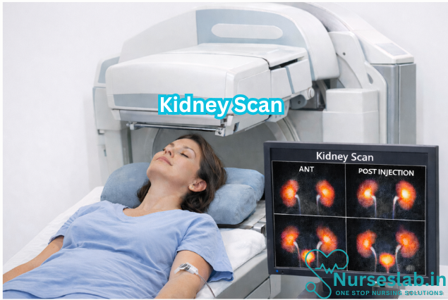
A renal scan, also known as a kidney scan, is a specialised diagnostic imaging procedure used to evaluate the function, structure, and perfusion of the kidneys. Renal scans play a pivotal role in the assessment and management of various renal disorders, offering vital information that complements other clinical and laboratory findings. With advancements in imaging technology, renal scans have become increasingly accurate, minimally invasive, and essential in both adult and paediatric nephrology.
Purpose and Clinical Indications
The primary purpose of a renal scan is to provide functional and anatomical information about the kidneys that may not be apparent on conventional imaging techniques such as ultrasonography or plain radiography. Unlike structural imaging, renal scans can assess renal perfusion, filtration, excretion, and drainage, making them indispensable in diagnosing and managing a range of renal pathologies.
Common Clinical Indications
- Evaluation of Renal Function: Quantification of split renal function, especially in patients with known or suspected unilateral renal disease.
- Detection of Obstruction: Assessment of urinary tract obstruction, especially in cases of hydronephrosis or suspected ureteric obstruction.
- Investigation of Hypertension: Identification of renovascular hypertension, particularly in young patients or those with refractory hypertension.
- Assessment of Renal Transplants: Monitoring perfusion, function, and possible complications in transplanted kidneys.
- Detection of Renal Scarring: Evaluation of chronic pyelonephritis or recurrent urinary tract infections in children.
- Preoperative Planning: Assessment of differential renal function prior to nephrectomy or renal surgery.
- Evaluation of Mass Lesions: Differentiation between functional and non-functional renal masses.
Conditions Commonly Assessed
- Renal artery stenosis
- Obstructive uropathy
- Congenital anomalies (e.g., duplex kidney, ectopic kidney)
- Renal transplant dysfunction
- Chronic kidney disease
- Renal trauma or infarction
Patient Preparation
Proper patient preparation is essential to ensure the accuracy and safety of a renal scan. The steps outlined below are generally followed, but specific protocols may vary depending on the institution and the type of scan performed.
Pre-Procedure Instructions
- Scheduling and Information: Patients are typically informed about the purpose of the scan, expected duration, and procedural steps. Written and verbal instructions should be provided.
- Hydration: Adequate hydration is encouraged prior to the scan to facilitate optimal renal function and radiotracer excretion. Patients may be asked to drink water before arriving at the imaging centre.
- Fasting: Fasting requirements depend on the type of renal scan. For most radionuclide scans, fasting is not necessary, but for scans involving contrast agents (e.g., CT or MRI), fasting for 4–6 hours may be required.
- Medication Review: Certain medications, such as angiotensin-converting enzyme inhibitors, diuretics, or antihypertensives, may need to be withheld or adjusted. The referring physician should provide guidance.
- Consent: Informed consent is obtained after explaining the risks, benefits, and alternatives of the procedure.
Contraindications
- Pregnancy (relative contraindication for radionuclide scans due to radiation exposure)
- Known allergy to contrast agents (for CT or MRI scans)
- Severe renal impairment (for scans involving nephrotoxic contrast)
- Inability to cooperate or remain still during the procedure
Special Considerations
- Paediatric patients may require sedation or parental presence for comfort.
- Diabetic patients should have their insulin or oral hypoglycaemic agents timed appropriately, especially if fasting is required.
- Patients with urinary catheters may need special handling to ensure accurate results.
Methodology
Renal scans utilise a variety of imaging modalities, each with its unique methodology and clinical applications. The most commonly used techniques are radionuclide (nuclear medicine) scans, but CT and MRI-based renal imaging are increasingly utilised for specific indications.
Types of Renal Scans
- Radionuclide Renal Scan (Nuclear Medicine Scan): Uses small amounts of radioactive tracers (radiopharmaceuticals), commonly Technetium-99m labelled agents. Provides dynamic and functional imaging of renal perfusion, filtration, and excretion.
- Most widely used agents: Tc-99m DTPA (for glomerular filtration rate), Tc-99m MAG3 (for tubular secretion and drainage), and Tc-99m DMSA (for cortical imaging and scarring).
- Computed Tomography (CT) Renal Scan:
- Uses X-rays and intravenous contrast agents to visualise renal anatomy and vascular structures.
- Primarily structural imaging but can assess perfusion with contrast-enhanced protocols.
- Magnetic Resonance Imaging (MRI) Renal Scan:
- Employs strong magnetic fields and radio waves, sometimes with gadolinium-based contrast, to produce detailed renal images.
- Useful in patients with contraindications to iodinated contrast or radiation.
Step-by-Step Procedure:
- Patient Arrival and Identification: Patient details are confirmed, and the procedure is explained again to ensure understanding.
- Positioning: The patient is asked to lie down on the imaging table, usually in a supine position. Proper positioning is important for accurate imaging.
- Intravenous Access: A small cannula is inserted into a peripheral vein, usually in the arm, for administration of the radiotracer.
- Tracer Injection: The radiopharmaceutical (e.g., Tc-99m MAG3 or DTPA) is injected intravenously. The timing and choice of agent depend on the suspected pathology.
- Imaging Acquisition: A gamma camera is positioned over the abdomen to capture serial images as the tracer is taken up by the kidneys, filtered, and excreted. Dynamic imaging typically lasts 20–40 minutes.
- Post-Processing: Images are processed to generate time-activity curves, calculate renal function parameters, and assess drainage patterns.
- Additional Interventions: Diuretic or ACE inhibitor challenges may be performed during the scan to differentiate obstructive from non-obstructive pathology.
- Completion: The cannula is removed, and the patient is advised to hydrate well to facilitate tracer excretion.
Equipment Used
- Gamma camera (for radionuclide scans)
- Radiopharmaceuticals (e.g., Tc-99m DTPA, MAG3, DMSA)
- Intravenous cannulation supplies
- Computer workstation for image processing and analysis
- CT or MRI scanner (for alternative modalities)
Interpretation of Results
The interpretation of renal scan results requires expertise in both nephrology and nuclear medicine. The findings are correlated with clinical history, laboratory data, and other imaging studies for accurate diagnosis and management.
Normal Findings
- Symmetrical tracer uptake by both kidneys
- Prompt and uniform perfusion
- Normal time-activity curves indicating efficient filtration and excretion
- Absence of areas of decreased uptake (cold spots) or delayed excretion
Abnormal Findings and Clinical Implications
- Reduced or Delayed Perfusion: May suggest renal artery stenosis, acute tubular necrosis, or hypoperfusion.
- Asymmetrical Function: Indicates differential renal function, seen in conditions like chronic pyelonephritis, congenital anomalies, or unilateral obstruction.
- Delayed Tracer Excretion: Suggests obstructive uropathy or impaired renal drainage (e.g., pelvi-ureteric junction obstruction).
- Areas of Decreased Uptake: Indicate renal scarring, infarction, or non-functioning renal tissue.
- Abnormal Drainage Patterns: May be seen in vesicoureteral reflux or urinary tract abnormalities.
Reporting
A standard renal scan report includes patient demographics, clinical indication, type and dose of radiotracer, imaging protocol, quantitative results (e.g., split renal function, glomerular filtration rate), qualitative findings (e.g., perfusion defects, drainage patterns), and an overall impression with recommendations.
Risks and Benefits
Potential Risks
- Radiation Exposure: Although the radiation dose from radionuclide scans is low, there is a small risk, particularly in children and pregnant women.
- Allergic Reactions: Rare with radiopharmaceuticals; more common with iodinated or gadolinium-based contrast agents used in CT/MRI scans.
- Contrast-Induced Nephropathy: Relevant for CT/MRI scans with contrast, especially in patients with pre-existing renal dysfunction.
- Injection Site Complications: Minor risks such as pain, bruising, or infection at the cannula site.
Benefits
- Minimally Invasive: Renal scans are less invasive than surgical or endoscopic diagnostic procedures.
- Functional Assessment: Provide unique information on renal function, perfusion, and drainage not available from other imaging modalities.
- Guidance for Management: Aid in treatment planning, monitoring therapeutic response, and preoperative evaluation.
- Early Detection: Facilitate early diagnosis and intervention for potentially reversible conditions.
Nursing Care of Patients Undergoing Renal Scan Procedure
Nursing care plays a pivotal role throughout the process, ensuring patient safety, comfort, and optimal outcomes.
Pre-procedural Nursing Care
1. Patient Assessment
Comprehensive assessment is fundamental before the procedure. This includes:
- Detailed medical history, especially renal disease, allergies (particularly to contrast agents and radiopharmaceuticals), and current medications.
- Assessment of renal function, hydration status, and recent laboratory results (e.g., serum creatinine, urea).
- Evaluation of the patient’s understanding and emotional state regarding the procedure.
2. Patient Preparation and Education
Patient education is crucial to reduce anxiety and improve cooperation. Nurses should:
- Explain the purpose, process, and expected duration of the renal scan.
- Discuss the use of radioactive substances, assuring the patient of their safety and minimal risk.
- Advise on the need to remain still during image acquisition.
- Inform about possible sensations, such as a cold feeling during injection.
- Clarify that the procedure is painless and usually takes 30–60 minutes.
- Answer any questions and address concerns empathetically.
In addition, ensure informed consent is obtained and documented as per institutional policy.
3. Physical Preparation
- Verify fasting requirements: Most renal scans do not require fasting, but confirm with the radiology department.
- Encourage adequate hydration before the test to facilitate renal excretion of the tracer.
- Remove metallic objects and jewelry that may interfere with imaging.
- Ensure the patient has voided prior to the procedure to improve image quality.
- Establish intravenous access using a suitable vein for tracer administration.
- Monitor baseline vital signs (blood pressure, heart rate, respiratory rate, temperature).
4. Safety Precautions
- Confirm patient identity and match with the scheduled procedure.
- Screen for pregnancy in women of childbearing age.
- Ensure all safety guidelines related to radiation exposure are in place.
- If the patient is a child or has cognitive impairment, arrange for appropriate support or sedation as needed.
Intra-procedural Nursing Care
1. Patient Positioning and Comfort
Proper positioning is necessary for optimal image acquisition. Nurses should:
- Assist the patient onto the scanning table, ensuring comfort and alignment.
- Provide pillows or supports as needed to prevent discomfort during prolonged immobility.
- Instruct the patient to remain still and breathe normally unless otherwise directed.
2. Monitoring During Procedure
- Observe for any signs of allergic reaction or discomfort following tracer injection (e.g., rash, itching, shortness of breath).
- Monitor vital signs if clinically indicated, especially in patients with comorbid conditions.
- Be alert for vasovagal responses, such as dizziness or fainting.
- Maintain communication with the patient, providing reassurance and instructions as needed.
3. Infection Control and Radiation Safety
- Practice strict aseptic technique during IV cannulation and tracer administration.
- Adhere to institutional protocols for handling and disposing of radioactive materials.
- Limit staff exposure to radiation by following time, distance, and shielding principles.
4. Documentation
- Record time of tracer administration, type and dose of radiopharmaceutical used.
- Document any adverse reactions or complications during the procedure.
- Note patient cooperation and any interventions performed.
Post-procedural Nursing Care
1. Immediate Post-Procedure Care
- Assist the patient off the scanning table safely, especially if sedation was used.
- Monitor for delayed reactions to the tracer (nausea, vomiting, allergic reactions).
- Check IV site for signs of extravasation or infection.
2. Patient Instructions
After the renal scan, nurses should provide clear instructions:
- Encourage increased oral fluid intake for 24–48 hours to facilitate excretion of the radioactive tracer.
- Advise the patient to void frequently to reduce radiation exposure to the bladder.
- Inform about any restrictions regarding contact with vulnerable populations (pregnant women, infants) as per local radiation safety guidelines.
- Educate about possible transient changes in urine color due to the tracer.
- Review any signs and symptoms that should prompt medical attention (e.g., persistent rash, fever, swelling at injection site).
3. Monitoring and Follow-up
- Monitor vital signs and general condition for a short period post-procedure, especially in high-risk patients.
- Arrange for follow-up visits or laboratory tests as ordered by the physician.
- Ensure that the patient is aware of when and how results will be communicated.
4. Documentation
- Complete procedural records, including tracer details, patient tolerance, and any adverse events.
- Document patient education and discharge instructions provided.
Special Considerations
1 Pediatric Patients
- Use age-appropriate communication and explanations.
- Arrange for parental presence and comfort measures.
- Consider sedation if necessary, under proper supervision.
2. Elderly and Cognitively Impaired Patients
- Provide extra support for mobility and comprehension.
- Monitor for confusion or agitation during and after the procedure.
- Involve caregivers in post-procedure instructions.
3. Patients with Renal Impairment
- Carefully assess hydration status and risk for fluid overload.
- Monitor renal function closely before and after the procedure.
- Report any unexpected changes in clinical status promptly.
Patient Advocacy and Emotional Support
Nurses serve as advocates for their patients, ensuring that their rights and preferences are respected throughout the renal scan process. Emotional support is vital, especially for anxious individuals or those with limited understanding of the procedure. Active listening, reassurance, and clear communication enhance patient satisfaction and reduce procedural stress.
REFERENCES
- Kidney Scan | Johns Hopkins Medicine, https://www.hopkinsmedicine.org/health/treatment-tests-and-therapies/kidney-scan
- Banker H, Sheffield EG, Cohen HL. Nuclear Renal Scan (https://www.ncbi.nlm.nih.gov/books/NBK562236/). 2023 Aug 28. In: StatPearls [Internet]. Treasure Island, FL: StatPearls Publishing; 2025 Jan.
- National Library of Medicine (U.S.). Renal Scan (https://medlineplus.gov/ency/article/003790.htm). Last reviewed 1/1/2023.
- Tests for kidney cancer. (2020).
https://www.cancer.org/cancer/kidney-cancer/detection-diagnosis-staging/how-diagnosed.html - Radiological Society of North America, Inc (RSNA). Renal (Kidney) Scintigraphy (https://www.radiologyinfo.org/en/info/renal). Last reviewed 7/15/2023.
- Urology Care Foundation. Kidney (Renal) Nuclear Medicine Scan (https://www.urologyhealth.org/urology-a-z/k/kidney-(renal%29-nuclear-medicine-scan). Last updated 12/2024.
Stories are the threads that bind us; through them, we understand each other, grow, and heal.
JOHN NOORD
Connect with “Nurses Lab Editorial Team”
I hope you found this information helpful. Do you have any questions or comments? Kindly write in comments section. Subscribe the Blog with your email so you can stay updated on upcoming events and the latest articles.


