


A pachymetry test is a quick, non‑invasive procedure that measures corneal thickness to help diagnose glaucoma, evaluate corneal diseases, and assess suitability for refractive surgery. It is vital in ophthalmology, optometry, and clinical eye‑care practice.
Introduction
Pachymetry is a vital diagnostic procedure in ophthalmology, used primarily to measure the thickness of the cornea. The cornea is the transparent, dome-shaped surface that covers the front of the eye, playing a crucial role in focusing vision. Accurate measurement of corneal thickness is essential in the diagnosis, management, and treatment of various ocular conditions, particularly glaucoma and corneal diseases.
Understanding the Cornea and Its Importance
The cornea comprises five distinct layers: the epithelium, Bowman’s layer, stroma, Descemet’s membrane, and endothelium. Each layer contributes to the overall function and integrity of the cornea. Corneal thickness varies from the centre to the periphery, with the central cornea typically being thinner than the peripheral regions. The average central corneal thickness (CCT) in healthy adults ranges from 520 to 550 micrometres (µm).
Maintaining optimal corneal thickness is critical for preserving visual acuity and protecting intraocular structures. Abnormalities in corneal thickness may indicate underlying pathology and can significantly influence intraocular pressure (IOP) measurements, which are fundamental in glaucoma assessment.
What is Pachymetry?
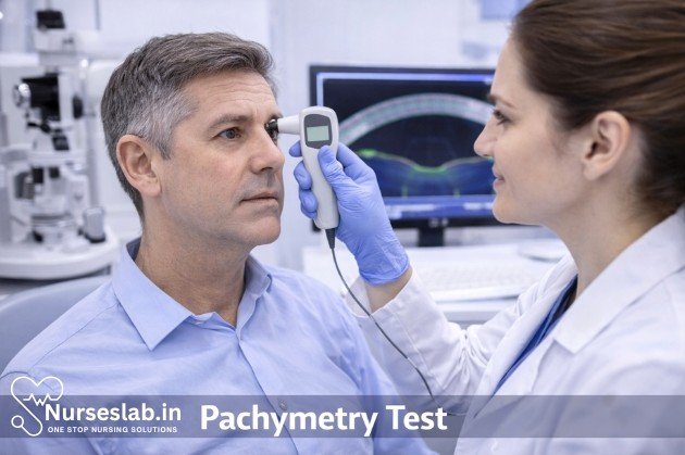
Pachymetry refers to the measurement of corneal thickness. The term derives from the Greek word ‘pachys’, meaning thick. Pachymetry tests are non-invasive, painless, and can be performed in a clinical setting within minutes. These tests are indispensable in preoperative evaluations for refractive surgery (such as LASIK), monitoring corneal diseases (like keratoconus), and refining glaucoma management.
Types of Pachymetry Techniques
Several technologies are available for measuring corneal thickness, each with distinct advantages and limitations. The main types of pachymetry are:
Ultrasound Pachymetry
This is the most widely used method for measuring corneal thickness. It employs a probe that emits high-frequency sound waves, which travel through the cornea and reflect back to the device, allowing calculation of thickness based on the time taken for the echo to return. The test is quick, reliable, and cost-effective. However, it requires contact with the cornea, which may pose a slight risk of infection or epithelial disruption if not performed correctly.
Optical Pachymetry
Optical methods utilise light rather than sound to measure corneal thickness. Techniques include:
- Specular Microscopy: Analyses the reflection of light from the corneal endothelium to determine thickness.
- Confocal Microscopy: Provides high-resolution images of corneal layers and precise thickness measurements.
- Optical Coherence Tomography (OCT): Uses low-coherence interferometry to create cross-sectional images of the cornea, enabling non-contact, high-precision pachymetry.
Non-contact Specular Pachymetry
This method uses specular reflection to measure corneal thickness without touching the eye. It is especially useful for patients who are sensitive to contact procedures or at risk of infection.
- Slit-Scanning Pachymetry: Devices like the Orbscan utilise slit-scanning technology to generate a three-dimensional map of the cornea, including thickness measurements across different zones.
Procedure:
The exact procedure varies depending on the technology used. Here is an outline of the typical steps for ultrasound pachymetry:
- The patient is seated comfortably in an examination chair.
- A topical anaesthetic drop is instilled into the eye to numb the cornea and prevent discomfort.
- The examiner gently places the probe perpendicular to the central cornea, avoiding excessive pressure.
- Multiple readings are taken to ensure accuracy, and the mean value is recorded as the central corneal thickness.
- The process is repeated for both eyes, and results are documented for further analysis.
For optical methods such as OCT, the patient rests their chin and forehead on a support while the device scans the cornea without any contact. The procedure is painless and requires minimal preparation.
Clinical Applications of Pachymetry
Pachymetry serves as a cornerstone in several ophthalmic evaluations:
Glaucoma Assessment:
- Accurate measurement of corneal thickness is crucial in interpreting intraocular pressure readings. Thicker corneas may result in overestimation of IOP, while thinner corneas may cause underestimation. This has significant implications for glaucoma diagnosis and management, as untreated elevated IOP can lead to irreversible vision loss.
Refractive Surgery Screening:
- Procedures like LASIK, PRK, and SMILE require a minimum corneal thickness to ensure safety and prevent postoperative complications such as ectasia (corneal bulging). Pachymetry helps surgeons determine eligibility and customise surgical plans.
Cornal Disease Monitoring:
- Diseases such as keratoconus, Fuchs’ endothelial dystrophy, and corneal oedema manifest with changes in corneal thickness. Regular pachymetry assists in tracking disease progression and therapeutic response.
Contact Lens Fitting:
- In specialised cases, pachymetry informs the choice and fitting of contact lenses, especially in patients with irregular corneas.
Postoperative Follow-up:
- After corneal surgery, pachymetry is used to monitor healing and detect complications such as thinning or swelling.
Interpreting Pachymetry Results
The central corneal thickness is expressed in micrometres (µm). Normal adult values range from 520 to 550 µm. Deviations from this range may indicate:
- Thin Cornea (<500 µm): Associated with increased risk of glaucoma progression, keratoconus, and may contraindicate certain refractive procedures.
- Thick Cornea (>580 µm): May result from corneal oedema, Fuchs’ dystrophy, or other pathologies.
It is important to interpret pachymetry results in the context of the patient’s overall ocular health, history, and associated findings. For instance, a thin cornea in a glaucoma suspect warrants closer monitoring and potentially more aggressive therapy.
Limitations and Sources of Error
While pachymetry is generally reliable, certain factors may affect accuracy:
- Operator Technique: Excessive pressure with the probe can artificially lower thickness readings.
- Corneal Pathology: Scarring, opacities, or irregularities may interfere with measurement.
- Device Calibration: Regular maintenance and calibration of equipment is essential for consistent results.
- Patient Cooperation: Movement or blinking during the test can affect readings.
It is recommended to take multiple readings and use the mean value to minimise variability.
Patient Experience and Preparation
For patients, the pachymetry test is straightforward and comfortable. There is no need for special preparation. The entire procedure typically takes less than five minutes per eye. Patients are advised to avoid touching their eyes immediately after the test, especially if a contact procedure was performed.
Risks are minimal, with rare cases of mild irritation or infection following ultrasound pachymetry. Non-contact methods eliminate these risks entirely.
Nursing Care for Patients Undergoing a Pachymetry Test
Pachymetry is a diagnostic test used to measure the thickness of the cornea, the transparent front part of the eye. This procedure is vital in the assessment of various ocular conditions, including glaucoma, corneal oedema, and before refractive surgery. Accurate corneal thickness measurements can influence decisions about treatment options and patient management, making the role of the nurse integral to ensuring a safe and successful testing experience.
Pre-Test Preparation
Effective pre-test preparation lays the foundation for a smooth and accurate pachymetry test. Nurses play a crucial role in assessing the patient’s suitability for the procedure and ensuring all necessary steps are completed before the test begins.
- Patient Assessment: Begin with a thorough review of the patient’s medical history, noting any ocular conditions, allergies (especially to topical anaesthetics), or recent eye surgeries. Assess for active eye infections, as these may require postponement of the test.
- Consent: Obtain informed consent in accordance with institutional policies. Ensure the patient understands the purpose, procedure, and any potential risks of the pachymetry test.
- Preparation Steps: Advise patients to avoid wearing contact lenses on the day of the test, as these can affect corneal measurements. Confirm the identity of the patient and verify the correct eye(s) to be tested. Prepare the necessary equipment, including the pachymeter, single-use covers or tips, and topical anaesthetic drops if required.
Patient Education
Educating patients about the pachymetry test is essential to alleviate anxiety and ensure cooperation during the procedure. Nurses should provide clear, concise information tailored to the patient’s level of understanding.
- Explaining the Procedure: Describe the test as a painless and quick measurement of the cornea’s thickness. Emphasise that topical anaesthetic drops may be used to minimise discomfort, and explain that the patient will need to keep their eye still for a few seconds.
- Addressing Concerns: Encourage patients to ask questions and express any worries, particularly if they are apprehensive about procedures involving the eyes. Reassure them about the safety of the test and the sterile technique used to prevent infection.
- Instructions: Advise patients to avoid touching their eyes before and after the test and to notify the nurse if they experience any discomfort or changes in vision following the procedure.
Procedural Support
Nurses provide vital support during the pachymetry test, ensuring patient comfort, safety, and the accuracy of results.
- Environment Preparation: Ensure the testing area is clean, well-lit, and equipped with hand hygiene supplies. Arrange the equipment within easy reach to minimise delays.
- Patient Positioning: Assist the patient into a comfortable seated position, with their chin and forehead resting securely if a slit lamp is used. Instruct the patient to look straight ahead and remain as still as possible.
- Monitoring Comfort: Observe the patient for signs of anxiety or discomfort throughout the procedure. Offer reassurance and gentle guidance as needed. If topical anaesthetic is administered, monitor for any adverse reactions.
- Assisting the Practitioner: Hand over sterile single-use tips or covers and help with the application of anaesthetic drops if required. Support the patient in maintaining the required position until the test is complete.
Post-Test Care
After the pachymetry test, nurses are responsible for immediate aftercare and providing patients with instructions for ongoing wellbeing.
- Immediate Observation: Monitor the patient for any immediate side effects, such as irritation, redness, or allergic reaction to the anaesthetic drops. Provide tissues or cotton pads if the patient experiences tearing.
- Discharge Instructions: Advise the patient that any numbness from the anaesthetic drops will wear off within 15–30 minutes. Caution them against rubbing their eyes until sensation returns fully, as this could cause accidental injury.
- When to Seek Help: Instruct patients to contact the clinic or their healthcare provider if they notice persistent pain, vision changes, or signs of infection, such as discharge or swelling.
Special Considerations
Nurses must adapt their approach for patients with unique needs, such as paediatric, elderly, or anxious individuals, to ensure a successful and comfortable experience.
- Paediatric Patients: Children may be fearful or unable to remain still during the procedure. Use age-appropriate language to explain the test, allow a parent or guardian to be present, and use distraction techniques, such as storytelling or visual aids, to maintain cooperation.
- Elderly Patients: Older adults may have mobility or cognitive challenges. Provide extra time for explanations, ensure clear communication, and assist with positioning. Be mindful of comorbidities that may affect their ability to remain still or follow instructions.
- Anxious Patients: Some individuals may experience anxiety related to eye procedures. Offer reassurance, explain each step of the process, and use calming techniques, such as deep breathing or guided imagery, to reduce distress.
Documentation and Communication
Accurate documentation and effective communication are crucial components of nursing care during a pachymetry test.
- Recording Findings: Document the corneal thickness measurements, the eye(s) tested, the type of pachymeter used, and any anaesthetic administered. Note any patient reactions or complications during the procedure.
- Communicating with the Care Team: Share relevant findings promptly with the ophthalmologist or optometrist. Report any concerns or abnormal results that may require further assessment or intervention.
- Patient Records: Ensure all information is accurately entered into the patient’s medical records in accordance with local policies and data protection regulations.
REFERENCES
- Ayala M, Strandås R. Accuracy of optical coherence tomography (OCT) in pachymetry for glaucoma patients https://pubmed.ncbi.nlm.nih.gov/26420690/ . BMC Ophthalmol. 2015 Sep 29:15;124.
- Phu J, Khuu SK, Yapp M, Assaad N, Hennessy MP, Kalloniatis M. The value of visual field testing in the era of advanced imaging: clinical and psychophysical perspectives. Clin Exp Optom. 2017;100(4):313–332. doi:10.1111/cxo.12551
- Centers for Medicare & Medicaid Services (U.S.). Medicare Coverage Database: Corneal Pachymetry https://www.cms.gov/medicare-coverage-database/view/lcd.aspx?LCDId=34512.
- Feizi S, Jafarinasab MR, Karimian F, et al. Central and peripheral corneal thickness measurement in normal and keratoconic eyes using three corneal pachymeters https://www.ncbi.nlm.nih.gov/pmc/articles/PMC4307658/). J Ophthalmic Vis Res. 2014;9(3):296-304.
- Lestak J, Lestak T, Fus M, Klimesova I. Temporal visual field border. Clin Ophthalmol. 2021;15:3241-3246. doi:10.2147/OPTH.S321110
- Glaucoma Research Foundation (U.S.). The Importance of Corneal Thickness https://glaucoma.org/articles/the-importance-of-corneal-thickness. Last reviewed 3/23/2022.
- Robson AG, Frishman LJ, Grigg J, et al. ISCEV Standard for full-field clinical electroretinography (2022 update). Doc Ophthalmol. 2022;144(3):165-177. doi:10.1007/s10633-022-09872-0
Stories are the threads that bind us; through them, we understand each other, grow, and heal.
JOHN NOORD
Connect with “Nurses Lab Editorial Team”
I hope you found this information helpful. Do you have any questions or comments? Kindly write in comments section. Subscribe the Blog with your email so you can stay updated on upcoming events and the latest articles.


