


Last Updated on August 21, 2026 by Nurseslab.in Editorial Team
Pulse Volume Recordings (PVR) measure blood flow in the arms and legs to help diagnose peripheral artery disease and vascular circulation problems.
Introduction
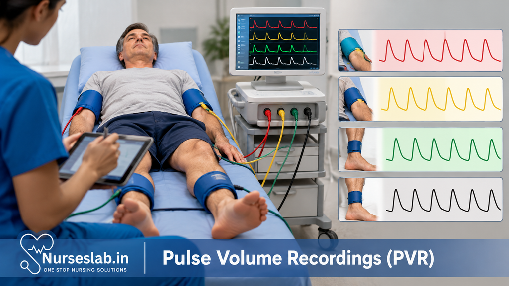
Pulse Volume Recordings (PVR), also known as volume plethysmography, are non-invasive vascular diagnostic procedures that play a pivotal role in the assessment of peripheral arterial disease and other circulatory disorders. Over the decades, PVR has evolved from rudimentary volume measurement techniques to sophisticated waveform analysis, providing clinicians with vital information about arterial blood flow and limb perfusion. In contemporary vascular diagnostics, PVR holds significant relevance due to its reliability, ease of use, and ability to complement other diagnostic modalities.
Historical Context and Relevance
The origins of plethysmography date back to the late 19th and early 20th centuries, when researchers first attempted to quantify changes in limb volume as a surrogate for blood flow. Early devices were cumbersome and limited by technical challenges. However, technological advancements, particularly in pressure transducers and data recording, have refined the PVR technique, making it a routine component of vascular laboratories worldwide. Its ongoing relevance is underscored by the increasing prevalence of peripheral vascular diseases, especially in ageing populations and those with diabetes mellitus.
Definition and Purpose of Pulse Volume Recordings
Pulse Volume Recordings are non-invasive tests that measure changes in blood volume within a limb segment during the cardiac cycle. By detecting and graphically representing these volume changes as waveforms, PVR provides an indirect assessment of arterial blood flow. The primary purpose of PVR is to evaluate the presence, location, and severity of arterial occlusive disease, especially in the lower extremities. Unlike Doppler ultrasound, which focuses on velocity and direction of blood flow, PVR assesses the volumetric changes associated with each pulse, offering a complementary perspective in vascular diagnostics.
Why is PVR Performed?
PVR is performed to assess arterial integrity and perfusion in patients with suspected or known peripheral arterial disease (PAD). It is particularly useful in the following scenarios:
- Screening for PAD in symptomatic patients (e.g., those with claudication, rest pain, or non-healing wounds)
- Localising the level of arterial obstruction or stenosis
- Monitoring disease progression or response to therapy
- Evaluating vascular status prior to surgical interventions or amputations
- Assessing graft patency and post-revascularisation outcomes
Conditions commonly assessed include atherosclerosis, thromboangiitis obliterans (Buerger’s disease), arterial embolism, and arterial trauma.
Clinical Applications of PVR
Pulse Volume Recordings have a broad range of clinical applications, serving both diagnostic and monitoring functions. Key applications include:
- Peripheral Arterial Disease (PAD) Diagnosis: PVR is routinely used to confirm and quantify the extent of PAD, particularly in lower limbs.
- Assessment in Diabetic Patients: Diabetics often have calcified vessels, making traditional ankle-brachial index (ABI) measurements unreliable. PVR offers valuable information in such cases.
- Preoperative Evaluation: Prior to lower limb surgeries or amputation, PVR can help determine the optimal level for intervention by assessing perfusion.
- Postoperative Monitoring: Following revascularisation or bypass procedures, PVR is used to monitor graft patency and detect restenosis or occlusion.
- Pediatric and Geriatric Assessments: Its non-invasive nature and minimal patient cooperation requirements make PVR suitable for vulnerable populations.
Indications for PVR include symptoms suggestive of arterial insufficiency (pain, pallor, pulselessness, paraesthesia, paralysis), non-healing ulcers, gangrene, and unexplained limb swelling.
Equipment and Preparation
Equipment Required
The standard PVR setup includes the following components:
- Pneumatic Cuffs: Similar to blood pressure cuffs, these are placed at various limb segments (thigh, calf, ankle, foot, or fingers/toes).
- Volume Plethysmograph: Device that detects and records volume changes within the cuff, typically using pressure transducers.
- Data Acquisition System: Hardware and software for real-time display and storage of waveform data.
- Chart Recorder/Computer: For graphical representation and analysis of waveforms.
- Patient Bed/Table: Comfortable surface to ensure patient relaxation during the procedure.
Patient Preparation
- Obtain informed consent and explain the procedure to the patient.
- Ensure the patient is relaxed and at a comfortable ambient temperature, as cold may induce vasoconstriction and affect results.
- Advise the patient to avoid caffeine, tobacco, and heavy exercise for at least 2 hours prior to the test.
- Position the patient supine, with limbs exposed and supported.
- Remove tight clothing, jewellery, or dressings from the limbs to be examined.
Step-by-Step Procedure
A methodical approach is essential for accurate and reproducible PVR measurements. The typical procedure involves the following steps:
- Limb Selection and Cuff Placement
- Identify the limb(s) to be examined based on clinical indications.
- Place pneumatic cuffs at standardised sites: thigh, above-knee, below-knee, ankle, and foot (or corresponding upper limb sites).
- Ensure the cuff width is approximately 20% greater than limb diameter to avoid artefacts.
- Apply cuffs snugly but not too tight; there should be no air leaks.
- Calibration and Zeroing
- Connect cuffs to the plethysmograph.
- Calibrate the system and zero the pressure transducers as per manufacturer guidelines.
- Baseline Pressure Inflation
- Inflate each cuff to a low baseline pressure (usually 40–65 mmHg), sufficient to detect volume changes but not occlude arterial flow.
- Data Acquisition
- Initiate recording. As arterial blood pulses through the limb, cyclic volume changes are transmitted to the cuff, generating waveforms.
- Obtain at least 3–5 cardiac cycles at each level for reliable interpretation.
- Repeat measurements at each limb segment sequentially.
- Waveform Assessment and Documentation
- Review real-time waveforms for quality, artefacts, and reproducibility.
- Document and save all relevant tracings for subsequent analysis and reporting.
- Special Maneuvers (if indicated)
- Toe or finger PVRs may be performed using smaller cuffs for distal perfusion assessment, especially in diabetics or patients with small vessel disease.
- Exercise or postural changes may be incorporated where appropriate to provoke symptoms or assess dynamic changes.
Interpretation of Results
The cornerstone of PVR interpretation lies in the analysis of waveform morphology, amplitude, and segmental changes. Understanding normal and abnormal patterns is crucial for accurate diagnosis.
Normal PVR Waveforms
- Rapid Upstroke: Steep ascent during systole, reflecting unimpeded arterial inflow.
- Sharp Peak: Well-defined apex at the top of the waveform.
- Dicrotic Notch: Secondary downward deflection in early diastole, indicating elastic recoil of healthy arteries.
- Gradual Downstroke: Smooth descent back to baseline during diastole.
Abnormal Findings
- Blunted/Flattened Waveform: Reduced amplitude and loss of dicrotic notch, seen in moderate to severe arterial obstruction.
- Delayed Upstroke: Sluggish ascent, indicating proximal or segmental stenosis.
- Decreased Amplitude: Lower pulse volume, often correlating with diminished arterial perfusion.
- Segmental Drop: Marked reduction in waveform amplitude between adjacent segments signifies the likely location of arterial disease.
Clinical Significance
Interpretation must consider patient symptoms, risk factors, and other diagnostic findings. A normal PVR may help exclude significant arterial disease, while abnormal patterns warrant further evaluation, such as duplex ultrasonography, computed tomography angiography, or magnetic resonance angiography. In diabetics, toe PVRs are particularly valuable as calcified tibial arteries may render standard cuffs less informative.
Advantages of Pulse Volume Recordings
- Non-Invasive and Painless: No needles or injections, minimising patient discomfort.
- Reliable in Calcified Vessels: Unlike ABI, PVR remains informative even in the presence of non-compressible arteries.
- Segmental Localisation: Ability to pinpoint the level of arterial obstruction across multiple limb segments.
- Rapid and Reproducible: Quick to perform and suitable for serial monitoring.
- Minimal Patient Cooperation Required: Particularly useful in elderly, paediatric, or non-communicative patients.
- Complementary to Other Modalities: Enhances diagnostic accuracy when used alongside Doppler studies.
Limitations and Challenges
Despite its utility, PVR has certain limitations and potential sources of error that must be acknowledged:
- Non-Specificity: PVR does not identify the precise vessel involved; it indicates the segmental level of disease.
- Influence of External Factors: Cold, anxiety, or improper cuff placement can alter waveforms and affect results.
- Obesity and Limb Oedema: Excess tissue may dampen waveform amplitude, leading to underestimation of disease severity.
- Movement Artefacts: Patient movement or tremors can introduce noise and compromise data quality.
- Limited Value in Certain Populations: Severely calcified vessels or microvascular disease (e.g., advanced diabetes) may necessitate adjunctive tests.
- Operator Dependence: Proper training and experience are essential for accurate performance and interpretation.
Safety and Patient Considerations
PVR is considered extremely safe, with no direct risks associated with the procedure. However, best practices should be followed to ensure patient comfort and optimal results:
- Pressure Injury: Avoid excessive cuff inflation to prevent discomfort or skin injury, especially in frail or elderly patients.
- Infection Control: Use clean equipment and adhere to infection prevention protocols, particularly with open wounds or ulcers.
- Patient Reassurance: Explain the procedure and address concerns to reduce anxiety-induced artefacts.
- Contraindications: Avoid PVR in limbs with acute deep vein thrombosis or severe cellulitis.
- Special Populations: Exercise caution in patients with severe pain, critical limb ischaemia, or recent vascular interventions.
Nursing Care for Patients Undergoing Pulse Volume Recordings (PVR) Diagnostic Procedures
A Comprehensive Guide for Nursing Professionals
Introduction
Pulse Volume Recording (PVR) is a non-invasive vascular diagnostic technique used to evaluate blood flow in the limbs and detect peripheral arterial disease (PAD). By assessing the volume changes in a limb during each cardiac cycle, PVR provides vital information about arterial patency and circulatory status. As the prevalence of vascular disorders rises, especially among ageing populations and patients with comorbidities such as diabetes and hypertension, the clinical significance of PVR continues to grow. Nursing professionals play an indispensable role in ensuring the procedure’s accuracy, patient safety, and overall comfort.
Pre-Procedure Nursing Responsibilities
Patient Education
Thorough patient education is a cornerstone of successful PVR procedures. Nurses should begin by explaining the purpose of the test, how it will be performed, and its importance in diagnosing vascular conditions. Clear communication helps demystify the process and alleviates anxiety. Patients should be informed that the procedure is painless, involves the application of blood pressure cuffs, and typically lasts between 30 to 60 minutes. Encourage patients to ask questions and clarify any doubts, ensuring they feel prepared and empowered for the experience.
Psychological Preparation
Additionally, the nurse should assess the patient’s psychological state and offer support to alleviate any fears or misconceptions. For patients who may be anxious or have had previous negative experiences with medical tests, providing extra reassurance and allowing time to ask questions can significantly improve the patient’s experience. Use active listening skills, maintain a calm tone, and encourage the patient to express any specific worries. Addressing psychological needs can improve cooperation and procedural outcomes.
Compliance with Pre-Procedure Instructions
The nurse should also confirm that the patient has complied with any specific pre-procedure instructions, such as fasting, medication adjustments, or wearing loose clothing to facilitate easy access to the limbs. Review the patient’s medical history for allergies, recent interventions, or medications that may influence the test. Ensure that the patient has removed jewellery and other items that may interfere with equipment. Document compliance and any deviations from instructions in the patient’s record.
Medical History Review
Prior to the procedure, a comprehensive review of the patient’s medical history is essential. Nurses should identify any underlying conditions, such as cardiovascular diseases, diabetes, or renal impairment, which may affect the PVR results or require specific precautions. Allergies, especially to latex or adhesives, should be noted to prevent adverse reactions. The nurse must also verify recent surgical interventions or angiographic studies, as these can influence vascular status. All relevant information should be accurately documented and communicated to the healthcare team.
Intra-Procedure Nursing Responsibilities
Monitoring and Patient Safety
During the PVR procedure, the nurse plays a pivotal role in ensuring patient safety and comfort. Continuous monitoring of the patient’s vital signs and overall well-being is essential, especially in patients with pre-existing cardiovascular conditions. Observe for signs of discomfort, anxiety, or adverse reactions such as dizziness or shortness of breath. Be prepared to intervene promptly should any complications arise, and communicate concerns to the vascular technologist or physician.
Equipment Assistance
The nurse assists the vascular technologist or physician by preparing and positioning the patient. This includes ensuring the patient is lying comfortably in the supine position, with limbs supported as needed. Blood pressure cuffs must be applied correctly to each limb, avoiding excessive tightness and ensuring proper placement to guarantee accurate measurements. The nurse should verify that all equipment is functioning correctly, and address any technical issues promptly to minimise procedural delays.
Patient Positioning and Comfort
Proper positioning is crucial for obtaining reliable PVR tracings. The nurse should ensure that the patient’s limbs are relaxed and supported, with the cuffs placed at the recommended anatomical sites. Pillows or supports may be used to enhance comfort, particularly for elderly or frail patients. Throughout the procedure, the nurse should regularly check for signs of discomfort and make necessary adjustments.
Communication with the Healthcare Team
Effective communication between the nurse, vascular technologist, and physician is vital throughout the procedure. The nurse should promptly relay any observations of patient distress, equipment malfunction, or unexpected findings. This collaborative approach ensures timely interventions and enhances the overall quality of care.
Post-Procedure Nursing Responsibilities
Patient Assessment
Following the PVR procedure, nurses must assess the patient for immediate post-procedural complications such as skin irritation from the cuffs, dizziness, or discomfort. Vital signs should be monitored and compared with baseline measurements to identify any significant changes. Instruct the patient to report any lingering symptoms or concerns before leaving the facility.
Monitoring for Complications
Although PVR is a low-risk procedure, vigilant observation for delayed reactions is necessary. In rare cases, patients may experience bruising, numbness, or allergic responses to materials used during the test. Nurses should educate patients on recognising these symptoms and provide clear instructions on seeking medical attention if needed.
Providing Post-Procedure Instructions
Patients should receive concise post-procedure guidance, including when to resume normal activities, any restrictions, and when to expect results. For those who have undergone medication adjustments or fasting, instructions regarding resumption of usual routines should be clearly communicated. Nurses should also address any questions related to follow-up appointments or further investigations.
Documentation
Accurate and thorough documentation is essential for continuity of care. The nurse should record all aspects of the procedure, including patient compliance, observations, interventions, and any deviations from standard protocols. Documenting patient responses and outcomes helps inform future care and supports interdisciplinary communication.
Special Considerations
Care for Patients with Comorbidities
Patients with chronic conditions such as diabetes, hypertension, or renal impairment may require tailored nursing interventions. For instance, diabetic patients are at increased risk for vascular complications and may need closer monitoring of limb integrity. Nurses should be aware of the specific needs of these patients and adjust care plans accordingly.
Pediatric and Geriatric Adaptations
Special populations, including children and the elderly, necessitate additional attention. In paediatric patients, age-appropriate explanations and parental involvement can help reduce anxiety and improve cooperation. For geriatric patients, considerations such as frailty, reduced mobility, and increased susceptibility to skin injury should guide nursing care. Use gentle techniques and provide extra support to ensure safety and comfort.
Infection Control
Strict adherence to infection control protocols is mandatory during PVR procedures. Nurses should ensure that all equipment is properly sanitised and that hand hygiene is maintained before and after patient contact. Single-use cuffs or protective barriers may be employed for patients with open wounds or compromised skin integrity.
Patient Education and Support
Ongoing patient education is fundamental to nursing care in PVR procedures. Nurses should continue to address patient concerns, clarify any misconceptions, and reinforce the importance of vascular health. Providing educational materials and resources can help patients understand their condition and the significance of test results. Emotional support, empathy, and encouragement are vital in promoting patient confidence and adherence to follow-up recommendations.
REFERENCES
- Sorber, R., Almeida, J.I., Raines, J.K., Abularrage, C.J. (2022). Pulse Volume Recording in the Diagnosis of Peripheral Vascular Disease. In: AbuRahma, A.F., Perler, B.A. (eds) Noninvasive Vascular Diagnosis. Springer, Cham. https://doi.org/10.1007/978-3-030-60626-8_23
- Mittleider D. Noninvasive Arterial Testing: What and When to Use (https://www.ncbi.nlm.nih.gov/pmc/articles/PMC6363559/). _Semin Intervent Radiol. _2018 Dec;35(5):384-392.
- National Library of Medicine. Doppler ultrasound exam of an arm or leg https://medlineplus.gov/ency/article/003775.htm.
- Society for Vascular Surgery. Ankle-Brachial Index or ABI Test https://vascular.org/patients-and-referring-physicians/conditions/ankle-brachial-index-or-abi-test.
- Centers for Disease Control and Prevention. Peripheral Arterial Disease (https://www.cdc.gov/heartdisease/PAD.htm).
Stories are the threads that bind us; through them, we understand each other, grow, and heal.
JOHN NOORD
Connect with “Nurses Lab Editorial Team”
I hope you found this information helpful. Do you have any questions or comments? Kindly write in comments section. Subscribe the Blog with your email so you can stay updated on upcoming events and the latest articles.


