


The Rheumatoid Factor (RF) test measures autoantibodies associated with rheumatoid arthritis and other autoimmune disorders. It supports diagnosis, disease assessment, and clinical decision‑making in rheumatology and laboratory practice.
Introduction
Rheumatoid Factor (RF) is an autoantibody that is often measured as part of the diagnostic process for various autoimmune diseases, particularly rheumatoid arthritis (RA). The RF diagnostic test has become a cornerstone in rheumatology, and its implications stretch beyond the mere detection of RA.
Biochemical Basis of Rheumatoid Factor
Rheumatoid Factor is an immunoglobulin, most commonly of the IgM class, that targets the Fc portion of IgG antibodies. This autoantibody is produced by the immune system in response to perceived threats but, in the case of autoimmune diseases, it mistakenly targets the body’s own tissues. The presence of RF is indicative of immune system dysregulation, and its detection can signal underlying pathology.
Types of Rheumatoid Factor
While IgM RF is the most prevalent and clinically significant, other classes such as IgG and IgA RF may also be present. The distribution and quantity of these autoantibodies can vary among individuals and disease states. In certain conditions, the presence of multiple types of RF may lend more specificity to the diagnosis.
Indications for Rheumatoid Factor Testing
- Suspected rheumatoid arthritis based on clinical symptoms (joint pain, swelling, stiffness)
- Evaluation of unexplained polyarthritis
- Assessment of other autoimmune disorders (e.g., Sjögren’s syndrome, systemic lupus erythematosus)
- Monitoring disease progression and response to therapy in established RA
- Screening for extra-articular manifestations in patients with chronic inflammatory symptoms
Methods of Rheumatoid Factor Testing
Several laboratory techniques are available for the detection and quantification of RF. The choice of method depends on resource availability, specificity, sensitivity, and clinical requirements.
1. Latex Agglutination Test
This is one of the most widely used screening tests in India for RF. It involves mixing patient serum with latex beads coated with human IgG. If RF is present, agglutination occurs, visible to the naked eye. The test is simple, rapid, and cost-effective, making it suitable for resource-limited settings. However, it is qualitative and may yield false positives due to other antibodies.
2. Nephelometry and Turbidimetry
These are quantitative methods that measure the amount of RF in the serum using light scattering principles. Nephelometry is highly sensitive and specific, providing numerical values that aid in monitoring disease activity and progression.
3. Enzyme-Linked Immunosorbent Assay (ELISA)
ELISA is another quantitative method that allows for the detection of different RF isotypes (IgM, IgG, IgA). It is highly specific and sensitive, making it valuable for both diagnosis and research purposes. However, it requires specialised equipment and trained personnel.
4. Other Techniques
Other advanced techniques such as radioimmunoassay and immunofluorescence are rarely used in clinical practice due to cost and complexity, but they may be employed in research settings to further characterise RF activity.
Clinical Interpretation
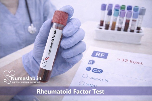
The interpretation of RF results must be contextualised within the patient’s clinical presentation, as the presence of RF is not exclusive to RA. The reference range for RF varies among laboratories, but generally, a value above 20 IU/mL is considered positive.
A positive RF test supports a diagnosis of RA when accompanied by typical symptoms and findings but does not confirm it in isolation. About 70-80% of patients with RA have a positive RF test, but its absence does not rule out the disease, as seronegative RA is a recognised entity.
False Positives and False Negatives
RF positivity can occur in a variety of other conditions, including:
- Other autoimmune diseases (Sjögren’s syndrome, systemic lupus erythematosus)
- Chronic infections (tuberculosis, hepatitis C, endocarditis)
- Ageing (elderly individuals may have low-level RF without disease)
- Other inflammatory conditions
Conversely, false negatives may occur in early RA or in seronegative subtypes. Thus, RF testing should be considered as part of a broader diagnostic workup.
Role of RF in Prognosis and Disease Monitoring
High RF titres are generally associated with more severe disease, greater risk of joint erosions, and extra-articular manifestations. RF status is used alongside other markers such as anti-cyclic citrullinated peptide (anti-CCP) antibodies to stratify risk and guide management decisions. In India, where access to advanced diagnostics may be limited, RF testing remains a valuable tool for prognostication and monitoring.
Limitations of Rheumatoid Factor Testing
Despite its widespread use, RF testing has notable limitations:
- Lack of specificity: RF is not exclusive to RA and can be positive in other diseases.
- Variable sensitivity: Not all patients with RA will have a positive RF test.
- Influence of age and chronic diseases: Elderly individuals and those with chronic infections may have elevated RF levels.
- Risk of overdiagnosis: Sole reliance on RF can lead to unnecessary treatment in patients without true RA.
- Technical variability: Different laboratories may use varying methods and reference ranges, affecting result interpretation.
Advances in Rheumatoid Factor Testing
With the advent of newer technologies, RF testing has become more accurate and reliable. The use of multiplex assays and molecular techniques allows for simultaneous detection of multiple autoantibodies, improving diagnostic yield. Research is ongoing to identify novel biomarkers that can complement RF, enhancing early diagnosis and personalised treatment.
Guidelines for Clinicians
- Always interpret RF results in conjunction with clinical findings and other laboratory tests.
- Consider anti-CCP antibody testing for increased specificity in diagnosing RA.
- Be aware of conditions that may cause false positives, such as infections and ageing.
- Educate patients about the meaning and limitations of RF testing.
- Use serial RF measurements to monitor disease activity and response to therapy.
Patient Perspective and Counselling
Patients in India often have limited understanding of autoimmune diseases and laboratory tests. It is essential for healthcare providers to explain the role of RF testing, its limitations, and the need for comprehensive evaluation. Counselling should emphasise that a positive RF test does not automatically mean RA, nor does a negative result rule it out. Patient education improves compliance and outcomes.
Nursing Care of Patients Undergoing Rheumatoid Factor Test
Rheumatoid factor (RF) testing is a vital diagnostic tool in the evaluation of patients suspected of having rheumatoid arthritis (RA) and other autoimmune disorders. As frontline healthcare providers, nurses play a critical role in ensuring the safe, efficient, and compassionate delivery of care throughout the RF testing process.
Nursing Responsibilities Before the Procedure
1. Patient Assessment
- Review the Medical History: Check for any previous diagnosis of autoimmune disorders, ongoing infections, or conditions that may affect test results.
- Current Medications: Identify medications that could interfere with the test or pose risks during blood collection (e.g., anticoagulants).
- Allergies: Confirm any allergies, especially to latex, antiseptics, or adhesives used during the procedure.
2. Patient Preparation
- Obtain Informed Consent: Ensure the patient understands the purpose, procedure, risks, and potential outcomes of the RF test. Answer any questions and obtain written consent as per hospital policy.
- Explain the Procedure: Use simple language to describe what the patient can expect. For instance, “A small sample of blood will be drawn from your arm to check for rheumatoid factor.”
- Address Anxiety: Assess the patient’s emotional state and provide reassurance. Patients may be anxious about the diagnosis or the blood draw itself.
- Fasting Requirements: Typically, RF testing does not require fasting. However, confirm with the laboratory if any special instructions are necessary.
- Verify Identity: Use two identifiers (e.g., name and date of birth) to ensure correct patient identification before the procedure.
3. Equipment Preparation
- Vacutainer tubes (as per lab protocol, usually red or yellow top)
- Needles and syringes or butterfly sets
- Tourniquet
- Alcohol swabs or antiseptic solution
- Sterile gauze and adhesive bandage
- Gloves and other personal protective equipment (PPE)
- Properly labeled specimen containers
- Request forms and documentation tools
During the Procedure
1. Ensuring Patient Comfort and Safety
- Position the Patient: Seat the patient comfortably, with the chosen arm extended and supported.
- Explain Each Step: Continue to communicate with the patient, describing each step to reduce anxiety.
- Apply PPE: Follow standard precautions to prevent cross-infection.
2. Blood Collection Technique
- Apply the tourniquet above the venipuncture site.
- Palpate and select an appropriate vein, usually in the antecubital fossa.
- Cleanse the site with an alcohol swab and allow it to air dry.
- Insert the needle at an appropriate angle and collect the required amount of blood.
- Release the tourniquet before withdrawing the needle.
- Apply gentle pressure with sterile gauze to the puncture site.
- Label the specimen tube in the presence of the patient, with full identifying details.
3. Monitoring for Complications
- Observe for Vasovagal Reactions: Watch for signs of fainting, dizziness, or pallor; respond appropriately by assisting the patient to a safe position and providing reassurance.
- Manage Bleeding: Apply pressure until bleeding stops and monitor for hematoma formation.
- Infection Control: Maintain aseptic technique throughout to minimize infection risk.
Post-Procedure Nursing Care
1. Immediate Care
- Ensure Hemostasis: Confirm that bleeding has stopped before applying an adhesive bandage.
- Assess the Site: Check for swelling, redness, or excessive bruising at the puncture site.
- Patient Instructions: Advise the patient to keep the bandage on for at least 15-30 minutes and to avoid heavy lifting with the affected arm for a few hours.
2. Specimen Handling and Documentation
- Transport: Send the labeled specimen to the laboratory promptly, following institutional protocols for sample handling and chain of custody.
- Documentation: Record the procedure details in the patient’s chart, including date, time, site of collection, patient response, and any complications.
3. Monitoring and Follow-up
- Monitor for Delayed Reactions: Instruct the patient to report any delayed swelling, pain, or signs of infection at the puncture site.
- Arrange Follow-up: Inform the patient when and how they will receive their test results, and schedule follow-up appointments as needed.
Patient Education and Psychosocial Support
1. Explaining Test Results
While nurses do not interpret laboratory results, they play a vital role in helping patients understand the next steps. Explain that RF testing is just one part of the diagnostic process, and results must be correlated with clinical symptoms and other tests. Encourage patients to discuss their results with the physician for a comprehensive interpretation.
2. Addressing Emotional Concerns
Undergoing tests for chronic conditions such as rheumatoid arthritis can be stressful. Provide emotional support by listening to patient concerns, offering reassurance, and connecting them with counseling services or support groups if needed.
3. Health Promotion and Lifestyle Guidance
Educate patients about the importance of early detection and management of autoimmune diseases. If the patient is diagnosed with RA or another related condition, provide resources on lifestyle modifications, medication adherence, and symptom monitoring.
Special Considerations
1. Pediatric Patients
- Use age-appropriate language and comfort measures such as distraction or parental presence.
- Consider smaller gauge needles and minimal blood volume collection.
2. Elderly Patients
- Be cautious of fragile veins and increased bleeding risk.
- Monitor for dehydration and ensure adequate hydration before the procedure.
3. Patients with Bleeding Disorders
- Consult with the healthcare team regarding the need for additional precautions, such as extended pressure on the puncture site or alternative sampling methods.
Legal and Ethical Considerations
- Confidentiality: Maintain strict confidentiality of patient information and test results.
- Informed Consent: Ensure that the patient is fully informed and consents voluntarily.
- Non-Discrimination: Provide equitable care regardless of age, gender, ethnicity, or socioeconomic status.
Documentation Checklist
- Patient identification and informed consent
- Date, time, and site of blood collection
- Type and number of specimens collected
- Patient response and any complications
- Education and instructions given to the patient
- Signature and designation of the nurse performing the procedure
REFERENCES
- National Library of Medicine (U.S.). Rheumatoid factor (RF) (https://medlineplus.gov/ency/article/003548.htm). Last reviewed 4/30/2023.
- National Library of Medicine (U.S.). Rheumatoid factor (RF) test https://medlineplus.gov/lab-tests/rheumatoid-factor-rf-test/). Last updated 9/28/2022.
- Nielsen SF, Bojesen SE, Schnohr P, Nordestgaard BG. Elevated rheumatoid factor and long term risk of rheumatoid arthritis: a prospective cohort study https://www.ncbi.nlm.nih.gov/pmc/articles/PMC3435445/). BMJ. 2012 Sep 6;345:e5244.
- Rheumatoid factor (RF) test. Testing.com. https://www.testing.com/tests/rheumatoid-factor-rf/.
- Firestein GS, et al. Autoantibodies in rheumatoid arthritis. In: Firestein & Kelley’s Textbook of Rheumatology. 11th ed. Elsevier; 2021. https://www.clinicalkey.com.
- Tiwari V, Jandu JS, Bergman MJ. Rheumatoid Factor (https://pubmed.ncbi.nlm.nih.gov/30422493/). 2023 Jul 24. In: StatPearls. Treasure Island (FL): StatPearls Publishing;2023 Jan.
Stories are the threads that bind us; through them, we understand each other, grow, and heal.
JOHN NOORD
Connect with “Nurses Lab Editorial Team”
I hope you found this information helpful. Do you have any questions or comments? Kindly write in comments section. Subscribe the Blog with your email so you can stay updated on upcoming events and the latest articles.


