


Last Updated on January 21, 2026 by Nurseslab.in Editorial Team
A skin biopsy is a minor procedure that removes a small sample of skin for microscopic examination. It helps diagnose rashes, infections, autoimmune diseases, suspicious moles, and skin cancers, supporting accurate dermatologic assessment and treatment planning.
Introduction
A skin biopsy is a fundamental diagnostic procedure in dermatology, providing essential information for the evaluation and management of a wide range of cutaneous disorders. Its role in the identification of neoplastic, inflammatory, infectious, and autoimmune conditions is unparalleled, making it a cornerstone for both clinical practice and research.
Definition and Purpose
A skin biopsy involves the removal of a segment of skin tissue for microscopic examination. The procedure is performed to obtain a definitive diagnosis when a clinical assessment alone is insufficient. Its primary purpose is to facilitate histopathological analysis, which can reveal cellular and structural details crucial for identifying the nature of skin lesions.
Skin biopsies are indispensable in diagnosing malignancies (such as melanoma and squamous cell carcinoma), chronic inflammatory conditions (like psoriasis or lupus erythematosus), infectious diseases (e.g., cutaneous tuberculosis), and other dermatological disorders.
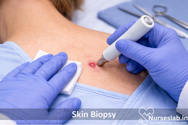
Types of Skin Biopsies
Several types of skin biopsies are utilised in clinical practice, each with distinct indications and technical considerations. The choice of biopsy type depends on the clinical scenario, lesion characteristics, and diagnostic objectives.
- Punch Biopsy: Utilises a circular blade to remove a full-thickness cylindrical core of skin, typically ranging from 2 to 8 mm in diameter. Punch biopsies are suitable for diagnosing inflammatory dermatoses, small neoplastic lesions, and for sampling representative areas of diffuse skin disease.
- Excisional Biopsy: Involves the complete removal of a skin lesion along with a margin of normal tissue. This technique is preferred for suspected malignancies, such as melanoma, where assessment of lesion depth and margins is critical.
- Incisional Biopsy: Removes only a portion of a larger lesion for diagnostic evaluation. Indicated when complete excision is impractical or unnecessary, such as in large tumours or when sampling specific areas of heterogenous lesions.
- Shave Biopsy: Employs a scalpel or razor blade to shave off a superficial section of skin. Ideal for raised, superficial lesions like basal cell carcinoma, seborrhoeic keratosis, or benign tumours, but not suitable for suspected melanomas due to inadequate depth assessment.
- Other Techniques: These include curettage (scraping), needle aspiration, and fine-needle techniques, which are less common but may be used in specific clinical contexts.
Indications for Each Type
- Punch biopsy: Inflammatory dermatoses, small neoplasms, bullous diseases.
- Excisional biopsy: Suspected melanoma, small tumours, diagnostic and therapeutic removal.
- Incisional biopsy: Large tumours, lesions with variable morphology, ulcerated or infiltrative diseases.
- Shave biopsy: Superficial lesions, benign growths, non-melanoma skin cancers.
Indications and Contraindications
Skin biopsy is indicated when:
- Clinical diagnosis is uncertain or ambiguous.
- Malignant neoplasms are suspected.
- Lesions are atypical in appearance, duration, or behaviour.
- Chronic, recurrent, or treatment-resistant dermatoses require histopathological confirmation.
- Infectious diseases (e.g., fungal, mycobacterial) need definitive identification.
- Autoimmune or bullous disorders require direct immunofluorescence studies.
Contraindications
- Active infection at the biopsy site (risk of spreading infection).
- Bleeding disorders or anticoagulant therapy without adequate control.
- Allergy to local anaesthetics (alternative agents may be considered).
- Patient refusal or inability to provide informed consent.
- Poor general condition where procedure risk outweighs benefit.
Pre-procedure Preparation
Patient Assessment
A thorough patient assessment is essential prior to performing a skin biopsy. This includes a detailed medical history (including bleeding diathesis, medication use, allergies), physical examination, and evaluation of the lesion for suitability of biopsy type and site selection. Informed consent must be obtained, with clear explanation of the procedure, risks, benefits, and possible outcomes.
Site Selection
Choosing the biopsy site is critical for diagnostic accuracy. The most representative area of the lesion should be selected, avoiding necrotic, ulcerated, or heavily excoriated regions. For conditions with widespread involvement, multiple biopsies from different sites may be warranted.
Anaesthesia
Local anaesthesia is administered using agents such as lignocaine (with or without adrenaline). The choice of anaesthetic, concentration, and method of infiltration should be tailored to the patient and the procedure. Topical anaesthetics may be employed for superficial biopsies in select cases.
Step-by-Step Technique
Punch Biopsy
- Cleanse the biopsy site with antiseptic solution.
- Mark the area and infiltrate local anaesthetic.
- Choose an appropriately sized punch instrument.
- Apply the punch perpendicular to the skin and rotate downward until subcutaneous fat is reached.
- Lift the specimen with forceps and cut the base with scissors.
- Control bleeding and close the wound with sutures if necessary.
Excisional Biopsy
- Prepare and anaesthetise the area.
- Mark the lesion with a margin of normal skin.
- Use a scalpel to excise the entire lesion, including a rim of healthy tissue.
- Undermine edges if required for wound closure.
- Achieve haemostasis and close with sutures.
Incisional Biopsy
- Identify and mark the area to be sampled.
- Administer local anaesthetic.
- Excise a representative portion of the lesion using a scalpel.
- Control bleeding and close or dress the wound as appropriate.
Shave Biopsy
- Clean and anaesthetise the area.
- Hold the blade parallel to the skin surface.
- Shave off the lesion with gentle, steady strokes.
- Apply haemostatic agent and dress the site.
Specimen Handling and Processing
Proper specimen handling is essential for accurate diagnosis. Immediately after removal, the tissue is placed in an appropriate fixative, usually 10% buffered formalin for routine histopathology. For immunofluorescence or microbiological studies, special media or transport conditions may be required (e.g., Michel’s transport medium for immunofluorescence).
The specimen must be labelled accurately with patient details, biopsy site, and clinical information. Timely transport to the laboratory ensures optimal tissue preservation. In the laboratory, the tissue is processed, sectioned, stained (e.g., haematoxylin and eosin), and examined by a pathologist.
Interpretation of Results
The pathology report is the culmination of the biopsy process. It includes a description of tissue architecture, cellular morphology, and the presence of pathological changes (e.g., dysplasia, malignancy, inflammation, infection). Correlation with clinical findings is essential for accurate diagnosis.
Common findings include:
- Malignant neoplasms: Cellular atypia, invasion, mitotic activity.
- Inflammatory dermatoses: Pattern of infiltrate (lymphocytic, neutrophilic, granulomatous), epidermal changes.
- Infectious diseases: Presence of organisms, granulomas, necrosis.
- Bullous disorders: Level of blister formation, immunofluorescence findings.
Clinical correlation is vital, as histopathological features may overlap among different conditions. Additional stains or molecular tests may be employed for further clarification.
Risks and Complications
While skin biopsy is generally safe, several risks and complications may arise:
- Infection: Localised cellulitis or abscess formation, minimised by aseptic technique.
- Bleeding: More common in vascular areas or patients on anticoagulant therapy.
- Scarring: Varies with biopsy type, site, and patient factors; keloid formation possible in predisposed individuals.
- Allergic reactions: To anaesthetic agents or dressings.
- Delayed wound healing: Especially in immunocompromised or diabetic patients.
- Nerve injury: Rare, but possible if biopsy performed near superficial nerves.
Aftercare and Follow-up
Post-procedure care is crucial for optimal healing and minimising complications. Patients should receive clear instructions regarding wound care:
- Keep the biopsy site clean and dry for the first 24 hours.
- Apply prescribed topical antibiotics or dressings as advised.
- Avoid strenuous activity or trauma to the area until healing is complete.
- Suture removal typically occurs after 5–14 days, depending on site and tension.
- Monitor for signs of infection, bleeding, or abnormal scarring.
- Follow up for pathology results and further management as indicated.
Patient education regarding expectations, signs of complications, and the importance of follow-up is essential for successful outcomes.
Nursing Care of Patients Undergoing Skin Biopsy Procedure
Nurses play a pivotal role in ensuring the safety, comfort, and well-being of patients before, during, and after the biopsy. Their responsibilities extend to patient education, procedural assistance, monitoring for complications, and holistic care throughout the process.
Pre-Procedural Nursing Care
Assessment
A thorough assessment helps identify risk factors and prepare the patient for the procedure. Key steps include:
- Reviewing the patient’s medical history, including allergies, bleeding disorders, and medications (especially anticoagulants and antiplatelet agents).
- Assessing the psychological readiness of the patient, addressing fears and anxieties.
- Evaluating the condition of the skin at the biopsy site for signs of infection or inflammation.
Patient Education
Education is fundamental to alleviate anxiety and ensure cooperation. Nurses should explain:
- The purpose of the biopsy and what the procedure entails.
- Potential risks and benefits.
- Expected sensations during the procedure (e.g., local anaesthetic injection, pressure, or mild discomfort).
- Post-procedure care and signs of complications to watch for.
Providing written instructions and answering questions can empower patients and improve outcomes.
Preparation
Preparatory steps include:
- Obtaining informed consent, ensuring the patient understands all aspects of the procedure.
- Ensuring the biopsy site is clearly marked, and the area is free from lotions, creams, or makeup.
- Preparing the necessary equipment: sterile gloves, biopsy instruments, local anaesthetic, syringes, dressings, and specimen containers.
- Assisting with positioning to provide access to the biopsy site and comfort for the patient.
Nurses should also verify that laboratory requisition forms are correctly filled out and ready for specimen handling.
Intra-Procedural Nursing Care
Assisting During the Procedure
Nurses play a supportive role during the biopsy, including:
- Maintaining aseptic technique to prevent infection.
- Assisting the physician by handing instruments and managing the sterile field.
- Monitoring the patient’s vital signs and observing for signs of discomfort, anxiety, or adverse reactions to anaesthesia.
- Providing verbal reassurance and distraction techniques to reduce anxiety.
- Documenting the procedure, including the site, type of biopsy, and any immediate complications.
Specimen Handling
Proper specimen management is crucial for accurate diagnosis. Nurses should:
- Label the specimen container accurately with patient details, biopsy site, date, and time.
- Ensure prompt delivery of the specimen to the laboratory.
- Record any observations about the specimen, such as size, appearance, or unusual features.
Post-Procedural Nursing Care
Immediate Post-Procedure
After the biopsy, nurses must focus on patient safety and comfort:
- Apply appropriate dressings and pressure to control bleeding.
- Monitor for immediate complications such as excessive bleeding, allergic reactions, or syncope.
- Assess pain levels and provide prescribed analgesics if necessary.
Patient Discharge Instructions
Clear discharge instructions are vital for preventing complications and ensuring proper healing. Nurses should advise on:
- Keeping the biopsy site clean and dry for the recommended period.
- Changing dressings as instructed and observing for signs of infection (redness, swelling, pus, increased pain).
- Avoiding strenuous activity or trauma to the area until healing is complete.
- When to resume medications that may have been withheld for the procedure.
- Contacting the healthcare provider if there are concerns about healing, bleeding, or signs of infection.
Providing written instructions and emergency contact information ensures the patient knows when and how to seek help.
Follow-Up Care
Nurses should arrange for appropriate follow-up, including:
- Scheduling return visits for wound assessment and removal of sutures, if applicable.
- Ensuring the patient receives biopsy results promptly and understands their implications.
- Coordinating multidisciplinary care if the biopsy reveals serious conditions (e.g., malignancy).
Complications and Nursing Interventions
Nurses must be vigilant for potential complications and intervene appropriately:
- Bleeding: Apply direct pressure, elevate the site, and notify the physician if bleeding persists.
- Infection: Monitor for signs, educate the patient, and ensure prompt treatment if infection develops.
- Allergic Reaction: Recognize signs of hypersensitivity to anaesthetic or dressings and respond immediately.
- Pain: Assess severity, administer analgesics as prescribed, and provide comfort measures.
- Scarring or Delayed Healing: Educate on wound care, encourage follow-up, and support psychological adjustment if cosmetic concerns arise.
Psycho-Social Support
The skin biopsy procedure may provoke anxiety, especially if cancer is suspected. Nurses should:
- Offer empathetic listening and reassurance.
- Address fears related to the procedure, diagnosis, and cosmetic outcomes.
- Refer to counselling or support services if needed.
Building trust and rapport can significantly improve the patient’s experience and cooperation.
Documentation
Accurate and thorough documentation is essential for continuity of care. Nurses should record:
- Pre-procedure assessment findings.
- Details of the procedure, including site, type, and specimen handling.
- Immediate and delayed complications.
- Patient education and discharge instructions provided.
- Follow-up arrangements and communications with other healthcare professionals.
REFERENCES
- Kelly AP, et al. Keloids. In: Taylor and Kelly’s Dermatology for Skin of Color. 2nd ed. McGraw-Hill Education; 2016. https://accessmedicine.mhmedical.com.
- American Cancer Society. Tests for Basal and Squamous Cell Skin Cancers (https://www.cancer.org/cancer/basal-and-squamous-cell-skin-cancer/detection-diagnosis-staging/how-diagnosed.html).
- Medline Plus. Skin Biopsy https://medlineplus.gov/lab-tests/skin-biopsy/.
- Alguire PC, et al. Skin biopsy techniques. https://www.uptodate.com/contents/search.
- Ramsey ML, Rostami S. Skin Biopsy (https://www.ncbi.nlm.nih.gov/books/NBK470457/). [Updated 2020 Jul 31]. In: StatPearls [Internet]. Treasure Island (FL): StatPearls Publishing; 2020 Jan-.
- Health Education & Content Services. Healing after your skin biopsy. Mayo Clinic; 2022.
- Pickett H. Shave and Punch Biopsy for Skin Lesions https://www.aafp.org/afp/2011/1101/p995.html. Am Fam Physician. 2011 Nov 1;84(9):995-1002.
Stories are the threads that bind us; through them, we understand each other, grow, and heal.
JOHN NOORD
Connect with “Nurses Lab Editorial Team”
I hope you found this information helpful. Do you have any questions or comments? Kindly write in comments section. Subscribe the Blog with your email so you can stay updated on upcoming events and the latest articles.


