


A SPECT scan (Single Photon Emission Computed Tomography) is a nuclear medicine imaging test that provides 3D views of blood flow and organ function. It helps diagnose heart disease, brain disorders, infections, and bone abnormalities with high functional accuracy.
Introduction
Single Photon Emission Computed Tomography (SPECT) has emerged as a pivotal diagnostic imaging technique in contemporary medicine. SPECT scans provide invaluable functional information about organs and tissues, enabling clinicians to diagnose, monitor, and manage a wide array of medical conditions. The content is structured to serve as a detailed reference for medical students, radiology trainees, and healthcare professionals seeking a thorough understanding of SPECT imaging.
What is a SPECT Scan?
Definition and Principle
A SPECT scan is a nuclear medicine imaging technique that utilises gamma rays to generate three-dimensional images of functional processes within the body. Unlike anatomical imaging modalities such as CT or MRI, which primarily depict structural details, SPECT provides insights into physiological and metabolic activities by tracking the distribution of radiotracers introduced into the patient’s body.
Technology and Mechanism
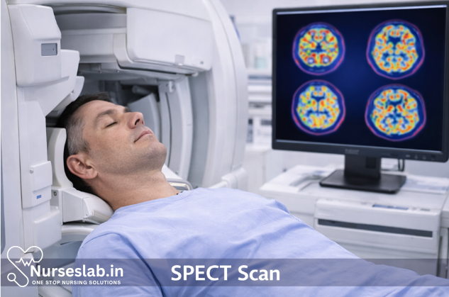
The core technology of SPECT involves the administration of a radiopharmaceutical—a compound labelled with a gamma-emitting radionuclide. After injection, this radiotracer accumulates in specific tissues based on their metabolic activity. A gamma camera, which rotates around the patient, detects the emitted photons and reconstructs cross-sectional images using computer algorithms. These images reflect the spatial distribution and concentration of the radiotracer, thereby indicating regional physiological function.
Comparison with Other Imaging Modalities
SPECT is often compared with Positron Emission Tomography (PET), another functional imaging technique. While both utilise radiotracers, PET employs positron-emitting isotopes and offers higher spatial resolution. However, SPECT is more widely available, less expensive, and uses longer-lived isotopes, making it suitable for a range of clinical scenarios. In contrast to CT and MRI, SPECT focuses on function rather than form, complementing these structural imaging modalities in comprehensive patient evaluation.
Indications and Clinical Uses
Cardiology
One of the most common applications of SPECT is in myocardial perfusion imaging to assess coronary artery disease. It helps in identifying areas of reduced blood flow to the heart muscle, evaluating the extent of ischaemia, and guiding revascularisation decisions.
Neurology
SPECT plays a significant role in the evaluation of cerebral blood flow in patients with stroke, epilepsy, dementia (such as Alzheimer’s disease), and other neurological disorders. It assists in localising seizure foci, differentiating types of dementia, and assessing brain perfusion abnormalities.
Oncology
In oncology, SPECT is utilised for tumour localisation, staging, and monitoring therapeutic response. Certain radiotracers target specific tumour types, enabling precise imaging of cancerous lesions and their metastases.
Orthopaedics and Musculoskeletal Imaging
SPECT is valuable in detecting bone infections, occult fractures, avascular necrosis, and bone metastases. It is particularly useful when conventional radiographs are inconclusive or when functional assessment is required.
Other Applications
Additional uses include renal imaging (for assessing split renal function), pulmonary imaging (for evaluating ventilation-perfusion mismatch), and endocrine imaging (such as parathyroid localisation).
Preparation for the Procedure
Patient Preparation
Proper patient preparation is essential for optimal SPECT scan results and patient safety. The specific instructions may vary depending on the type of scan and the radiotracer used, but general guidelines include:
- Fasting: For certain scans, such as myocardial perfusion imaging, patients may be required to fast for several hours before the procedure.
- Medication Review: Some medications can interfere with radiotracer uptake (e.g., beta-blockers, nitrates, caffeine-containing drugs). Patients should inform the healthcare team about all medications and supplements they are taking.
- Hydration: Adequate hydration is encouraged before and after the scan to facilitate radiotracer elimination.
- Clothing and Accessories: Patients are advised to wear comfortable clothing and remove any metal objects, as these can interfere with imaging quality.
Specific Instructions and Contraindications
Women of childbearing age should inform the radiology team if there is any possibility of pregnancy, as exposure to ionising radiation may pose risks to the foetus. Breastfeeding mothers may be instructed to suspend breastfeeding for a specific period following radiotracer administration, depending on the isotope used.
Absolute contraindications are rare but may include known hypersensitivity to the radiopharmaceutical or inability to cooperate with the scan (e.g., severe claustrophobia or inability to remain still).
Step-by-Step Procedure
1. Radiotracer Administration
The procedure begins with the intravenous injection (occasionally oral or inhaled administration) of a radiotracer appropriate for the target tissue or organ. The choice of radiotracer depends on the clinical indication; for example, technetium-99m (Tc-99m) labelled compounds are commonly used for cardiac and bone scans.
2. Uptake Period
After administration, the patient waits for a predetermined period to allow the radiotracer to localise within the target tissue. This uptake time varies from a few minutes to several hours, depending on the tracer and the organ being imaged.
3. Imaging Acquisition
The patient is positioned on a scanning table, and the gamma camera is aligned accordingly. The camera rotates around the patient, capturing multiple two-dimensional projections from various angles. The process is generally painless but requires the patient to remain still for 30 to 60 minutes to minimise motion artefacts and ensure image clarity.
4. Post-Scan Instructions
Upon completion, patients are typically encouraged to drink fluids to expedite the elimination of the radiotracer from the body. Most individuals can resume normal activities immediately unless otherwise advised.
Patient Experience
The SPECT scan is non-invasive and generally well-tolerated. Patients may experience mild discomfort from the intravenous injection. Claustrophobic individuals may find the enclosed space of the scanner challenging, but most modern SPECT systems are designed to maximise patient comfort.
Risks, Safety, and Contraindications
Radiation Exposure
SPECT scans involve exposure to a small amount of ionising radiation from the radiotracer. The effective dose varies with the type of scan and radiopharmaceutical used, but it is generally within the acceptable limits for diagnostic imaging. The benefits of accurate diagnosis and management typically outweigh the minimal radiation risks.
Potential Side Effects
- Allergic Reactions: Hypersensitivity to the radiotracer is extremely rare but may manifest as mild skin reactions or, in very rare cases, anaphylaxis.
- Injection Site Reactions: Mild pain or redness at the injection site may occur but usually resolves spontaneously.
- Radiation Risks: There is a theoretical risk of cancer with cumulative exposure to ionising radiation, but the risk from a single SPECT scan is negligible.
Safety Measures
Strict protocols are followed to ensure patient safety, including:
- Using the lowest effective dose of radiotracer.
- Screening for pregnancy and breastfeeding status.
- Monitoring for adverse reactions during and after the scan.
Contraindications
Absolute contraindications are rare. Relative contraindications may include:
- Pregnancy or suspected pregnancy (unless benefits outweigh risks).
- Severe allergy to the radiopharmaceutical.
- Inability to cooperate with the procedure, such as severe mobility issues or claustrophobia unresponsive to reassurance or sedation.
Interpretation of Results
Image Reconstruction and Analysis
After data acquisition, specialised software reconstructs the two-dimensional projections into three-dimensional images. Radiologists or nuclear medicine physicians analyse these images to assess the distribution of radiotracer within the target organ.
Common Findings and Their Significance
- Areas of Increased Uptake: Indicate regions of heightened metabolic activity, which may correspond to tumours, infection, or inflammation.
- Areas of Decreased Uptake: Suggest reduced perfusion or function, often seen in ischaemic tissue, infarcts, or necrosis.
Reporting and Follow-up
The interpreting physician generates a detailed report, correlating SPECT findings with clinical history and other investigations. Further diagnostic tests or therapeutic interventions may be recommended based on the scan results.
Advantages and Limitations
Advantages
- Functional Imaging: SPECT provides unique insights into physiological and metabolic processes, complementing structural imaging techniques.
- Wide Availability: SPECT scanners are more widely available and less costly compared to PET.
- Versatility: A broad range of radiotracers allows assessment of various organs and disease processes.
- Non-Invasive: The procedure is minimally invasive and generally well-tolerated by patients.
Limitations
- Lower Resolution: SPECT images have lower spatial resolution compared to PET or CT scans.
- Radiation Exposure: Although minimal, there is still exposure to ionising radiation.
- Motion Artefacts: Patient movement can degrade image quality.
- Limited Anatomical Detail: SPECT is primarily functional; anatomical correlation often requires hybrid imaging (e.g., SPECT/CT).
Nursing Care of Patients Undergoing SPECT Scan Procedure
The procedure involves the administration of a radioactive tracer and the use of a gamma camera to detect emitted photons. Due to the complexity of the procedure and the involvement of radioactive materials, nurses play a vital role in ensuring patient safety, comfort, and optimal outcomes throughout the SPECT scan process.
Pre-Procedure Nursing Care
Patient Assessment and Preparation
Before the SPECT scan, thorough assessment and preparation are essential. The nurse should review the patient’s medical history, current medications, allergies, and prior reactions to contrast agents or radioactive tracers. It is important to identify any contraindications, such as pregnancy or breastfeeding, as radioactive substances may pose risks to the fetus or infant. Nurses should also assess the patient’s understanding of the procedure and address any concerns or anxiety.
- Medical History Review: Check for chronic illnesses (e.g., renal impairment, diabetes), current medications, and allergies.
- Pregnancy and Lactation: Confirm pregnancy status; SPECT is generally contraindicated in pregnant or breastfeeding women unless absolutely necessary.
- Consent: Ensure informed consent is obtained after explaining the procedure, risks, and benefits.
- Patient Education: Explain the purpose of the scan, the use of radioactive tracers, and the steps involved. Reassure the patient regarding radiation safety and address any misconceptions.
Pre-Procedure Instructions
- Fasting: Some SPECT scans, especially cardiac scans, may require fasting for several hours before the procedure. Confirm specific instructions with the physician.
- Medication Management: Advise on withholding certain medications (e.g., beta-blockers, caffeine) as directed. Document any changes to the medication regimen.
- Clothing and Accessories: Instruct the patient to wear comfortable clothing and remove jewelry, metal objects, or accessories that may interfere with imaging.
- Hydration: Encourage adequate hydration unless contraindicated, as it can aid in tracer distribution and post-procedure elimination.
Psychological Support
Many patients experience anxiety before diagnostic procedures. Nurses should provide emotional support, answer questions, and offer reassurance about the safety and necessity of the scan. If the patient is claustrophobic, discuss coping strategies and, if appropriate, arrange for mild sedation as prescribed.
Intra-Procedure Nursing Care
Administration of Radioactive Tracer
The nurse is responsible for the safe handling and administration of the radioactive tracer, which is typically injected intravenously. Adherence to radiation safety protocols is paramount to protect both the patient and healthcare personnel.
- Tracer Preparation: Verify the correct tracer, dose, and patient identity prior to administration. Use appropriate shielding and follow institutional guidelines for handling radioactive materials.
- Injection: Perform the injection using aseptic technique. Monitor for immediate adverse reactions, such as allergic responses or extravasation.
- Documentation: Record the type, dose, time of administration, and any observed reactions in the patient’s chart.
Patient Positioning and Monitoring
Proper positioning is essential for optimal image acquisition. The nurse assists the patient onto the scanning table and ensures they are comfortable, as the scan may take 30 to 60 minutes.
- Comfort Measures: Provide pillows and blankets as needed. Encourage the patient to remain still during imaging.
- Vital Signs: Monitor blood pressure, heart rate, and oxygen saturation, especially if the patient has cardiovascular or neurological conditions.
- Emergency Preparedness: Keep emergency equipment (oxygen, suction, resuscitation kit) readily available in case of adverse reactions.
- Communication: Maintain verbal contact with the patient during the scan. Use intercom systems to provide reassurance and instructions as needed.
Radiation Safety
Nurses must follow strict radiation safety protocols to minimize exposure. This includes wearing personal protective equipment (PPE), using lead shields, and limiting time spent near the patient after tracer administration. Educate the patient about radiation precautions and reassure them that the exposure is minimal and controlled.
Post-Procedure Nursing Care
Immediate Post-Scan Care
- Observation: Monitor for delayed adverse reactions to the tracer, such as nausea, dizziness, or allergic responses.
- Hydration and Elimination: Encourage the patient to drink fluids to facilitate the excretion of the radioactive tracer through urine. Advise frequent voiding and proper handwashing after using the restroom.
- Radiation Precautions: Instruct the patient to avoid close contact with pregnant women and young children for a specified period (usually 24 hours or as per hospital protocol).
- Discharge Instructions: Provide written instructions regarding post-procedure care, signs of complications, and when to seek medical attention.
Patient Education and Follow-Up
Effective patient education is critical for safety and compliance. The nurse should explain the importance of hydration, the expected duration of radioactive tracer in the body, and any required follow-up appointments.
- Results Communication: Inform the patient about when and how results will be communicated. Advise them to contact the healthcare provider with any concerns or symptoms.
- Symptom Monitoring: Instruct the patient to report signs of infection, allergic reactions, or unusual symptoms promptly.
Documentation
Accurate documentation is essential for continuity of care. Record all aspects of the procedure, including patient assessment, tracer administration, vital signs, adverse reactions, patient education, and discharge instructions.
Special Considerations in SPECT Scan Nursing Care
Pediatric Patients
Children may require additional support, including age-appropriate explanations, distraction techniques, and parental presence during the scan. Dosage of the tracer should be carefully calculated based on body weight, and radiation precautions should be strictly followed.
Geriatric Patients
Older adults may have comorbidities, limited mobility, or cognitive impairment. Nurses should assess fall risk, provide extra comfort measures, and ensure clear communication. Monitor for confusion or agitation, which may be exacerbated by unfamiliar environments or procedures.
Patients with Disabilities
Patients with physical or cognitive disabilities may require tailored approaches, such as assistance with positioning, communication aids, and collaboration with family or caregivers.
Collaboration and Interdisciplinary Communication
Nursing care during SPECT scan procedures involves coordination with radiologists, nuclear medicine technologists, and physicians. Effective communication ensures patient safety, timely intervention in case of complications, and accurate interpretation of results.
REFERENCES
- American Heart Association. Single Photon Emission Computed Tomography (SPECT) https://www.heart.org/en/health-topics/heart-attack/diagnosing-a-heart-attack/single-photon-emission-computed-tomography-spect. Last reviewed 7/31/2015.
- ACR–SPR Practice parameter for the performance of single photon emission computed tomography (SPECT) brain perfusion imaging, including brain death examinations. American College of Radiology and the Society for Pediatric Radiology. Revised 2016. https://www.acr.org/Clinical-Resources/Practice-Parameters-and-Technical-Standards/Practice-Parameters-by-Modality.
- National Cancer Institute (U.S.). Molecular and Nuclear Imaging (PET and SPECT https://imaging.cancer.gov/imaging_basics/cancer_imaging/nuclear_imaging.htm. Last reviewed 12/22/2016.
- Jankovic J, et al., eds. Functional and molecular neuroimaging. In: Bradley and Daroff’s Neurology in Clinical Practice. 8th ed. Elsevier; 2022. https://www.clinicalkey.com.
- Yandrapalli S, Puckett Y. SPECT Imaging https://www.ncbi.nlm.nih.gov/books/NBK564426/). 2022 Oct 3. In: StatPearls [Internet]. Treasure Island (FL): StatPearls Publishing.
- General Nuclear Medicine. Radiological Society of North America. https://www.radiologyinfo.org/en/info/gennuclear
Stories are the threads that bind us; through them, we understand each other, grow, and heal.
JOHN NOORD
Connect with “Nurses Lab Editorial Team”
I hope you found this information helpful. Do you have any questions or comments? Kindly write in comments section. Subscribe the Blog with your email so you can stay updated on upcoming events and the latest articles.


