


Atrial tachycardia (AT) is a type of supraventricular tachycardia—a fast heart rhythm originating above the heart’s ventricles, arising specifically from an abnormal electrical focus within the atria. Though less common than other arrhythmias like atrial fibrillation (AF) or atrial flutter, atrial tachycardia holds clinical significance due to its potential impact on cardiac function and its diagnostic and therapeutic challenges.
What is Atrial Tachycardia?
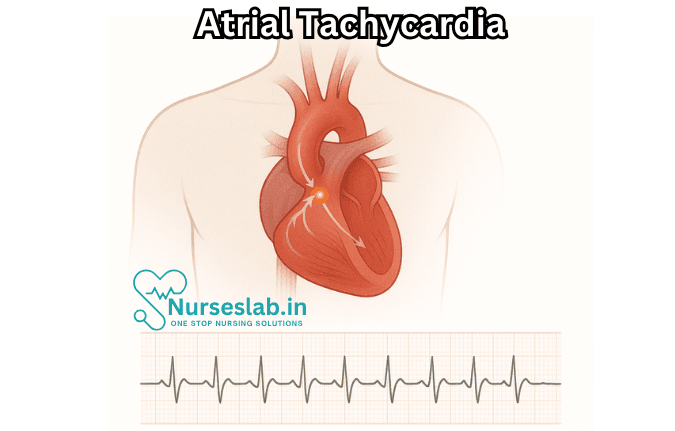
Atrial tachycardia is a form of abnormal heart rhythm in which the atria (the upper chambers of the heart) contract at a rapid rate, typically between 100 and 250 beats per minute. Unlike sinus tachycardia, which is the heart’s normal response to stress or exercise, atrial tachycardia is caused by an ectopic focus an area of the atrium other than the sinoatrial (SA) node firing electrical impulses at a faster rate.
Types of Atrial Tachycardia
- Focal Atrial Tachycardia: Originates from a single point in the atrium, producing regular, rapid atrial activity.
- Multifocal Atrial Tachycardia: Multiple abnormal foci produce varying P-wave morphologies, leading to more irregular rhythms, often seen in patients with underlying lung disease.
- Macroreentrant Atrial Tachycardias: Large circuits of abnormal electrical activity, sometimes seen after cardiac surgery, ablation, or in the setting of scarring within the atria.
Epidemiology and Risk Factors
Atrial tachycardia accounts for approximately 5-15% of supraventricular tachycardias. It can occur in individuals of all ages but is more frequently observed in older adults, especially those with structural heart disease. Risk factors include:
- Previous heart surgery (especially involving the atria)
- Congenital heart disease
- Chronic lung disease (COPD, pulmonary hypertension)
- Coronary artery disease
- Use of certain medications (theophylline, digitalis)
- Electrolyte imbalances (low potassium or magnesium)
- Alcohol intoxication or withdrawal
Pathophysiology
The mechanisms underlying atrial tachycardia usually fall into one of two categories:
- Enhanced Automaticity: Cells outside the SA node develop the ability to generate impulses faster than the natural pacemaker.
- Triggered Activity: Abnormal afterdepolarizations within atrial cells trigger extra beats, often due to electrolyte imbalances or drug effects.
- Reentry Circuits: An electrical impulse travels in a circular pathway within the atria, re-exciting the tissue and causing rapid firing.
Clinical Presentation
Symptoms can range from mild to severe, depending on the rate, duration, and underlying cardiac function. Many individuals with atrial tachycardia experience:
- Palpitations—a rapid, fluttering, or pounding heart sensation
- Dizziness or lightheadedness
- Shortness of breath
- Fatigue or exercise intolerance
- Chest discomfort or pain
- In severe cases: syncope (fainting) or signs of heart failure
In some cases, atrial tachycardia is discovered incidentally on an electrocardiogram (ECG) performed for another reason.
Diagnosis
A careful diagnosis of atrial tachycardia involves a combination of clinical assessment, ECG evaluation, and sometimes advanced electrophysiological studies.
Electrocardiogram (ECG) Findings
Atrial tachycardia is characterized by:
- Atrial rate of 100–250 beats per minute
- P-waves with abnormal morphology (different from sinus P-waves) preceding each QRS complex
- Regular or irregular rhythm, depending on conduction through the AV node
- Ventricular rate that may vary depending on AV conduction
Other Diagnostic Tools
- Holter monitor or event recorder: For intermittent symptoms, these devices capture arrhythmias over 24 hours or longer.
- Electrophysiological study (EPS): Invasive testing to map the source and mechanism of tachycardia, often used prior to ablation.
- Echocardiography: Evaluates structural heart disease or atrial enlargement.
- Blood tests: Assess for electrolyte imbalances, thyroid dysfunction, and drug levels.
Complications
Persistent or recurrent atrial tachycardia can lead to:
- Worsening of heart failure in susceptible individuals
- Cardiomyopathy due to tachycardia-induced ventricular dysfunction (“tachycardia-mediated cardiomyopathy”)
- Increased risk of thromboembolism (though less than in atrial fibrillation)
- Syncope or near-syncope
Management Strategies
Management of atrial tachycardia focuses on controlling symptoms, preventing recurrence, and addressing underlying causes.
Acute Management
- Vagal Maneuvers: Simple physical maneuvers (such as Valsalva or carotid sinus massage) may slow conduction through the AV node and terminate the tachycardia.
- Pharmacological Therapy: Administration of antiarrhythmic drugs such as beta-blockers, calcium channel blockers (verapamil, diltiazem), or adenosine may be effective in slowing or terminating the arrhythmia.
- Direct Current Cardioversion: For unstable patients or refractory tachycardia, synchronized electrical shock may be needed to restore sinus rhythm.
Long-Term Management
- Catheter Ablation: Radiofrequency ablation of the arrhythmogenic focus offers a high success rate and is considered first-line therapy for recurrent or drug-resistant cases.
- Medication: Long-term use of antiarrhythmic drugs (e.g., beta-blockers, calcium channel blockers, Class IC or III agents) may be indicated for some patients, especially those who are not candidates for ablation.
- Addressing Underlying Conditions: Correcting electrolyte disturbances, treating structural heart disease, or discontinuing pro-arrhythmic drugs is crucial.
- Lifestyle Modifications: Reducing alcohol intake, managing stress, treating sleep apnea, and optimizing overall cardiovascular health can decrease recurrence.
Prognosis
The prognosis for atrial tachycardia is generally favorable, especially with prompt identification and management. Catheter ablation can be curative in many cases. However, persistent, untreated AT can lead to significant morbidity, particularly in patients with existing heart disease.
Special Considerations
Atrial Tachycardia in Children
AT in pediatric populations is relatively rare but can be seen in congenital heart disease or as an isolated electrical abnormality. Symptoms often include palpitations, poor feeding (in infants), and failure to thrive. Management principles are similar but tailored to age and comorbidities.
Atrial Tachycardia After Cardiac Surgery
Postoperative AT may arise due to atrial scarring and inflammation. It tends to be more resistant to medications and often requires ablation or other advanced interventions.
Nursing Care of Patients with Atrial Tachycardia
Assessment and Monitoring
- Vital Signs Monitoring: Regularly monitor heart rate, blood pressure, respiratory rate, and oxygen saturation. Be alert for signs of haemodynamic instability such as hypotension, syncope, or chest pain.
- Electrocardiogram (ECG): Obtain and interpret ECGs to identify atrial tachycardia and monitor for changes or complications.
- Observation for Symptoms: Watch for palpitations, dizziness, breathlessness, fatigue, and signs of heart failure.
- Fluid and Electrolyte Balance: Monitor intake and output, and check for electrolyte disturbances, especially potassium and magnesium levels.
Acute Management
- Ensure Safety: Place the patient in a safe, comfortable environment and provide bed rest if haemodynamically unstable.
- Oxygen Therapy: Administer supplemental oxygen if the patient is hypoxic.
- Medication Administration: Administer prescribed antiarrhythmic drugs (e.g., beta-blockers, calcium channel blockers) as per physician’s orders. Monitor for adverse effects.
- Preparation for Procedures: Prepare the patient for possible interventions such as electrical cardioversion or catheter ablation if indicated.
Patient Education
- Understanding the Condition: Explain the nature of atrial tachycardia, possible triggers, and risk factors in simple language.
- Medication Adherence: Educate the patient on the importance of taking medications regularly and not stopping them without consulting the doctor.
- Lifestyle Modifications: Advise on reducing caffeine, avoiding tobacco and alcohol, and managing stress to prevent recurrence.
- Recognising Warning Signs: Instruct the patient and family to seek immediate medical attention if symptoms worsen or new symptoms appear.
Prevention of Complications
- Monitor for Thromboembolism: Assess for signs of stroke or embolic events, especially in patients with persistent arrhythmia.
- Prevent Heart Failure: Monitor for signs such as peripheral oedema, breathlessness, and weight gain.
- Skin Integrity: Prevent pressure sores in bedridden patients by regular repositioning and skin care.
Psychosocial Support
- Emotional Support: Listen to patient concerns, provide reassurance, and involve family members in care.
- Counselling: Refer to counselling services if anxiety or depression is noted due to chronic illness.
Documentation
- Record all assessments, interventions, and patient responses in the nursing notes.
- Document medication administration and any adverse reactions observed.
- Note patient education provided and understanding demonstrated.
REFERENCES
- American Heart Association. Tachycardia: Fast Heart Rate. https://www.heart.org/en/health-topics/arrhythmia/about-arrhythmia/tachycardia–fast-heart-rate Last reviewed 9/24/2024.
- Lee BK. Supraventricular Tachycardias. In: Crawford MH, eds. Current Diagnosis & Treatment: Cardiology. 6th ed. McGraw-Hill Education; 2023.
- Liwanag M, Willoughby C. Atrial Tachycardia. https://pubmed.ncbi.nlm.nih.gov/31194392/. 2023 Jun 26. In: StatPearls [Internet]. Treasure Island (FL): StatPearls Publishing; 2025 Jan.
- Sohinki D, Obel OA. Current trends in supraventricular tachycardia management. https://pubmed.ncbi.nlm.nih.gov/25598724/. Ochsner J. 2014;14(4):586-595.
- Spragg D, Calkins H. Supraventricular Tachycardia: Atrial Tachycardia, Atrioventricular Nodal Reentry, and Wolff-Parkinson-White Syndrome. In: Fuster V, Narula J, Vaishnava P, et al., eds. Fuster and Hurst’s The Heart. 15th ed. McGraw-Hill Education; 2022.
- AskMayoExpert. Supraventricular tachycardia. Mayo Clinic; 2024.
- Liwanag M, Willoughby C. Atrial Tachycardia. [Updated 2023 Jun 26]. In: StatPearls [Internet]. Treasure Island (FL): StatPearls Publishing; 2025 Jan-. Available from: https://www.ncbi.nlm.nih.gov/books/NBK542235/
- Ferri FF. Supraventricular tachycardia. In: Ferri’s Clinical Advisor 2025. Elsevier; 2025. https://www.clinicalkey.com.
- Brugada J, et al. 2019 ESC guidelines for the management of patients with supraventricular tachycardia: The Task Force for the management of patients with supraventricular tachycardia of the European Society of Cardiology (ESC). European Heart Journal. 2020; doi:10.1093/eurheartj/ehz467.
Stories are the threads that bind us; through them, we understand each other, grow, and heal.
JOHN NOORD
Connect with “Nurses Lab Editorial Team”
I hope you found this information helpful. Do you have any questions or comments? Kindly write in comments section. Subscribe the Blog with your email so you can stay updated on upcoming events and the latest articles.


