


Last Updated on October 21, 2026 by Nurseslab.in Editorial Team
Breast cancer is the most commonly diagnosed cancer worldwide, and almost every nurse will care for a patient living with it — on an oncology unit, in surgery, in the community, or incidentally while the patient is admitted for something else entirely.
Anatomy and physiology
The breast is a modified sweat gland lying over the pectoralis major, extending from the second to sixth rib and from the sternal edge to the midaxillary line. The tail of Spence extends into the axilla, which is why breast tissue — and breast cancer — can appear in the armpit.
Internally, each breast contains 15 to 20 lobes, subdivided into lobules containing the acini (alveoli) that produce milk. Milk drains through lactiferous ducts converging at the nipple. Cooper’s ligaments are fibrous bands suspending the breast from the chest wall — when a tumour invades and shortens these ligaments, it produces the characteristic skin dimpling seen on inspection.
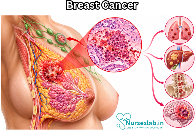
Lymphatic drainage is the key anatomical concept for staging. Roughly 75% of breast lymph drains to the axillary nodes, with the remainder going to internal mammary and supraclavicular nodes. This is why axillary assessment determines nodal staging, and why axillary surgery carries the risk of lymphedema.
Clinically: most breast cancers arise in the ducts rather than the lobules, and the upper outer quadrant is the most common site, because it contains the greatest volume of glandular tissue.
Pathophysiology
Breast cancer begins when genetic damage in epithelial cells of the terminal duct lobular unit disrupts normal regulation of cell growth and apoptosis. Accumulated mutations in proto-oncogenes and tumour suppressor genes — including TP53, PIK3CA, BRCA1, and BRCA2 — produce a clone of cells that proliferates without normal constraint.
The progression follows a recognizable sequence:
- Hyperplasia — increased cell number with normal architecture
- Atypical hyperplasia — abnormal cells, still confined
- Carcinoma in situ — malignant cells that have not breached the basement membrane
- Invasive carcinoma — cells penetrate the basement membrane into surrounding stroma
- Metastasis — spread via lymphatics and bloodstream
The basement membrane is the critical boundary. In situ disease cannot metastasize, because it has no access to lymphatics or vessels. Once that membrane is breached, the disease becomes systemic in potential — which is the entire rationale for systemic adjuvant therapy even after apparently complete surgical removal.
Common metastatic sites: bone, lung, liver, and brain. A useful mnemonic is that breast cancer metastasizes to Bone, Brain, Lung, Liver. Bone is the most frequent, which is why new back pain in a patient with a breast cancer history is never dismissed as musculoskeletal without thought.
Hormonal influence matters pathophysiologically: oestrogen stimulates proliferation in hormone receptor–positive tumours, which explains why lifetime oestrogen exposure is a risk factor and why endocrine blockade is a treatment.
Classification
Non-invasive (in situ)
Ductal carcinoma in situ (DCIS). Malignant cells confined within the duct. Often detected as microcalcifications on mammography rather than as a palpable mass. Considered a true precursor lesion — treated to prevent progression to invasive disease.
Lobular carcinoma in situ (LCIS). Now regarded less as a cancer and more as a marker of increased risk in both breasts. Typically an incidental biopsy finding, usually managed with surveillance rather than resection.
Invasive
Invasive ductal carcinoma (IDC) — approximately 70–80% of cases. The commonest type; presents as a hard, irregular, fixed, usually painless mass.
Invasive lobular carcinoma (ILC) — roughly 10–15%. Grows in a single-file, diffuse pattern rather than as a discrete lump, which makes it harder to detect on mammography and on palpation. More often bilateral and multicentric. A patient describing “thickening” rather than a lump deserves attention.
Special clinical presentations
Inflammatory breast cancer (IBC). Uncommon but aggressive. Tumour emboli obstruct dermal lymphatics, producing redness, warmth, oedema, and peau d’orange (orange-peel skin) — frequently without a palpable mass. It is routinely misdiagnosed as mastitis. Rule to carry: mastitis in a non-lactating woman, or mastitis not resolving on antibiotics, requires urgent referral.
Paget disease of the breast. Malignant cells invade the nipple epidermis, producing eczematous, scaling, crusting or ulcerating changes of the nipple and areola, often with itching or burning. Frequently treated as dermatitis for months. Key distinguishing feature: Paget disease typically starts at the nipple and spreads outward, while eczema usually spares the nipple.
Molecular subtypes — the framework that drives treatment
This is the single most important modern classification, and it’s where many students lose marks.
| Subtype | Receptors | Approximate share | Treatment backbone |
|---|---|---|---|
| Luminal A | ER+/PR+, HER2−, low Ki-67 | ~40% | Endocrine therapy; best prognosis |
| Luminal B | ER+/PR±, HER2±, high Ki-67 | ~20% | Endocrine ± chemotherapy |
| HER2-enriched | ER−/PR−, HER2+ | ~15% | HER2-targeted therapy + chemotherapy |
| Triple-negative (TNBC) | ER−, PR−, HER2− | ~15% | Chemotherapy ± immunotherapy; worst prognosis |
Why it matters clinically: receptor status determines whether a patient receives endocrine therapy, HER2-directed therapy, chemotherapy, or a combination. A nurse who understands that a triple-negative patient cannot be given tamoxifen — because there is no receptor to block — understands the logic of the entire treatment plan.
HER2-low and HER2-ultralow. A significant recent development. Tumours once simply called “HER2-negative” are now recognized as a spectrum, and those with low-level HER2 expression (<cite index=”2-1″>immunohistochemistry 1+, or 2+ with negative in situ hybridization</cite>) can respond to antibody–drug conjugates. <cite index=”2-2″>An even lower category, HER2-ultralow, describes faint, incomplete staining in 10% or fewer of tumour cells</cite>. Trials in this space — notably the DESTINY-Breast series — have extended targeted therapy to patients previously considered ineligible for it.
Risk factors
Non-modifiable
- Female sex — the strongest risk factor; male breast cancer accounts for roughly 1% of cases
- Increasing age
- Family history of breast or ovarian cancer
- Inherited mutations, particularly BRCA1 and BRCA2 (substantially elevated lifetime risk; BRCA1 is associated with triple-negative disease)
- Early menarche (<12 years) and late menopause (>55 years) — greater lifetime oestrogen exposure
- Nulliparity or first full-term pregnancy after age 30
- Dense breast tissue
- Previous chest wall radiation, especially in adolescence
- Personal history of breast cancer or atypical hyperplasia
Modifiable
- Alcohol intake — dose-dependent
- Overweight and obesity, particularly post-menopausal (adipose tissue produces oestrogen)
- Physical inactivity
- Combined hormone replacement therapy
- Never having breastfed
Teach this honestly. Most women diagnosed with breast cancer have no identifiable risk factor beyond being female and growing older. Over-emphasizing lifestyle implies patients caused their disease.
Clinical manifestations
The classic presentation is a painless, hard, irregular, fixed, non-mobile mass, most often in the upper outer quadrant. But fixating on the lump is the most harmful simplification in breast cancer teaching.
Teach patients and students to report:
- A new lump or thickening in the breast or axilla
- Change in breast size, shape, or contour
- Skin dimpling or puckering (Cooper’s ligament retraction)
- Peau d’orange — oedema with exaggerated pores
- Nipple retraction or inversion that is new
- Nipple discharge, especially bloody, unilateral, spontaneous, or from a single duct
- Persistent redness, warmth, or swelling of the breast
- Nipple or areolar scaling, crusting, or ulceration
- Persistent, localized, non-cyclical breast pain
- Palpable axillary or supraclavicular lymph nodes
Benign versus malignant on examination (classic exam distinction):
| Feature | Benign (e.g. fibroadenoma) | Malignant |
|---|---|---|
| Consistency | Soft to firm, rubbery | Hard, stony |
| Borders | Well-defined | Irregular, poorly defined |
| Mobility | Freely mobile | Fixed to skin or chest wall |
| Tenderness | Often tender, cyclical | Usually painless |
| Skin changes | Absent | Dimpling, peau d’orange, retraction |
Late and metastatic signs: unexplained weight loss, bone pain, pathological fracture, hypercalcaemia, dyspnoea, jaundice, headache or neurological deficit.
Diagnostic evaluation
Triple assessment
The standard diagnostic approach combines three elements, and a discordant result in any one mandates further investigation:
- Clinical examination
- Imaging — mammography and/or ultrasound
- Pathology — needle biopsy
Imaging
- Mammography — primary screening tool; detects microcalcifications and masses. Less sensitive in dense breast tissue, which is why it performs less well in younger women.
- Ultrasound — distinguishes solid from cystic lesions; preferred as first-line imaging in women under about 30, in pregnancy, and for guiding biopsy.
- MRI — high sensitivity; used for high-risk screening (including BRCA carriers), assessing extent of disease, evaluating implants, and monitoring response to neoadjuvant therapy.
- Staging imaging — CT, bone scan, or PET-CT where metastatic disease is suspected.
Biopsy
Core needle biopsy is the standard, because it preserves tissue architecture and allows receptor testing. Fine needle aspiration yields cytology only and cannot reliably distinguish in situ from invasive disease. Excisional biopsy is reserved for cases where needle biopsy is non-diagnostic.
Pathology and molecular testing
Every invasive cancer should have:
- ER, PR, HER2 status
- Histological grade (1–3) and Ki-67 proliferation index
- Genomic assays — Oncotype DX or MammaPrint — in selected ER-positive, node-negative disease, to determine whether chemotherapy adds benefit beyond endocrine therapy
- Genetic testing for BRCA1/2 and other mutations where family history, young age, triple-negative disease, or male sex suggests hereditary predisposition
Tumour markers (CA 15-3, CA 27.29, CEA) are used for monitoring in advanced disease, not for diagnosis or screening.
Staging
Staging uses the TNM system: tumour size, nodal involvement, and distant metastasis. Current AJCC staging incorporates both anatomic factors and prognostic factors — grade and receptor status — so two tumours of identical size can be staged differently based on biology.
Simplified for teaching:
- Stage 0 — carcinoma in situ (DCIS)
- Stage I — tumour ≤2 cm, node-negative
- Stage II — larger tumour and/or limited nodal involvement
- Stage III — locally advanced; extensive nodal involvement, chest wall or skin involvement, or inflammatory breast cancer
- Stage IV — distant metastasis
Tumour size thresholds worth memorizing: T1 ≤2 cm, T2 >2–5 cm, T3 >5 cm, T4 = chest wall or skin involvement (T4d is inflammatory breast cancer).
Five-year survival falls steeply with stage, which is the entire argument for early detection — and the basis of the WHO target that at least 60% of invasive cancers be diagnosed at stage I or II.
Treatment modalities
Surgery
Breast-conserving surgery (lumpectomy) plus radiation offers survival equivalent to mastectomy in appropriately selected patients. This is a frequently tested point and an important one for patient counselling — many patients assume mastectomy is inherently safer.
Mastectomy types include total (simple), modified radical (breast plus axillary nodes, preserving pectoralis major), and skin- or nipple-sparing approaches where reconstruction is planned. Radical mastectomy, which removed the pectoral muscles, is now of historical interest only.
Axillary management:
- Sentinel lymph node biopsy (SLNB) — the first draining node is identified with radioisotope and/or blue dye. If negative, further axillary surgery is avoided, substantially reducing lymphedema risk.
- Axillary lymph node dissection (ALND) — reserved for proven significant nodal disease; carries considerably higher lymphedema and nerve injury risk.
Radiation therapy
Standard after breast-conserving surgery; also used post-mastectomy for large tumours or significant nodal involvement, and palliatively for bone or brain metastases. Side effects include fatigue, skin reaction (erythema, dry then moist desquamation), and, uncommonly, pneumonitis or lymphedema.
Systemic therapy
Chemotherapy. Common regimens combine anthracyclines (doxorubicin) with cyclophosphamide and a taxane (paclitaxel, docetaxel). May be neoadjuvant — given before surgery to downstage a tumour, enable breast conservation, and assess in-vivo response — or adjuvant.
Endocrine therapy (hormone receptor–positive disease only):
- Tamoxifen — a selective oestrogen receptor modulator; used in pre- and post-menopausal women
- Aromatase inhibitors — anastrozole, letrozole, exemestane; post-menopausal women only, because they block peripheral conversion of androgens and do not suppress ovarian oestrogen production
- Ovarian suppression — GnRH agonists such as goserelin in premenopausal patients
- Duration is typically 5 to 10 years
HER2-targeted therapy: trastuzumab, pertuzumab, and the antibody–drug conjugates T-DM1 (ado-trastuzumab emtansine) and trastuzumab deruxtecan (T-DXd). T-DXd in particular has <cite index=”2-1″>extended benefit to HER2-low and HER2-ultralow tumours</cite>, redrawing the line between “HER2-positive” and “HER2-negative” disease.
CDK4/6 inhibitors — palbociclib, ribociclib, abemaciclib — combined with endocrine therapy in HR-positive, HER2-negative advanced disease, and increasingly in high-risk early disease.
Immunotherapy — pembrolizumab, in combination with chemotherapy, for triple-negative disease.
PARP inhibitors — olaparib, talazoparib — for BRCA-mutated disease, exploiting synthetic lethality in cells with defective DNA repair.
Nursing Care of the Patient with Breast Cancer
Breast cancer care is not a single episode. A patient may move through diagnosis, surgery, chemotherapy, radiation, and years of endocrine therapy — each phase with its own risks, its own teaching priorities, and its own version of what “good nursing care” looks like.
Nursing assessment
Health history
Presenting concern
- Onset, duration, and progression of the lump or change
- Associated symptoms: pain, nipple discharge, skin change, axillary swelling
- Whether the change was self-detected or screen-detected
Risk factor review
- Age, menarche and menopause history, parity, age at first full-term pregnancy
- Breastfeeding history
- Family history of breast or ovarian cancer; known BRCA1/2 or other mutations
- Previous breast biopsies or atypical hyperplasia
- Hormone replacement or oral contraceptive use
- Previous chest irradiation
- Alcohol intake, activity level, weight history
General history
- Comorbidities, particularly cardiac disease (relevant to anthracyclines and trastuzumab)
- Current medications, supplements, and herbal products
- Allergies, including contrast and latex
- Previous surgical and anaesthetic history
Physical assessment
- Inspection with the patient’s arms at the sides, raised overhead, and pressed on hips: symmetry, contour, skin dimpling, peau d’orange, erythema, nipple retraction or discharge, nipple or areolar scaling or ulceration
- Palpation of all four quadrants, the tail of Spence, and the nipple-areolar complex; document any mass by location (clock position and distance from the nipple), size, consistency, borders, mobility, and tenderness
- Regional nodes: axillary, supraclavicular, infraclavicular
- Systemic review for metastatic signs: bone pain or tenderness, respiratory status, abdominal examination, neurological assessment, weight trend
Psychosocial assessment
- Understanding of the diagnosis and treatment plan
- Emotional response: fear, anger, denial, grief
- Body image and sexual concerns
- Support system, caregiving responsibilities, and employment impact
- Financial concerns, transport, and ability to attend appointments
- Cultural and religious considerations affecting care decisions
- Health literacy and preferred language
- Screen for depression and anxiety — both are markedly elevated in this population
Diagnostic data to review
Imaging reports and BI-RADS category; core biopsy histology; ER, PR, HER2 status; grade and Ki-67; genomic assay results where performed; baseline CBC, renal and liver function; baseline LVEF (echocardiogram or MUGA) where cardiotoxic agents are planned; and genetic testing results where applicable.
Priority nursing diagnoses
- Anxiety / Fear related to cancer diagnosis, treatment, and prognosis
- Acute Pain related to surgical incision and tissue trauma
- Risk for Infection related to surgical wound and treatment-induced immunosuppression
- Impaired Skin Integrity related to surgery and radiation
- Risk for Peripheral Neurovascular Dysfunction / Lymphedema related to lymph node dissection and radiation
- Impaired Physical Mobility related to surgical pain and restricted shoulder movement
- Disturbed Body Image related to loss of a breast and treatment effects
- Ineffective Sexuality Pattern related to body image change and treatment-induced oestrogen deprivation
- Fatigue related to disease process and treatment
- Imbalanced Nutrition: Less than Body Requirements related to nausea, vomiting, and mucositis
- Grieving related to loss of body part, fertility, and perceived future
- Deficient Knowledge regarding disease, treatment, self-care, and surveillance
- Risk for Ineffective Health Management related to long-duration endocrine therapy
Phase-specific nursing management
Pre-operative
- Verify informed consent and that the surgical site is correctly marked — critical in a bilateral organ
- Confirm understanding of the planned procedure, including whether axillary surgery and reconstruction are involved
- Record baseline bilateral arm circumference
- Teach post-operative arm exercises, drain care, and deep breathing
- Explain expected appearance of the incision and the presence of drains
- Discuss fertility preservation before treatment begins where relevant — this conversation is frequently omitted and patients report it as a major regret
- Address anxiety and arrange specialist nurse contact
Immediate post-operative
- Position in semi-Fowler’s with the affected arm elevated on a pillow
- Assess the dressing and check beneath the patient — blood tracks posteriorly and pooling is easily missed
- Monitor vital signs; observe for haematoma, excessive bleeding, and seroma
- Assess circulation, sensation, and movement of the affected limb
- Manage pain proactively; begin early mobilization of hand and elbow
- Place the “no BP / no venipuncture” alert on the affected side
- Encourage deep breathing and early ambulation
Drain management
- Maintain compression suction; empty and record output each shift
- Expect serosanguineous drainage progressing to serous over several days
- Teach the patient to strip tubing, empty, measure, and record at home
- Teach reporting of: sudden cessation of drainage, bright red bleeding, foul odour, purulent drainage, or fever
- Drains are generally removed when output falls below the surgeon’s threshold over 24 hours — commonly around 30 mL, though protocols vary
- Reassure that seroma after drain removal is common and can be aspirated
During chemotherapy
- Verify baseline LVEF before anthracyclines or trastuzumab, and monitor serially; teach reporting of breathlessness, orthopnoea, oedema, and palpitations
- Ensure patent venous access before vesicants such as doxorubicin; monitor the site continuously and know the extravasation protocol
- Warn that red-tinged urine after doxorubicin is expected and harmless
- Hydrate and encourage frequent voiding with cyclophosphamide; report haematuria
- Assess for peripheral neuropathy with taxanes at every visit; teach foot inspection, safe footwear, and caution with hot water
- Premedicate for taxane hypersensitivity per protocol and monitor closely during the first infusions
- With trastuzumab deruxtecan, teach urgent reporting of new cough, breathlessness, or fever — interstitial lung disease is the key toxicity
- With immune checkpoint inhibitors, teach immune-related effects: persistent diarrhoea, rash, jaundice, breathlessness, and symptoms of thyroid or adrenal dysfunction
- With CDK4/6 inhibitors, monitor neutrophils; expect diarrhoea with abemaciclib and QT effects with ribociclib
During radiation therapy
- Teach gentle washing with lukewarm water and mild soap; pat dry, do not rub
- Avoid perfumed products, deodorant on the treated side unless approved, and adhesive tape on the field
- Advise loose cotton clothing and avoidance of underwire bras
- Do not remove skin markings
- Avoid extremes of temperature — heating pads, ice packs, direct sun
- Apply only approved moisturizers, and not within the hours before treatment
- Assess for erythema, dry and moist desquamation; escalate moist desquamation
- Anticipate and manage fatigue, which accumulates over the course
Survivorship
- Reinforce surveillance: clinical follow-up, mammography of remaining tissue, and adherence to endocrine therapy
- Teach breast awareness and signs of recurrence: new lumps or chest wall nodules, persistent bone pain, unexplained weight loss, persistent cough, neurological symptoms
- Address bone health where aromatase inhibitors are used
- Support return to work, exercise, alcohol reduction, and weight management — framed forward-looking, never as blame
- Screen for ongoing anxiety, depression, and fear of recurrence, which commonly intensifies once active treatment ends and contact with the team reduces
Advanced and metastatic disease
- Shift emphasis to symptom control and quality of life
- Assess and manage pain comprehensively, including bone pain and pathological fracture risk
- Recognize oncological emergencies: spinal cord compression (new back pain with neurological signs — a medical emergency), hypercalcaemia, superior vena cava obstruction, and febrile neutropenia
- Support advance care planning and goals-of-care conversations at the patient’s pace
- Involve palliative care early — it improves both quality of life and symptom burden
- Remember that patients with metastatic disease will not “finish treatment”; avoid survivorship framing that excludes them
Evaluation criteria
Care is effective when the patient:
- Reports pain and nausea within an acceptable range
- Remains free of wound infection and sepsis
- Maintains baseline arm circumference and demonstrates lymphedema precautions
- Achieves the expected shoulder range of motion
- Participates in incision care and verbalizes an adjusting body image
- Maintains weight and adequate hydration
- Verbalizes the treatment plan, side effects to report, and when to seek urgent help
- Continues prescribed endocrine therapy and attends surveillance appointments
- Uses available support systems and reports manageable emotional distress
Documentation essentials
Record: baseline and serial bilateral arm circumference; incision and drain site appearance; drain output volume and character each shift; pain scores before and after intervention; range of motion achieved; temperature and ANC trends during chemotherapy; LVEF results; all teaching delivered with evidence of teach-back; psychosocial assessment and referrals made; and the patient’s stated goals and concerns in her own words.
REFERENCES
- Abdulrahman Bin Sumaida, Nandan Maruti Shanbhag,Breast Cancer: A Comprehensive Clinical Guide, Published: 15 October 2026 https://doi.org/10.1007/978-981-92-5842-0
- American Society of Clinical Oncology. Breast Cancer: Introduction https://www.cancer.net/cancer-types/breast-cancer/introduction). Multiple pages reviewed. Last updated 10/2022.
- Breastcancer.org. Metastatic Breast Cancer. https://www.breastcancer.org/types/metastatic#section-what-is-metastatic-breast-cancer.
- Centers for Disease Control and Prevention (U.S.). Breast Cancer. https://www.cdc.gov/cancer/breast/. Multiple pages reviewed. Last reviewed 9/26/2022.
- National Cancer Institute. Breast Cancer. https://www.cancer.gov/types/breast?redirect=true-Patient Version.
Stories are the threads that bind us; through them, we understand each other, grow, and heal.
JOHN NOORD
Connect with “Nurses Lab Editorial Team”
I hope you found this information helpful. Do you have any questions or comments? Kindly write in comments section. Subscribe the Blog with your email so you can stay updated on upcoming events and the latest articles.


