


Gram‑negative bacilli infections caused by Enterobacterales and Vibrio cholerae are major contributors to gastrointestinal illness worldwide. This topic explores important pathogens, routes of transmission, clinical manifestations, diagnostic methods, and nursing interventions.
Introduction
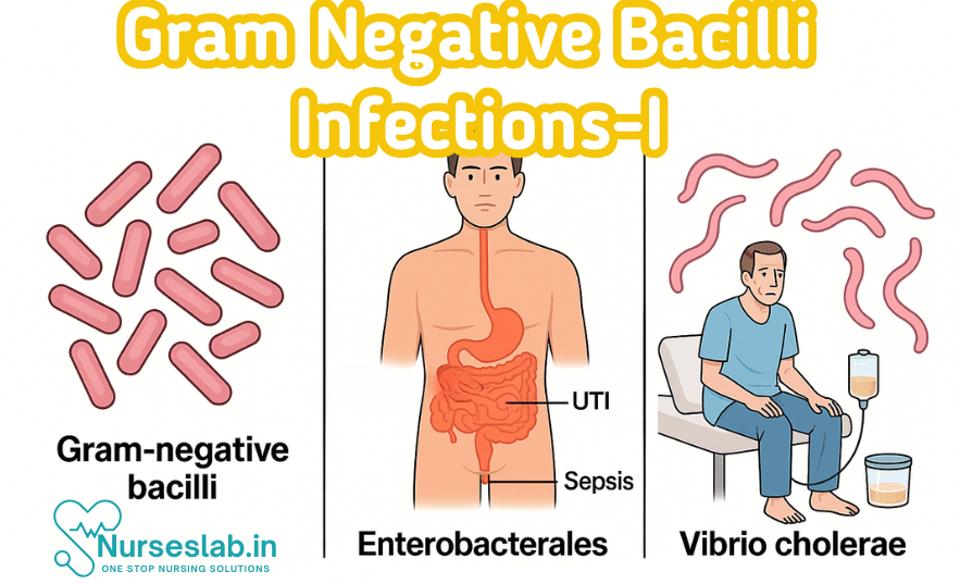
Gram negative bacilli (GNB) are a diverse group of rod-shaped bacteria characterised by their thin peptidoglycan layer and the presence of an outer membrane containing lipopolysaccharide (LPS). These organisms are of immense clinical significance due to their propensity to cause a wide range of infections, their ability to develop antimicrobial resistance, and their impact on global health. Among GNB, Enterobacterales and Vibrio cholerae are particularly noteworthy for their epidemiological relevance, variety of clinical manifestations, and challenges in diagnosis and management.
Enterobacterales
Definition and Classification
Enterobacterales is an order of Gram negative, facultatively anaerobic bacilli within the class Gammaproteobacteria. Traditionally known as Enterobacteriaceae, this group has undergone taxonomic revisions, now encompassing several genera that are clinically significant. Members of Enterobacterales are ubiquitous in nature, commonly colonising the human gastrointestinal tract, and are notorious for causing both community-acquired and nosocomial infections.
Major genera and species of clinical importance include:
- Escherichia coli
- Klebsiella species (notably K. pneumoniae, K. oxytoca)
- Enterobacter species (e.g., E. cloacae, E. aerogenes)
- Proteus species (P. mirabilis, P. vulgaris)
- Serratia (S. marcescens)
- Citrobacter (C. freundii, C. koseri)
- Salmonella (S. enterica, S. typhi)
- Shigella (S. dysenteriae, S. flexneri, S. sonnei, S. boydii)
- Yersinia (Y. enterocolitica, Y. pestis)
These organisms share certain characteristics: they are oxidase-negative, ferment glucose, and reduce nitrate to nitrite. Their identification relies on a combination of biochemical assays and molecular techniques.
Types of Infections Caused by Enterobacterales
Enterobacterales are responsible for a broad spectrum of infections affecting various organ systems. The most common types include:
- Urinary Tract Infections (UTIs): E. coli is the leading cause of community-acquired UTIs, while Klebsiella and Proteus are significant in hospital settings.
- Respiratory Tract Infections: Klebsiella pneumoniae is a known cause of pneumonia, especially in immunocompromised individuals and alcoholics.
- Gastrointestinal Infections: Salmonella, Shigella, and certain strains of E. coli (e.g., EHEC, EPEC, ETEC) are important agents of diarrhoeal diseases.
- Sepsis and Bloodstream Infections: E. coli, Klebsiella, and Enterobacter species frequently cause bacteraemia, especially in hospitalised patients.
- Wound and Soft Tissue Infections: Proteus and Klebsiella may infect wounds, burns, and surgical sites, often as part of polymicrobial flora.
- Meningitis: E. coli and Citrobacter are notable causes of neonatal meningitis.
- Other Infections: Yersinia pestis (plague), Y. enterocolitica (gastroenteritis), and Serratia marcescens (opportunistic infections).
Clinical Manifestations
The clinical presentation varies by species and site of infection. Below is a summary of key features for major genera:
Escherichia coli
- UTI: Dysuria, frequency, urgency, suprapubic pain; pyelonephritis presents with fever, flank pain.
- Gastroenteritis: Watery or bloody diarrhoea, abdominal cramps (depending on pathotype: ETEC, EPEC, EHEC, etc.).
- Neonatal Meningitis: Fever, irritability, poor feeding, seizures.
- Sepsis: Fever, hypotension, multi-organ dysfunction.
Klebsiella pneumoniae
- Pneumonia: High-grade fever, productive cough with “currant jelly” sputum, chest pain, dyspnoea.
- UTI: Similar to E. coli, often associated with underlying urinary tract abnormalities.
- Liver abscess: Fever, right upper quadrant pain, jaundice (invasive strains).
Proteus species
- UTI: Often associated with struvite stone formation, alkaline urine due to urease activity.
- Wound infections: Purulent discharge, delayed healing, malodorous wounds.
Enterobacter and Citrobacter
- Nosocomial infections: Pneumonia, sepsis, UTIs, often in patients with indwelling devices or immunosuppression.
- Neonatal meningitis (Citrobacter): Severe neurological sequelae.
Salmonella and Shigella
- Gastroenteritis: Fever, diarrhoea (may be bloody), abdominal pain.
- Enteric fever (Salmonella Typhi): Prolonged fever, rose spots, hepatosplenomegaly.
- Dysentery (Shigella): Acute onset bloody diarrhoea, tenesmus.
Yersinia
- Plague (Y. pestis): Buboes, high fever, septicaemia.
- Gastroenteritis (Y. enterocolitica): Abdominal pain mimicking appendicitis, diarrhoea.
Serratia marcescens
- Opportunistic infections: Pneumonia, UTI, bacteraemia in hospitalised patients.
Laboratory Diagnosis
Accurate laboratory diagnosis is crucial for guiding effective management and infection control. The process involves:
Specimen Collection
- Urine (for UTIs)
- Blood (for sepsis, bacteraemia)
- Stool (for gastrointestinal infections)
- CSF (for meningitis)
- Respiratory secretions (for pneumonia)
- Wound swabs or pus (for soft tissue infections)
Microscopy
- Gram stain: Reveals Gram negative rods; direct visualisation may be limited except in urine or pus samples.
Cultural Methods
- Media: MacConkey agar (lactose fermenters vs. non-fermenters), blood agar, selective media for Salmonella and Shigella.
- Colony morphology: E. coli typically produces pink colonies on MacConkey agar (lactose fermenter); Proteus shows swarming on blood agar.
Biochemical Tests
- IMViC tests (Indole, Methyl Red, Voges-Proskauer, Citrate): Differentiates E. coli (Indole +, MR +, VP -, Citrate -) from Klebsiella (Indole -, MR -, VP +, Citrate +).
- Urease test: Positive in Proteus and Klebsiella.
- Triple Sugar Iron (TSI) agar: Assesses fermentation of glucose, lactose, sucrose, and H2S production.
Serotyping and Molecular Methods
- Serotyping (O, H, K antigens) for E. coli, Salmonella, Shigella.
- PCR-based detection of specific virulence genes (e.g., stx for EHEC).
- Automated systems (VITEK, MALDI-TOF MS) for rapid identification.
Antimicrobial Susceptibility Testing
- Disc diffusion (Kirby-Bauer), automated systems, broth microdilution for Minimum Inhibitory Concentration (MIC) determination.
- Detection of Extended-Spectrum Beta-Lactamases (ESBLs), carbapenemases (e.g., KPC, NDM), and other resistance mechanisms.
Preventive Measures
Prevention of Enterobacterales infections requires multifaceted strategies:
- Infection Control: Strict hand hygiene, use of personal protective equipment (PPE), isolation of infected patients, and environmental cleaning in healthcare settings.
- Antibiotic Stewardship: Rational use of antibiotics to curb resistance, regular audits, and adherence to guidelines.
- Vaccination: Vaccines are available for Salmonella Typhi (typhoid), but not for most Enterobacterales.
- Proper Food Handling: Cooking meat thoroughly, avoiding cross-contamination, and safe food storage.
- Safe Water: Ensuring potable water to prevent transmission of Salmonella, Shigella, E. coli (especially EHEC).
- Catheter Care: Minimising catheterisation and prompt removal of unnecessary devices to prevent UTIs and bloodstream infections.
- Patient Education: Informing patients about hygiene, especially in high-risk groups such as diabetics and immunocompromised individuals.
Vibrio cholerae
Classification and Serotypes
Vibrio cholerae is a Gram negative, comma-shaped bacillus belonging to the family Vibrionaceae. It is motile due to a single polar flagellum. V. cholerae is classified into over 200 serogroups based on the O antigen, but only O1 and O139 serogroups are associated with epidemic cholera.
- V. cholerae O1: Further divided into two biotypes—Classical and El Tor; each with Inaba, Ogawa, and Hikojima serotypes.
- V. cholerae O139: Emerged in the early 1990s as a significant cause of cholera in Asia.
- Non-O1, non-O139: Rarely cause epidemic cholera, but may lead to sporadic diarrhoea and extraintestinal infections.
Types of Infections Caused by Vibrio cholerae
- Cholera (Epidemic diarrhoea): The hallmark infection, characterised by profuse watery diarrhoea.
- Extraintestinal infections: Rare, but can include wound infections, septicaemia, and otitis media, particularly in immunocompromised individuals or those exposed to contaminated water.
Clinical Manifestations
Cholera is classically characterised by:
- Incubation period: 2–3 days (range: few hours to 5 days)
- Acute onset of diarrhoea: Sudden, painless, voluminous watery stools (“rice water stools”) with flecks of mucus.
- Vomiting: Typically follows diarrhoea, may be severe.
- Dehydration: Rapid loss of fluids leads to severe dehydration, sunken eyes, dry mouth, decreased skin turgor, hypotension, tachycardia.
- Electrolyte imbalance: Hypokalaemia, metabolic acidosis.
- Complications: Hypovolaemic shock, acute renal failure, death if untreated.
Milder infections may present with moderate diarrhoea and vomiting without severe dehydration. Extraintestinal infections are rare and typically present as localised wound infections or septicaemia.
Laboratory Diagnosis
Specimen Collection
- Stool sample (fresh, preferably before antibiotic therapy)
- Rectal swab (in resource-limited settings)
Microscopy
- Direct dark-field or phase-contrast microscopy reveals motile, comma-shaped bacilli in “rice water” stool.
Cultural Methods
- Enrichment media: Alkaline Peptone Water (APW) enhances recovery.
- Solid media: Thiosulfate-Citrate-Bile Salts-Sucrose (TCBS) agar—V. cholerae produces large yellow colonies due to sucrose fermentation.
- Other media: MacConkey agar (non-lactose fermenter), blood agar.
Biochemical Tests
- Oxidase positive (distinguishing feature from Enterobacterales).
- Ferments glucose, sucrose, and mannitol; does not ferment lactose.
Serotyping and Rapid Diagnostic Tests
- Slide agglutination with O1 and O139 antisera for serogroup identification.
- Rapid immunochromatographic tests for field diagnosis.
Molecular Diagnosis
- PCR assays targeting ctxA (cholera toxin gene) and other virulence markers.
Antimicrobial Susceptibility Testing
- Disc diffusion, MIC determination for guiding therapy (resistance to tetracyclines, fluoroquinolones emerging).
Preventive Measures
- Water Sanitation: Ensuring supply of safe drinking water, proper sewage disposal, and protection of water sources from faecal contamination.
- Food Hygiene: Thorough cooking of seafood, safe handling and storage of food.
- Personal Hygiene: Hand washing with soap, especially before eating and after defecation.
- Vaccination: Oral cholera vaccines (e.g., Dukoral, Shanchol, Euvichol) are recommended for high-risk populations and during outbreaks.
- Public Health Interventions: Surveillance, early case detection, mass awareness campaigns, and rapid response to outbreaks.
- Case Management: Rapid rehydration therapy, appropriate use of antibiotics in severe cases.
Comparative Table
| Feature | Enterobacterales | Vibrio cholerae |
| Classification | Order: Enterobacterales; multiple genera (E. coli, Klebsiella, etc.) | Family: Vibrionaceae; Genus: Vibrio, species: V. cholerae |
| Shape & Motility | Rod-shaped; variable motility | Comma-shaped; motile (single polar flagellum) |
| Oxidase Reaction | Negative | Positive |
| Types of Infections | UTI, sepsis, pneumonia, gastroenteritis, meningitis, wound infections | Cholera (epidemic diarrhoea), rare extraintestinal infections |
| Clinical Features | Site-specific (dysuria, fever, cough, diarrhoea, etc.) | Profuse watery diarrhoea, vomiting, dehydration, shock |
| Laboratory Diagnosis | Culture on MacConkey, blood agar; biochemical tests, PCR | TCBS agar, enrichment in APW; oxidase test, PCR, rapid tests |
| Preventive Measures | Infection control, antibiotic stewardship, typhoid vaccine, hygiene | Water sanitation, oral cholera vaccine, public health interventions |
| Antimicrobial Resistance | ESBLs, carbapenemases, MDR strains common | Emerging resistance to tetracyclines, fluoroquinolones |
| Global Impact | Nosocomial and community infections; significant morbidity and mortality | Major cause of epidemic diarrhoea in developing countries |
Recent Advances
Emerging Resistance Patterns
Antimicrobial resistance among Gram negative bacilli, especially Enterobacterales, is escalating globally. Extended-Spectrum Beta-Lactamases (ESBLs), carbapenemase-producing organisms (e.g., KPC, NDM, OXA-48), and multidrug-resistant (MDR) strains complicate treatment. Resistance to fluoroquinolones and aminoglycosides is also rising. In Vibrio cholerae, resistance to tetracyclines and fluoroquinolones has been reported, necessitating alternative therapeutic options.
Novel Diagnostic Techniques
- MALDI-TOF Mass Spectrometry: Rapid identification of bacterial species directly from clinical specimens.
- Multiplex PCR: Simultaneous detection of multiple pathogens and resistance genes.
- Next-Generation Sequencing (NGS): Offers insights into epidemiology, virulence, and resistance mechanisms.
- Point-of-Care Rapid Tests: Immunochromatographic assays for quick detection of cholera in field settings.
New Preventive Strategies
- Improved Vaccines: Development of conjugate vaccines for typhoid and newer oral vaccines for cholera.
- Enhanced Infection Control: Use of antimicrobial-impregnated catheters, improved hospital surveillance, and targeted hand hygiene campaigns.
- Public Health Campaigns: Mass education drives, community engagement, and improved water/sanitation infrastructure.
- Global Surveillance: Real-time monitoring of outbreaks, resistance trends, and implementation of international guidelines.
Conclusion
Gram negative bacilli, particularly Enterobacterales and Vibrio cholerae, continue to pose significant challenges in clinical practice due to their diverse clinical manifestations, evolving resistance patterns, and public health impact. Early recognition, accurate laboratory diagnosis, and implementation of preventive measures are paramount in reducing morbidity and mortality. Healthcare professionals must remain vigilant, adopt rational antimicrobial practices, and promote infection control to curb the burden of these infection.
REFERENCES
- Apurba S Sastry, Essential Applied Microbiology for Nurses including Infection Control and Safety, First Edition 2022, Jaypee Publishers, ISBN: 978-9354659386
- Joanne Willey, Prescott’s Microbiology, 11th Edition, 2019, Innox Publishers, ASIN- B0FM8CVYL4.
- Anju Dhir, Textbook of Applied Microbiology including Infection Control and Safety, 2nd Edition, December 2022, CBS Publishers and Distributors, ISBN: 978-9390619450
- Gerard J. Tortora, Microbiology: An Introduction 13th Edition, 2019, Published by Pearson, ISBN: 978-0134688640
- Durrant RJ, Doig AK, Buxton RL, Fenn JP. Microbiology Education in Nursing Practice. J Microbiol Biol Educ. 2017 Sep 1;18(2):18.2.43. https://pmc.ncbi.nlm.nih.gov/articles/PMC5577971/
Stories are the threads that bind us; through them, we understand each other, grow, and heal.
JOHN NOORD
Connect with “Nurses Lab Editorial Team”
I hope you found this information helpful. Do you have any questions or comments? Kindly write in comments section. Subscribe the Blog with your email so you can stay updated on upcoming events and the latest articles.


