


Last Updated on January 10, 2026 by Nurseslab.in Editorial Team
Haematocrit, ESR estimation, and peripheral blood smear examination are essential hematology tests used to assess anemia, inflammation, infections, and blood disorders. These investigations support accurate diagnosis and guide evidence‑based nursing and medical care.
Introduction
Haematological investigations are fundamental to the practice of modern medicine, serving as indispensable tools for diagnosis, prognosis, and monitoring of a wide array of diseases. Among the most crucial of these tests are the haematocrit, erythrocyte sedimentation rate (ESR) estimation, and peripheral blood smear examination. Each of these investigations offers unique insights into the patient’s haematological and systemic health, providing information that is often pivotal in clinical decision-making.
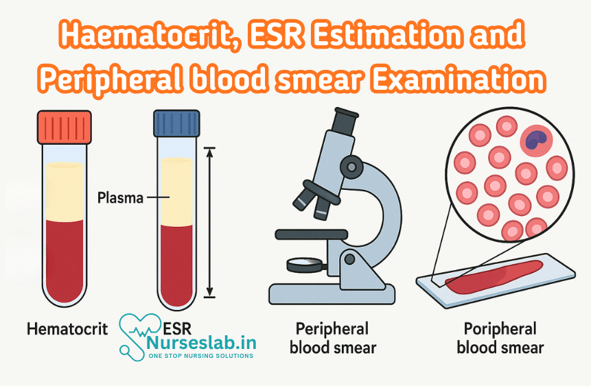
Haematocrit
Definition and Principle
Haematocrit, also known as packed cell volume (PCV), represents the proportion of blood volume that is occupied by red blood cells (RBCs). It is expressed as a percentage or fraction of the total blood volume. The principle of haematocrit measurement is based on the centrifugation of anticoagulated whole blood, which causes separation of blood components by their density. The red cell mass settles at the bottom, followed by a thin layer of white blood cells and platelets (buffy coat), and plasma forms the uppermost layer.
Measurement Techniques
- Microhaematocrit Method: This is the most commonly used technique in clinical laboratories. Capillary blood is collected in a heparinised capillary tube, sealed at one end, and centrifuged at high speed (usually 10,000–12,000 rpm) for 5 minutes. The proportion of RBCs is read using a haematocrit reader.
- Wintrobe Tube Method: A graduated Wintrobe tube is filled with anticoagulated blood and centrifuged at 3,000 rpm for 30 minutes. The packed cell volume is read directly from the scale on the tube.
- Automated Cell Counters: Modern laboratories use automated haematology analysers which calculate haematocrit based on RBC count and mean corpuscular volume (MCV) using the formula: Haematocrit (%) = RBC count × MCV / 10.
Clinical Significance
Measurement of haematocrit is a simple yet vital investigation in clinical practice. It is used to assess the oxygen-carrying capacity of blood, evaluate anaemia or polycythaemia, and monitor patients with acute or chronic blood loss. Haematocrit values guide transfusion decisions, fluid management, and are crucial in perioperative and critical care settings.
Pathological Variations and Interpretation
- Low Haematocrit (Anaemia): This may result from decreased RBC production (e.g., iron deficiency, megaloblastic anaemia, bone marrow failure), increased RBC destruction (haemolytic anaemias), or blood loss (acute or chronic haemorrhage). Interpretation should be correlated with clinical findings and other haematological parameters.
- High Haematocrit (Polycythaemia): Elevated haematocrit may be seen in primary polycythaemia (polycythaemia vera), secondary polycythaemia (chronic hypoxia, high altitude, erythropoietin-secreting tumours), or due to dehydration (relative polycythaemia).
- Interfering Factors: False elevation can occur with prolonged tourniquet application, dehydration, or capillary leak syndrome. False low values may be observed in overhydration or improper sample handling.
Interpretation of Results
Normal haematocrit values vary with age, sex, and altitude. Adult males typically have values between 40–54%, while adult females range from 36–48%. Interpretation of abnormal results must consider the clinical context, hydration status, and concurrent laboratory findings. Serial measurements are often more informative than isolated values.
ESR Estimation
Principle
The erythrocyte sedimentation rate (ESR) measures the rate at which red blood cells settle in a column of anticoagulated blood over a specified period (usually one hour). The test reflects the presence of acute phase reactants (mainly fibrinogen) in plasma, which promote the aggregation of RBCs into rouleaux, thereby increasing their sedimentation rate. ESR is a non-specific marker of inflammation and tissue injury.
Methods of Estimation
- Westergren Method: The gold standard for ESR estimation, this method uses a Westergren tube (30 cm long, 2.5 mm internal diameter) filled with 1:4 diluted blood (with 3.8% sodium citrate). The tube is placed vertically in a rack, and the fall of RBCs is measured in mm after one hour.
- Wintrobe Method: A shorter tube (11 cm) is used, filled with undiluted anticoagulated blood (usually with EDTA). The tube is kept upright, and the sedimentation is read at one hour. The Wintrobe method is less sensitive to higher ESR values.
- Automated ESR Analysers: These instruments use optical or mechanical methods to estimate ESR rapidly, improving standardisation and reducing manual errors.
Factors Affecting ESR
- Plasma Factors: Increased levels of fibrinogen, immunoglobulins, and other acute phase reactants accelerate ESR. Hypergammaglobulinaemia (e.g., in multiple myeloma) also increases ESR.
- Red Cell Factors: Anaemia increases ESR due to reduced cell concentration. Abnormal RBC shapes (sickle cells, spherocytes) hinder rouleaux formation, reducing ESR.
- Technical Factors: Tube angle, room temperature, anticoagulant ratio, and timing can affect results. Tubes must be kept vertical and readings taken precisely at one hour.
Clinical Relevance
ESR is a non-specific indicator of inflammation, infection, and tissue damage. It is widely used in the diagnosis and monitoring of chronic inflammatory conditions such as rheumatoid arthritis, temporal arteritis, polymyalgia rheumatica, and tuberculosis. ESR is also useful in assessing the response to therapy in these conditions.
Pathological Changes and Interpretation
- Elevated ESR: Seen in acute and chronic infections, autoimmune diseases, malignancies, chronic kidney disease, and pregnancy. Markedly high ESR (>100 mm/hr) is suggestive of serious pathology such as multiple myeloma, temporal arteritis, or severe infections.
- Decreased ESR: Polycythaemia, sickle cell disease, hereditary spherocytosis, and conditions with abnormal RBC morphology may cause low ESR.
- Non-Specificity: Normal ESR does not rule out disease; elevated ESR must be interpreted in conjunction with clinical findings and other investigations.
Interpretation of Results
Reference ranges vary by age and sex. In adults, normal values for the Westergren method are up to 15 mm/hr for men and 20 mm/hr for women. Values increase with age and may be mildly elevated in the elderly. It is essential to interpret ESR trends over time rather than isolated values.
Peripheral Blood Smear Examination
Preparation and Staining Techniques
- Preparation: A drop of fresh anticoagulated blood is placed on a clean glass slide and spread using another slide at a 30–45° angle to create a thin, even film. The smear is air-dried and fixed (usually with methanol).
- Staining: Romanowsky stains (such as Leishman, Giemsa, or Wright’s stain) are commonly used. These stains differentiate cellular components by imparting distinct colours to the cytoplasm, nucleus, and granules.
Morphological Assessment
Examination of the peripheral blood smear under a microscope provides invaluable information regarding the morphology of RBCs, white blood cells (WBCs), and platelets. The assessment includes:
- Red Blood Cells: Evaluation for size (anisocytosis), shape (poikilocytosis), colour (hypochromia, polychromasia), inclusions (Howell-Jolly bodies, basophilic stippling), and abnormal forms (sickle cells, spherocytes, schistocytes).
- White Blood Cells: Differential count, identification of abnormal cells (blasts, atypical lymphocytes, toxic granulation), and changes in nuclear or cytoplasmic features.
- Platelets: Assessment for number (thrombocytopenia or thrombocytosis), size (giant platelets), and clumping.
Common Pathological Findings
- Anaemias: Microcytic hypochromic RBCs in iron deficiency anaemia; macrocytic RBCs in megaloblastic anaemia; target cells in thalassaemia; sickle cells in sickle cell anaemia.
- Leukaemias: Presence of immature blast cells, increased lymphocytes, or abnormal mononuclear cells.
- Infections: Reactive lymphocytes in viral infections; toxic granulation in neutrophils in bacterial infections.
- Platelet Disorders: Reduced platelet count in immune thrombocytopenic purpura; giant platelets in myeloproliferative disorders.
- Other Disorders: Schistocytes in microangiopathic haemolytic anaemia; spherocytes in hereditary spherocytosis; rouleaux formation in multiple myeloma.
Interpretation of Results
Peripheral blood smear examination is a cornerstone in the diagnosis of haematological disorders, often providing the first clue to underlying pathology. It aids in classifying anaemias, diagnosing leukaemias and lymphomas, identifying infections, and recognising inherited red cell disorders. Interpretation should always be integrated with clinical findings and other laboratory data.
Comparison and Correlation
Haematocrit, ESR, and peripheral blood smear examination are complementary investigations in the diagnostic approach to haematological and systemic diseases. Each test provides unique information:
- Haematocrit: Quantifies the red cell mass, helping to identify and classify anaemias and polycythaemias.
- ESR: Serves as a sensitive, albeit non-specific, marker of inflammation, infection, and neoplastic processes.
- Peripheral Blood Smear: Offers detailed morphological assessment, enabling identification of specific cellular abnormalities.
Combined interpretation enhances diagnostic accuracy. For instance, a low haematocrit with microcytic hypochromic RBCs on smear suggests iron deficiency anaemia, while a high ESR in the presence of rouleaux formation may point towards multiple myeloma. Conversely, a normal ESR with significant anaemia may indicate an inherited red cell disorder rather than an inflammatory process.
Case Examples
Case 1: Iron Deficiency Anaemia
A 32-year-old woman presents with fatigue and pallor. Laboratory findings reveal:
- Haematocrit: 28% (low)
- ESR: 35 mm/hr (mildly elevated)
- Peripheral Blood Smear: Microcytic, hypochromic RBCs with anisopoikilocytosis
Interpretation: The findings are consistent with iron deficiency anaemia, commonly seen in women of reproductive age due to menstrual blood loss. The mildly elevated ESR suggests a chronic process.
Case 2: Multiple Myeloma
A 65-year-old man complains of bone pain and weakness. Laboratory results:
- Haematocrit: 32% (low)
- ESR: 120 mm/hr (markedly elevated)
- Peripheral Blood Smear: Rouleaux formation, normocytic normochromic RBCs
Interpretation: The combination of low haematocrit, marked ESR elevation, and rouleaux formation on smear is highly suggestive of multiple myeloma, prompting further evaluation for monoclonal gammopathy.
Case 3: Polycythaemia Vera
A 54-year-old male presents with headache and pruritus. Investigations show:
- Haematocrit: 58% (high)
- ESR: 2 mm/hr (low)
- Peripheral Blood Smear: Increased RBC mass, normal morphology
Interpretation: Raised haematocrit with low ESR is characteristic of polycythaemia vera, a myeloproliferative disorder. The normal blood smear morphology supports this diagnosis.
Case 4: Acute Leukaemia
A 20-year-old female presents with fever and bleeding gums. Her reports are:
- Haematocrit: 25% (low)
- ESR: 65 mm/hr (elevated)
- Peripheral Blood Smear: Presence of blasts, thrombocytopenia
Interpretation: The findings are highly suggestive of acute leukaemia, requiring urgent haematology referral and further diagnostic workup.
REFERENCES
- Ramadas Nayak, Textbook of Pathology and Genetics for Nurses, 2nd Edition,2024, Jaypee Publishers, ISBN: 978-93-5270-031-8.
- Suresh Sharma, Textbook of Pharmacology, Pathology & Genetics for Nurses II, 2nd Edition, 31 August 2022, Jaypee Publishers, ISBN: 978-9354655692.
- Kumar, V., Abbas, A.K., & Aster, J.C. (2020). Robbins and Cotran Pathologic Basis of Disease. 10th Edition. Elsevier.
- McCance KL, Huether SE. Pathophysiology: the biologic basis for disease in adults and children, 8th edn. St Louis (MI): Mosby; 2018, https://www.britishjournalofnursing.com/content/clinical-new-series/pathophysiology-applied-to-nursing-the-basis-for-disease-and-illness
Stories are the threads that bind us; through them, we understand each other, grow, and heal.
JOHN NOORD
Connect with “Nurses Lab Editorial Team”
I hope you found this information helpful. Do you have any questions or comments? Kindly write in comments section. Subscribe the Blog with your email so you can stay updated on upcoming events and the latest articles.


