


Last Updated on January 10, 2026 by Nurseslab.in Editorial Team
Pathology of blood chemistry and other lab tests focuses on analyzing biochemical markers to diagnose metabolic, renal, hepatic, and endocrine disorders. These tests guide clinical decisions, monitor treatment, and support evidence‑based nursing and medical practice.
Introduction
Pathology, the scientific study of disease, is fundamentally intertwined with laboratory medicine. Laboratory tests serve as the cornerstone of modern diagnostics, offering objective data that guide clinicians in making informed decisions about patient care. Among these, blood chemistry and other laboratory investigations provide critical insights into physiological and pathological processes.
Importance of Laboratory Tests in Diagnosis
Laboratory tests bridge the gap between clinical suspicion and definitive diagnosis. While history and physical examination remain essential, laboratory investigations validate, clarify, or even refute clinical hypotheses. They enable early detection of diseases, monitor progression, assess response to therapy, and predict outcomes. In certain instances, laboratory findings may reveal subclinical abnormalities, allowing for timely intervention and improved prognosis.
Principles of Blood Chemistry
Blood Components and Their Functions
Blood comprises plasma (the liquid component) and formed elements (cells). Plasma contains water, electrolytes, proteins, hormones, nutrients, and waste products. The cellular components include:
- Red blood cells (erythrocytes): Carry oxygen and carbon dioxide via haemoglobin.
- White blood cells (leukocytes): Defend against infections and mediate immune responses.
- Platelets (thrombocytes): Essential for blood clotting and maintaining vascular integrity.
Normal Ranges and Reference Values
Laboratory results are interpreted against established reference ranges, which represent the values found in healthy individuals. These ranges may vary based on age, sex, ethnicity, and laboratory methods. A value outside the reference range may indicate disease, but must be interpreted in clinical context.
| Parameter | Normal Range | Unit |
| Haemoglobin (male) | 13.5–17.5 | g/dL |
| Haemoglobin (female) | 12.0–15.5 | g/dL |
| White Blood Cell Count | 4,000–11,000 | cells/μL |
| Platelet Count | 1.5–4.5 lakh | cells/μL |
| Serum Sodium | 135–145 | mmol/L |
| Serum Potassium | 3.5–5.0 | mmol/L |
| Blood Urea | 15–45 | mg/dL |
| Serum Creatinine | 0.6–1.2 | mg/dL |
| Fasting Blood Glucose | 70–100 | mg/dL |
Test Methodologies
Laboratory tests employ various methodologies, including spectrophotometry, immunoassays, chromatography, and molecular techniques. Automation, quality control, and standardisation are integral to ensuring accuracy and reproducibility. Pre-analytical (specimen collection and handling), analytical (actual testing), and post-analytical (result reporting) phases all influence the reliability of laboratory data.
Common Blood Chemistry Tests
Blood chemistry encompasses a broad spectrum of tests evaluating different physiological systems. The following are some of the most frequently ordered investigations:
Complete Blood Count (CBC)
The CBC quantifies and characterises blood cells. Key parameters include:
- Haemoglobin (Hb): Reflects oxygen-carrying capacity. Low Hb denotes anaemia; high Hb may indicate polycythaemia.
- Haematocrit (Hct): Percentage of blood volume occupied by RBCs.
- Total and Differential WBC Count: Evaluates immune status and detects infections, inflammation, or haematological malignancies.
- Platelet Count: Assesses bleeding risk and bone marrow function.
- Red Cell Indices (MCV, MCH, MCHC): Aid in anaemia classification.
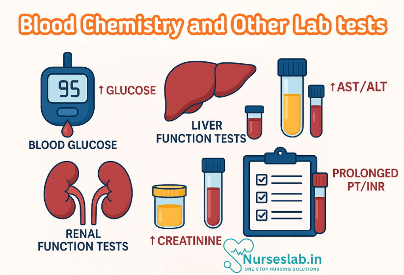
Liver Function Tests (LFTs)
LFTs assess hepatic integrity, synthetic ability, and excretory function. Common components:
- Serum Bilirubin: Elevated in jaundice, haemolysis, or liver dysfunction.
- Aminotransferases (ALT, AST): Raised in hepatocellular injury (e.g., viral hepatitis, toxins).
- Alkaline Phosphatase (ALP): Increased in cholestasis, bone disorders.
- Albumin: Marker of synthetic function; low in chronic liver disease, malnutrition.
- Prothrombin Time (PT): Prolonged in severe liver dysfunction.
Renal Function Tests (RFTs)
RFTs evaluate kidney function and include:
- Blood Urea and Serum Creatinine: Elevated in renal impairment.
- Estimated Glomerular Filtration Rate (eGFR): Calculated from creatinine levels; indicates overall kidney function.
- Electrolytes (Na+, K+, Cl–, HCO3–): Monitor fluid, acid-base, and electrolyte balance.
Blood Glucose and Glycated Haemoglobin (HbA1c)
Blood glucose testing is vital for diagnosing and monitoring diabetes mellitus. Random, fasting, and postprandial glucose levels are measured. HbA1c reflects average blood glucose over the preceding 2-3 months, aiding long-term glycaemic control assessment.
Lipid Profile
Lipid parameters assess cardiovascular risk and metabolic status:
- Total Cholesterol, LDL, HDL: LDL (“bad” cholesterol) is atherogenic; HDL (“good” cholesterol) is protective.
- Triglycerides: Elevated in metabolic syndrome, diabetes, and pancreatitis risk.
Other Key Blood Chemistry Tests
- Serum Calcium, Phosphate, Magnesium: Abnormalities indicate parathyroid, bone, or renal disorders.
- Thyroid Function Tests: TSH, T3, T4 for thyroid dysfunction screening.
- Serum Proteins and Electrophoresis: Assess immune disorders, paraproteinaemias.
Other Laboratory Tests
Urinalysis
Urinalysis is a non-invasive, informative investigation for renal and systemic diseases. Components include:
- Physical Examination: Colour, clarity, specific gravity.
- Chemical Analysis: pH, protein, glucose, ketones, blood, bilirubin.
- Microscopic Examination: Cells, casts, crystals, organisms.
Abnormal findings suggest urinary tract infection, glomerular disease, diabetes, or metabolic disorders.
Coagulation Profiles
These tests evaluate the integrity of the clotting system:
- Prothrombin Time (PT): Assesses extrinsic pathway.
- Activated Partial Thromboplastin Time (aPTT): Evaluates intrinsic pathway.
- International Normalised Ratio (INR): Standardises PT for warfarin monitoring.
- Fibrinogen, D-dimer: Used in disseminated intravascular coagulation (DIC), thrombosis.
Immunological Assays
Immunological tests detect antibodies, antigens, or immune complexes, aiding in diagnosis of infections, autoimmune diseases, and allergies. Examples include:
- Enzyme-Linked Immunosorbent Assay (ELISA): For HIV, hepatitis, dengue, and autoimmune markers.
- Rheumatoid Factor (RF), Anti-Nuclear Antibody (ANA): For autoimmune conditions.
- Allergy Panels: Identify allergen-specific IgE.
Molecular Diagnostics
Molecular techniques, such as PCR (Polymerase Chain Reaction), detect genetic material from pathogens or host. Applications include:
- Detection of Infectious Agents: Tuberculosis, COVID-19, hepatitis viruses.
- Genetic Testing: Thalassaemia, sickle cell anaemia, hereditary cancers.
- Pharmacogenomics: Tailoring drug therapy based on genetic profile.
Interpretation of Results
Understanding Reference Ranges
Reference ranges are not absolute boundaries between health and disease. Values may be influenced by physiological (age, pregnancy), pathological, or technical factors. Minor deviations may be benign, while significant changes often warrant further evaluation.
Principles of Interpretation
- Clinical Correlation: Laboratory results must be interpreted in conjunction with clinical history, symptoms, and findings.
- Pattern Recognition: Clusters of abnormal results (e.g., elevated AST, ALT, bilirubin) suggest specific pathologies (e.g., hepatitis).
- Trend Analysis: Serial measurements reveal disease progression or response to therapy.
- Pre-analytical and Analytical Errors: Haemolysis, delayed processing, or improper storage can produce spurious results.
Significance of Deviations
A single abnormal value may not confirm disease, but persistent or pronounced deviations often indicate pathology. For example, mild isolated hyperglycaemia may be stress-induced, while repeated elevations suggest diabetes mellitus.
Clinical Relevance: Correlation with Disease States
Examples of Pathological Findings
- Anaemia: Low haemoglobin, low RBC count, altered red cell indices. Further workup (iron studies, B12, folate) distinguishes causes such as iron deficiency, megaloblastic anaemia, or chronic disease.
- Liver Disease: Elevated bilirubin, aminotransferases, ALP; prolonged PT; low albumin. Patterns differentiate hepatitis, cirrhosis, or cholestasis.
- Renal Failure: Raised serum urea and creatinine; electrolyte imbalances (hyperkalaemia, acidosis); abnormal urinalysis (proteinuria, casts).
- Diabetes Mellitus: Elevated fasting and postprandial glucose, high HbA1c, glycosuria.
- Myocardial Infarction: Raised cardiac enzymes (CK-MB, troponin), ECG changes, suggest acute coronary syndrome.
- Infections: Leukocytosis with neutrophilia (bacterial), lymphocytosis (viral), positive cultures or serology.
Case Examples
Case 1: Anaemia Workup
A 25-year-old woman presents with fatigue and pallor. CBC reveals:
- Haemoglobin: 8.5 g/dL (low)
- MCV: 70 fL (microcytic)
- Serum iron: low; ferritin: low
Interpretation: Microcytic, hypochromic anaemia due to iron deficiency, likely from menstrual blood loss. Further evaluation may include stool occult blood to rule out gastrointestinal causes.
Case 2: Acute Liver Injury
A 40-year-old man with jaundice and malaise. LFTs show:
- ALT: 900 U/L (elevated)
- AST: 750 U/L (elevated)
- Total bilirubin: 6.5 mg/dL (elevated)
- ALP: 120 U/L (mildly elevated)
- Prothrombin time: prolonged
Interpretation: Marked hepatocellular injury, suggestive of acute viral hepatitis. Prolonged PT indicates significant hepatic dysfunction.
Case 3: Renal Dysfunction
A 60-year-old patient with hypertension and swelling. RFTs reveal:
- Serum creatinine: 2.4 mg/dL (elevated)
- Blood urea: 65 mg/dL (elevated)
- eGFR: reduced
- Urinalysis: proteinuria, RBC casts
Interpretation: Chronic kidney disease, likely secondary to hypertensive nephropathy. Proteinuria and casts confirm glomerular involvement.
Case 4: Diabetes Mellitus
A 50-year-old with weight loss and polyuria. Blood chemistry:
- Fasting glucose: 180 mg/dL
- HbA1c: 9.2%
- Urinalysis: glycosuria, ketonuria
Interpretation: Poorly controlled diabetes mellitus. Presence of ketones may indicate risk of diabetic ketoacidosis.
Recent Advances in Laboratory Diagnostics
Automation and High-Throughput Testing
Automated analysers have revolutionised laboratory medicine, enabling rapid, precise, and reproducible measurements. High-throughput platforms allow simultaneous processing of multiple samples, essential for large-scale screening and epidemiological studies.
Point-of-Care Testing (POCT)
Point-of-care devices enable testing at the bedside or in remote settings, providing immediate results for glucose, cardiac markers, coagulation, and more. This facilitates prompt clinical decision-making, especially in emergencies.
Molecular and Genomic Technologies
Real-time PCR, next-generation sequencing, and microarray technologies have expanded the scope of diagnostics. These tools detect pathogens, genetic mutations, and molecular signatures of disease with high sensitivity and specificity.
Biomarkers and Personalised Medicine
Novel biomarkers (e.g., procalcitonin, NT-proBNP, troponin subtypes) improve diagnostic accuracy and prognostication. Integration of laboratory data with genomic and proteomic information paves the way for personalised medicine, tailoring therapy to individual patient profiles.
Quality Assurance and Informatics
Stringent quality control, accreditation, and laboratory information systems ensure data integrity, traceability, and effective communication between clinicians and laboratories. Advances in artificial intelligence and machine learning enhance pattern recognition and predictive analytics.
REFERENCES
- Ramadas Nayak, Textbook of Pathology and Genetics for Nurses, 2nd Edition,2024, Jaypee Publishers, ISBN: 978-93-5270-031-8.
- Suresh Sharma, Textbook of Pharmacology, Pathology & Genetics for Nurses II, 2nd Edition, 31 August 2022, Jaypee Publishers, ISBN: 978-9354655692.
- Kumar, V., Abbas, A.K., & Aster, J.C. (2020). Robbins and Cotran Pathologic Basis of Disease. 10th Edition. Elsevier.
- McCance KL, Huether SE. Pathophysiology: the biologic basis for disease in adults and children, 8th edn. St Louis (MI): Mosby; 2018, https://www.britishjournalofnursing.com/content/clinical-new-series/pathophysiology-applied-to-nursing-the-basis-for-disease-and-illness
Stories are the threads that bind us; through them, we understand each other, grow, and heal.
JOHN NOORD
Connect with “Nurses Lab Editorial Team”
I hope you found this information helpful. Do you have any questions or comments? Kindly write in comments section. Subscribe the Blog with your email so you can stay updated on upcoming events and the latest articles.


