


Last Updated on October 21, 2026 by Nurseslab.in Editorial Team
Breast cancer remains one of the most prevalent malignancies affecting women worldwide, with significant implications for morbidity and mortality. Early detection and appropriate risk stratification are crucial to improving outcomes. Risk assessment for breast cancer involves a combination of clinical evaluation, advanced imaging, genetic testing, and risk prediction models to identify individuals at heightened risk and to tailor prevention and management strategies accordingly.
Introduction
Breast cancer is a major public health concern, accounting for a significant proportion of cancer diagnoses and deaths among women globally. In India, the incidence of breast cancer is on the rise, with increasing awareness and improved diagnostic facilities contributing to higher rates of detection. The heterogeneity of breast cancer risk across populations necessitates accurate and personalised risk assessment. Such evaluation not only guides screening and surveillance strategies but also informs preventive interventions, genetic counselling, and therapeutic decision-making.
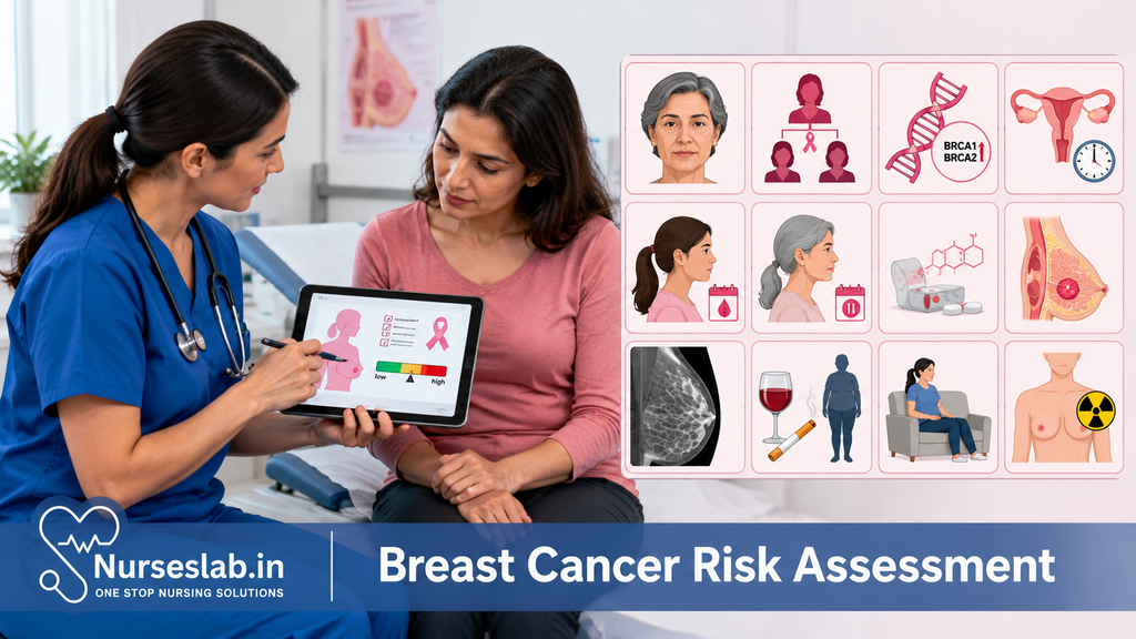
Risk assessment for breast cancer seeks to identify women who would benefit from enhanced surveillance, lifestyle modifications, chemoprevention, or prophylactic surgery. The process integrates clinical evaluation, imaging modalities, genetic risk profiling, and the application of validated risk prediction models. Each of these components plays a distinct yet complementary role in the comprehensive assessment of breast cancer risk.
Clinical Evaluation
1.1 Patient History
A detailed patient history forms the cornerstone of breast cancer risk assessment. The process involves eliciting information about personal and family health, reproductive factors, and lifestyle choices:
- Family history: A history of breast or ovarian cancer in first-degree relatives (mother, sister, daughter) or multiple family members increases risk, especially if diagnoses occurred at a young age.
- Personal history: Prior breast conditions, such as atypical hyperplasia or lobular carcinoma in situ, and previous breast cancer diagnoses elevate risk.
- Reproductive history: Early menarche (before age 12), late menopause (after age 55), nulliparity, late age at first childbirth, and not breastfeeding are associated with increased risk.
- Hormonal factors: Use of hormone replacement therapy, especially combined oestrogen-progestin regimens, is linked to higher risk.
- Lifestyle and environmental factors: Obesity, excessive alcohol consumption, sedentary lifestyle, and exposure to ionising radiation contribute to risk.
1.2 Physical Examination
The clinical breast examination (CBE) is performed to identify palpable masses, skin changes, nipple discharge, or lymphadenopathy. While the CBE has limitations in sensitivity, it remains a valuable component of comprehensive risk assessment, particularly in resource-limited settings where advanced imaging may not be readily available.
1.3 Risk Factors: Summary Table
| Category | Risk Factor |
| Genetic | BRCA1/2 mutations, family history |
| Reproductive | Early menarche, late menopause, nulliparity, late first childbirth |
| Hormonal | Hormone replacement therapy |
| Lifestyle | Obesity, alcohol, physical inactivity |
| Previous Breast Disease | Atypical hyperplasia, lobular carcinoma in situ |
1.4 Advantages and Limitations
Advantages: Non-invasive, cost-effective, provides holistic insight into individual risk, and is universally applicable.
Limitations: Relies on patient recall, may miss subclinical disease, and does not quantify risk precisely.
Section 2: Imaging Techniques
Imaging plays a critical role in both the detection of breast lesions and the assessment of risk, especially in individuals with dense breast tissue or family history of breast cancer. The choice of imaging modality is guided by patient age, risk profile, and clinical findings.
2.1 Mammography
Mammography is the gold standard for breast cancer screening and risk assessment in women above 40 years of age. It involves low-dose X-rays to visualise breast tissue, enabling the detection of microcalcifications, masses, and architectural distortions.
Purpose: Early detection of malignancy and risk stratification, especially in asymptomatic women.
Process: The breast is compressed between two plates and imaged from multiple angles.
Advantages: Proven to reduce mortality, widely available, and effective for detecting early-stage cancers.
Limitations: Reduced sensitivity in dense breasts, potential for false positives and negatives, and exposure to ionising radiation.
2.2 Ultrasound
Ultrasound uses high-frequency sound waves to create images of breast tissue. It is particularly useful as an adjunct to mammography, especially in younger women and those with dense breasts.
Purpose: Characterisation of palpable masses, assessment of cystic versus solid lesions, and guidance for biopsies.
Process: A transducer is moved over the breast, emitting sound waves that are converted into real-time images.
Advantages: No radiation exposure, excellent for differentiating cystic from solid lesions, portable, and affordable.
Limitations: Operator-dependent, limited in detecting microcalcifications, and may miss small or deep-seated lesions.
2.3 Magnetic Resonance Imaging (MRI)
Breast MRI is a highly sensitive imaging modality that uses magnetic fields and contrast agents to visualise breast tissue. It is recommended for high-risk individuals, such as those with BRCA mutations or strong family history.
Purpose: Screening high-risk women, evaluating extent of disease, and assessing response to therapy.
Process: Patients lie prone in an MRI scanner, and images are obtained before and after intravenous contrast administration.
Advantages: High sensitivity, no radiation, superior for detecting multifocal and multicentric disease.
Limitations: Expensive, limited availability, prone to false positives, and contraindicated in patients with certain implants or renal dysfunction.
2.4 Emerging Technologies
Newer imaging techniques, such as digital breast tomosynthesis (3D mammography), molecular breast imaging, and automated whole breast ultrasound, are being explored to improve sensitivity and specificity. While promising, these technologies are yet to replace standard modalities in routine risk assessment due to cost and limited long-term data.
Section 3: Genetic Testing
Genetic factors account for a significant proportion of hereditary breast cancer cases. Identification of pathogenic mutations enables targeted risk reduction and informs management for both patients and their relatives.
3.1 BRCA1 and BRCA2 Testing
Mutations in the BRCA1 and BRCA2 genes confer a markedly increased lifetime risk of breast and ovarian cancers. Testing is indicated for individuals with:
- Strong family history of breast or ovarian cancer
- Early-onset breast cancer (diagnosed before 50 years)
- Bilateral breast cancer
- Male breast cancer in the family
- Known familial mutation
Purpose: Identification of carriers for enhanced surveillance and preventive strategies.
Process: Blood or saliva samples are collected for DNA analysis using sequencing or targeted mutation detection.
Advantages: Enables personalised risk assessment, informs family members, and guides prophylactic interventions.
Limitations: Psychological impact, potential for insurance or employment discrimination, and variants of uncertain significance.
3.2 Other Gene Panels
Multi-gene panels now include additional genes such as TP53, PTEN, PALB2, CHEK2, and ATM, which also contribute to hereditary breast cancer risk. These tests are useful when BRCA mutations are absent but clinical suspicion remains high.
Purpose: Broader risk identification beyond BRCA genes.
Process: Next-generation sequencing technologies allow simultaneous analysis of multiple genes.
Advantages: Comprehensive, may identify additional at-risk individuals.
Limitations: Increased likelihood of detecting variants of uncertain significance, complex interpretation, and need for specialised counselling.
3.3 Genetic Counselling
Pre- and post-test genetic counselling is mandatory to ensure informed decision-making, address psychological concerns, and interpret results accurately. Counselling should cover implications for the individual and family, preventive strategies, and limitations of testing.
Section 4: Risk Prediction Models
Risk prediction models integrate clinical, epidemiological, and genetic data to estimate an individual’s absolute or relative risk of developing breast cancer over a specified timeframe. These tools are instrumental in guiding screening and preventive decisions.
4.1 Gail Model
The Gail Model estimates five-year and lifetime breast cancer risk based on factors such as age, reproductive history, family history, prior breast biopsies, and race. It is best suited for women without a strong family history or known genetic mutations.
Purpose: Identifying women eligible for chemoprevention or enhanced surveillance.
Process: Inputting patient data into a validated online calculator.
Advantages: Easy to use, widely validated, provides quantifiable risk.
Limitations: Underestimates risk in women with strong family history or genetic mutations, limited applicability in non-Western populations.
4.2 Tyrer-Cuzick Model (IBIS Tool)
The Tyrer-Cuzick Model incorporates a broader range of risk factors, including detailed family history, genetic mutations, hormonal factors, and benign breast disease. It is considered more comprehensive than the Gail Model.
Purpose: Stratifying risk in women with complex family histories.
Process: Detailed questionnaire and algorithm-based risk calculation.
Advantages: Highly comprehensive, includes genetic and personal risk factors, validated in diverse populations.
Limitations: More complex to administer, requires detailed information, and may overestimate risk in some populations.
4.3 Other Risk Assessment Tools
- Claus Model: Focuses on family history of breast cancer.
- BOADICEA: Incorporates genetic and non-genetic risk factors, suitable for BRCA mutation carriers.
- BRCAPRO: Bayesian model for estimating probability of carrying BRCA mutations.
Each model has specific indications, strengths, and weaknesses, and should be selected based on the clinical context and available data.
Section 5: Integrating Assessment Results
The integration of clinical, imaging, genetic, and risk model data enables a nuanced understanding of individual risk. Stratification may categorise patients as average, moderate, or high risk, guiding further management. Multidisciplinary discussions are often warranted, especially for high-risk individuals.
Interpreting Findings: Results must be contextualised based on patient preferences, comorbidities, and local resources. Discordant findings require careful reconciliation, and repeat assessments may be necessary as risk factors evolve.
Risk Stratification:
- Average risk: No significant personal or family history, no genetic mutations, normal imaging.
- Moderate risk: Limited family history, benign breast disease, or certain reproductive risk factors.
- High risk: BRCA mutation carriers, strong family history, prior chest irradiation, or previous breast cancer.
Section 6: Impact on Patient Management
Risk assessment directly informs patient management, influencing screening schedules, preventive interventions, and follow-up protocols.
6.1 Screening Recommendations
- Average risk: Annual or biennial mammography starting at age 40–50 years, with clinical breast examination as per guidelines.
- Moderate risk: Earlier initiation of screening, possible adjunct imaging (ultrasound or MRI) based on breast density and clinical findings.
- High risk: Annual MRI and mammography starting as early as 25–30 years, consideration of risk-reducing surgery, and enhanced surveillance for associated malignancies.
6.2 Preventive Strategies
- Lifestyle modifications: Weight management, regular physical activity, limiting alcohol, and breastfeeding.
- Chemoprevention: Selective oestrogen receptor modulators (e.g., tamoxifen) or aromatase inhibitors for eligible women.
- Prophylactic surgery: Bilateral mastectomy and/or oophorectomy for selected high-risk individuals.
6.3 Follow-up and Counselling
Continuous follow-up is essential, with periodic reassessment of risk as new family or personal history emerges. Counselling should address emotional, psychological, and social implications, ensuring that patients are empowered to make informed choices.
Section 7: Limitations and Challenges
While breast cancer risk assessment has advanced considerably, several limitations and challenges persist:
- False Positives/Negatives: Imaging and risk models may over- or under-estimate risk, leading to unnecessary interventions or missed diagnoses.
- Accessibility: Advanced imaging and genetic testing may not be widely available or affordable, particularly in rural or underprivileged settings in India.
- Ethical Considerations: Genetic testing raises concerns about confidentiality, discrimination, and psychological impact.
- Cultural Sensitivity: Stigma and lack of awareness may hinder acceptance of screening and genetic counselling.
- Population Diversity: Many risk models are validated in Western populations and may not fully capture risk profiles in Indian or other Asian women.
Nursing Care of Patients Undergoing Breast Cancer Risk Assessment
Nurses play an indispensable role throughout this process, providing holistic and evidence-based care that addresses not only physical but also psychological, social, and educational needs of patients.
Pre-Procedure Nursing Care
1. Patient Assessment
Medical and Family History: Gather comprehensive information about the patient’s medical history, family history of breast or ovarian cancer, previous breast biopsies, menstrual and reproductive history, and any existing comorbidities.
Physical Assessment: Perform baseline vital signs, assess for any breast abnormalities, and evaluate the patient’s overall health status.
Psychosocial Evaluation: Assess the patient’s emotional state, support systems, cultural beliefs, and understanding of the procedure. Indian patients may have specific cultural or societal concerns regarding breast health, privacy, and cancer stigma, which should be sensitively addressed.
2. Patient Education and Counselling
Procedure Explanation: Clearly explain the nature, purpose, and steps of the diagnostic procedure, using language that is easy to understand and culturally appropriate. For example, when explaining mammography, describe the sensations the patient may experience and reassure them regarding the safety and necessity of the test.
Preparation Instructions: Provide specific instructions such as avoiding deodorants, powders, or perfumes on the day of a mammogram, fasting requirements for certain tests, or wearing comfortable clothing.
Addressing Fears and Misconceptions: Many patients may harbour fears about cancer diagnosis, pain, or the implications of genetic testing. Offer empathetic listening and factual information to dispel myths and alleviate anxiety.
Informed Consent: Ensure the patient (and family, if appropriate) understands the risks, benefits, and alternatives to the procedure. Obtain written informed consent as per hospital policy.
3. Physical Preparation
Fasting and Medication: Advise the patient regarding any required fasting or medication adjustments, especially if sedation or contrast dye is to be used.
Allergy Assessment: Screen for allergies to contrast agents, latex, or local anaesthetics.
Skin Preparation: For procedures such as biopsies, prepare the skin aseptically to reduce infection risk.
Intra-Procedure Nursing Care
1. Providing Emotional Support
Presence and Reassurance: Stay with the patient during the procedure (when permitted), offering calm reassurance and answering any last-minute queries.
Pain Management: Monitor for signs of discomfort or pain. For procedures like core needle biopsy, ensure appropriate local anaesthesia is administered and assess pain levels regularly.
2. Assisting with the Procedure
Positioning: Assist in correct positioning for imaging or biopsy. For example, during mammography, help the patient place the breast on the plate and ensure comfort.
Maintaining Sterility: Adhere strictly to aseptic techniques during invasive procedures to prevent infection.
Monitoring Vital Signs: Observe for any adverse reactions, especially during procedures involving sedation or contrast media (e.g., allergic reactions, hypotension).
3. Communication
Clear Instructions: Provide step-by-step guidance during the procedure, informing the patient what to expect at each stage.
Addressing Discomfort: Encourage the patient to report any discomfort or unusual sensations immediately.
Post-Procedure Nursing Care
1. Immediate Care
Observation: Monitor the patient for any immediate complications such as bleeding, haematoma, infection, or allergic reactions.
Pain Relief: Administer prescribed analgesics if needed and provide comfort measures such as ice packs for swelling.
Wound Care: For biopsies, ensure the biopsy site is clean, dry, and covered with a sterile dressing. Educate the patient on signs of infection and wound care at home.
2. Patient Education and Discharge Planning
Post-Procedure Instructions: Clearly explain activity restrictions, wound care, signs of complications, and when to seek medical attention.
Follow-Up: Inform the patient about follow-up appointments, reporting of results, and further management if needed.
Emotional Support: Provide psychological support, especially if the patient is anxious about results. Offer referrals to counsellors, breast cancer support groups, or helplines as appropriate.
3. Documentation
Accurate Records: Document the procedure details, patient’s response, vital signs, education provided, and any complications encountered.
Special Considerations in Indian Context
Cultural Sensitivity: Recognise and respect traditional beliefs, modesty concerns, and the potential stigma associated with breast cancer. Use female staff for intimate procedures where possible, and ensure privacy and dignity are maintained at all times.
Family Involvement: In many Indian families, decision-making is collective. Involve family members (with the patient’s consent) in education and counselling.
Language and Literacy: Provide information in the patient’s preferred language and use visual aids or interpreters if required. Use simple, non-medical terms.
Accessibility and Affordability: Be aware of the financial implications of diagnostic tests. Direct patients to government schemes, NGOs, or hospital social workers for support if needed.
Role of the Nurse in Multidisciplinary Teams
Nurses act as a bridge between patients and the healthcare system, collaborating closely with doctors, radiologists, pathologists, genetic counsellors, and social workers. They coordinate appointments, facilitate communication, and advocate for the patient’s needs.
Common Challenges and Solutions
Anxiety and Fear: Address through empathetic communication, accurate information, and support services.
Non-Compliance: Mitigate by involving family, simplifying instructions, and addressing barriers such as transport or cost.
Delayed Results: Keep patients informed about expected timelines and provide interim support.
Language Barriers: Use translators, pictorial aids, and repeat instructions as necessary.
REFERENCES
- Patel BK, et al. Prospective study of supplemental screening with contrast-enhanced mammography in women with elevated risk of breast cancer: Results of the prevalence round. Journal of Clinical Oncology. 2024; doi:10.1200/JCO.22.02819.
- Klassen CL, et al. Breast cancer risk evaluation for the primary care physician. Cleveland Clinic Journal of Medicine. 2022; doi:10.3949/ccjm.89a.21023.
- Ikonopedia. IBIS (International Breast Cancer Intervention Study) Online Tyrer-Cuzick Model Breast Cancer Risk Evaluation Tool (https://ibis.ikonopedia.com/).
- Kurian AW, Hughes E, Simmons T, et al. Performance of the IBIS/Tyrer-Cuzick model of breast cancer risk by race and ethnicity in the Women’s Health Initiative (https://pubmed.ncbi.nlm.nih.gov/34228814/). Cancer. 2021 Oct 15;127(20):3742-3750.
- McCarthy AM, Guan Z, Welch M, et al. Performance of Breast Cancer Risk-Assessment Models in a Large Mammography Cohort (https://www.ncbi.nlm.nih.gov/pmc/articles/PMC7225681/). J Natl Cancer Inst. 2020 May;112(5):489-497.
- National Cancer Institute (U.S.) Risk Factors for Cancer https://www.cancer.gov/about-cancer/causes-prevention/risk. Updated 12/23/2015.
Stories are the threads that bind us; through them, we understand each other, grow, and heal.
JOHN NOORD
Connect with “Nurses Lab Editorial Team”
I hope you found this information helpful. Do you have any questions or comments? Kindly write in comments section. Subscribe the Blog with your email so you can stay updated on upcoming events and the latest articles.


