


Peritonitis is a serious condition that results from a generalized or localized inflammatory process in the peritoneum (membrane lining the abdominal cavity) with a primary or secondary cause.
Primary peritonitis is characterized by blood-borne organisms entering the peritoneal cavity. Secondary peritonitis, the more common type, occurs when abdominal organs rupture or perforate and release their contents into the peritoneal cavity. This is often caused by a ruptured appendix, perforated gastric ulcer, or a severely inflamed gallbladder.
When bacterial and other intestinal contents enter the peritoneum, they can irritate the sterile peritoneal cavity leading to chemical peritonitis. Inflammation causes massive fluid shifts and adhesions as the body will react to defend itself against infection.
Clinical manifestations can vary depending on the underlying condition’s severity and acuity. Peritonitis can be fatal if treatment is delayed. Complications include sepsis, hypovolemic shock, paralytic ileus, intraabdominal abscess formation, and acute respiratory distress syndrome.
Nursing Process
Patients with peritonitis require emergent treatment and skilled supportive care. The overall goal of peritonitis management and care includes resolving the inflammation, relieving abdominal pain, treating any bacterial infection, preserving skin and tissue integrity, and preventing complications like sepsis and hypovolemic shock.
Nursing Assessment
The first step of nursing care is the nursing assessment, during which the nurse will gather physical, psychosocial, emotional, and diagnostic data. In this section, we will cover subjective and objective data related to peritonitis.
Review of Health History
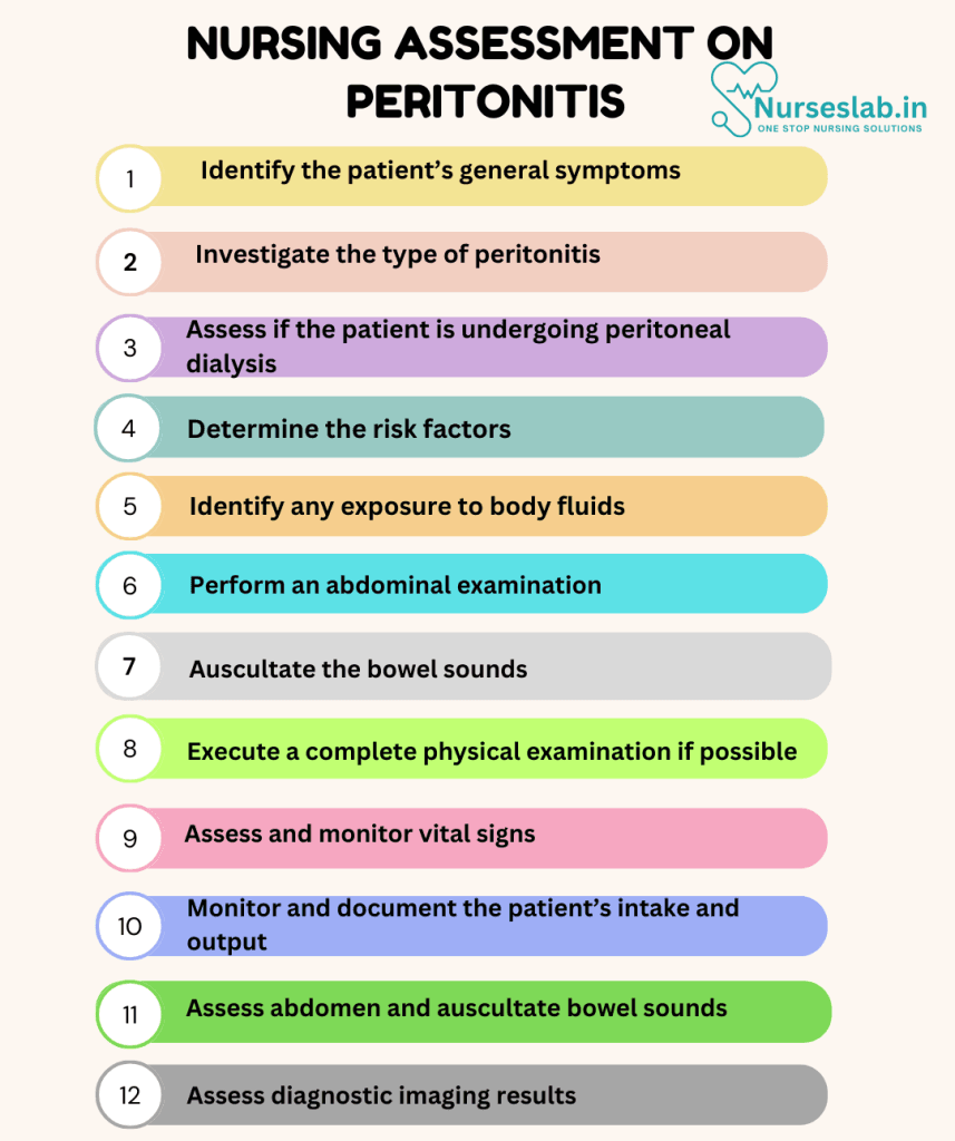
1. Identify the patient’s general symptoms.
Clinical manifestations of peritonitis include the following:
- Rebound tenderness in the affected area
- Abdominal pain
- Abdominal distension
- Fever
- Tachypnea
- Tachycardia
- Nausea
- Vomiting
- Altered bowel habits
2. Investigate the type of peritonitis.
Peritonitis typically results from a bacterial infection and can be primary or secondary.
- Spontaneous bacterial peritonitis occurs in patients with ascites from liver or kidney disease
- Secondary peritonitis results from an intra-abdominal rupture of an organ
3. Assess if the patient is undergoing peritoneal dialysis.
Peritoneal dialysis, a treatment for renal failure, uses a catheter inserted in the abdomen to filter and drain fluid via the peritoneum. A contaminated PD catheter is a source of infection-causing peritonitis.
4. Determine the risk factors.
The most prevalent risk factors are:
- Primary spontaneous peritonitis
- Liver disease with cirrhosis
- Ascites
- Kidney failure on peritoneal dialysis
- Contamination of implanted catheter in the peritoneum
- Secondary Peritonitis
- Inflammatory bowel disease (such as Crohn’s disease and diverticulitis)
- Pancreatitis
- Pelvic inflammatory disease
- Perforation of abdominal organs (such as the bowel and stomach)
- Ruptured ulcer, appendix, or diverticulum
- Abdominal surgery
- Ruptured ectopic pregnancy
- Abdominal trauma (such as an injury from a stabbing or gunshot wound)
5. Identify any exposure to body fluids.
While infection is a common source of inflammation, it can also result from an internal chemical interaction. This includes:
- Bile from the gallbladder
- Pancreatic enzymes from the pancreas
- Stomach acid from the stomach
- Ruptured tumor in the abdomen
Physical Assessment
1. Perform an abdominal examination.
Patients will exhibit a distended abdomen, increased abdominal wall rigidity, and palpable tenderness on an abdominal examination. Patients may avoid moving while maintaining their hips flexed to reduce the tension in the abdomen. There could be liver failure symptoms, including jaundice or angiomata.
2. Auscultate the bowel sounds.
Bowel sounds are sluggish to absent in peritonitis due to abdominal wall rigidity and inflammation of the peritoneum.
3. Execute a complete physical examination if possible.
A thorough physical examination is crucial for ruling out other conditions whose symptoms can be mistaken for peritonitis. The signs and symptoms of peritonitis may be confused with the following:
- Thoracic processes with diaphragm irritation (such as empyema- pus in the body cavity)
- Extraperitoneal (outside the peritoneum) processes (such as cystitis and pyelonephritis)
- External hernias to rule out intestinal incarceration
Diagnostic Procedures
1. Send blood samples to the laboratory.
- Blood chemistry findings indicate dehydration and acidosis.
- Complete blood cell (CBC) count reveals leukocytosis (high WBC), indicating infection.
- Blood cultures exhibit positive findings for gram-stain bacteria
2. Prepare the patient for paracentesis.
Assist the provider in performing a paracentesis, a procedure where a needle removes fluid directly from the abdominal cavity for fluid analysis. It can detect both primary spontaneous peritonitis and secondary peritonitis.
3. Visualize the peritoneum.
Peritoneoscopy uses a light source and scope to look within the peritoneal cavity and some of its internal organs. In peritonitis, the peritoneum has numerous adhesions between the intestinal loops, liver capsule, and abdominal walls.
4. Review peritoneal fluid analysis.
Peritoneal fluid analysis is crucial for diagnosis, with a neutrophil count larger than 250 cells/microliter as the most sensitive marker. The following should be examined in the fluid sample:
- Glucose
- Protein
- LDH
- Cell count
- Gram stain
- Aerobic and anaerobic cultures
5. Consider a rapid diagnosis approach.
The proposed use of bedside reagent strips analyzed by portable spectrophotometric equipment has advanced the quick detection of spontaneous bacterial peritonitis (SBP)—the shorter time between paracentesis and diagnosis, the faster treatment can begin. However, using the reagent strip as a stand-alone test is not advisable.
6. Complete imaging scans.
Imaging allows for better visualization of the peritoneum and its enclosed organs.
- CT scan of the abdomen and pelvis is used when a diagnosis cannot be made based on clinical factors and the results of plain x-ray films. It is still the preferred diagnostic test for peritoneal abscess and concomitant visceral disease.
- Magnetic resonance imaging (MRI) is utilized when diagnosing possible intra-abdominal abscesses.
- Plain x-rays are used as a first imaging test to visualize free air in the peritoneum.
- Ultrasound identifies volumes of less than 100 mL and is limited. Transabdominal ultrasonography does not visualize the middle peritoneal cavity well.
- Ultrasound-guided aspiration and placement of drains are valuable tools to determine and manage abdominal fluid collections.
Nursing Interventions
Nursing interventions and care are essential for the patients recovery. In the following section, you’ll learn more about possible nursing interventions for a patient with peritonitis.
Treat According to The Etiology
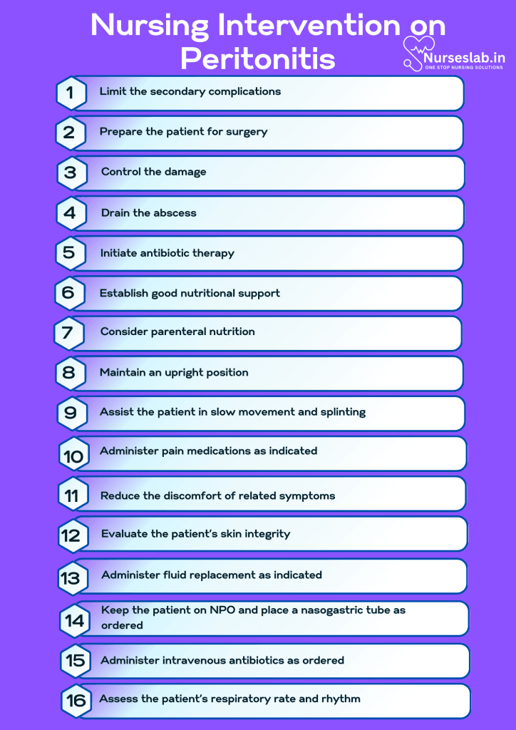
1. Limit the secondary complications.
Peritonitis and peritoneal abscesses are managed by correcting the underlying cause, administering systemic antibiotics, and using supportive therapy to stop or lessen additional complications brought on by failing organ systems.
2. Prepare the patient for surgery.
Surgery aims to eliminate bacteria and toxins while controlling the cause of peritonitis. The underlying disease process affects the kind of surgery.
3. Control the damage.
Damage control surgeries (DCS) temporarily drain the infection, rapidly limit the visceral leak, and postpone any definitive repair until the patient has stabilized.
4. Drain the abscess.
Percutaneous drainage is an effective and safe initial treatment option when an abscess can be entirely drained, and debridement and restoration of the anatomic organs are not required.
5. Initiate antibiotic therapy.
The use of antibiotic therapy decreases complications and the spread of infection. Broad-spectrum antibiotics are used until a specific organism is identified.
6. Establish good nutritional support.
After exploration, patients with peritonitis experience some gastrointestinal dysfunction (such as ileus). Because patients may have inadequate intake due to surgery or symptoms, it is beneficial to establish nutritional support early on in the course of treatment.
7. Consider parenteral nutrition.
Enteral feeding has been reported to have fewer issues in patients who are critically ill. Parenteral nutrition should be started if enteral feeding is not recommended or tolerated.
Prevent Infection
1. Prevent transmission of infection.
Microbes near the catheter frequently cause peritoneal dialysis-related peritonitis. It is easier to prevent infection than treat it. The nurse can instruct the patient on properly caring for their PD catheter by keeping the site clean and dry and covered with sterile gauze and tape.
2. Promote handwashing.
Most commonly, Staphylococcus species, which are linked to contact contamination, cause peritonitis. Before performing a dialysis exchange, patients are advised to clean their hands to lower the danger of contact contamination. Handwashing reduces the risk of infection.
3. Disinfect the port correctly.
The peritoneal dialysis port is an entry point for microorganisms that may cause infection. Use friction to scrub the hub, the sides, and the port’s end, using an antiseptic pad or solution.
4. Maintain asepsis.
The packaged peritoneal dialysis supplies should be handled carefully to maintain their sterility. To prevent infection, maintain sterile technique as required. Use aseptic technique when handling dialysate bags and throughout the PD process.
5. Teach the patient when to seek immediate medical attention.
Peritonitis could be fatal if left untreated. If there is a presence of intense abdominal discomfort or tenderness, bloating, or a feeling of fullness coupled with any of the following, seek immediate medical attention:
- Fever
- Nausea
- Vomiting
- Oliguria (less urine output)
- Constipation
- Abnormal dialysis fluid in patients receiving peritoneal dialysis (such as cloudy or unusual smell and color)
- Signs of severe abdominal trauma
- PD catheter site that is red, swollen, bulging, or draining pus
Nursing Care Plans
Once the nurse identifies nursing diagnoses for peritonitis, nursing care plans help prioritize assessments and interventions for both short and long-term goals of care. In the following section, you will find nursing care plan examples for peritonitis.
Acute Pain
Patients with peritonitis initially exhibit dull abdominal pain and tenderness. The abdominal pain can quickly become severe and persistent.
Nursing Diagnosis: Acute Pain
Related to:
- Biological injury agent
- Physical injury agent
- Peritoneal trauma
- Chemical irritation of the parietal peritoneum
- Fluid accumulation in the peritoneal cavity
- Inflammatory process
- Infection
As evidenced by:
- Expressive behavior
- Guarding behavior
- Tenderness on palpation
- Protective behavior
- Positioning to ease pain
- Reports pain intensity
- Reports pain characteristics
Expected outcomes:
- Patient will report pain relief.
- Patient will demonstrate interventions to help relieve pain and promote comfort.
Assessment:
1. Assess pain characteristics.
Pain characteristics, including intensity and location, may indicate the progression of the patient’s condition and possible complications. Peritonitis pain is often dull but becomes persistent and severe and is worsened by movement. A decreased appetite and nausea may accompany the pain.
2. Assess vital signs.
Alterations in vital signs can indicate worsening pain and possible complications.
Interventions:
1. Maintain an upright position.
Semi-Fowler’s position can help facilitate fluid drainage by gravity and reduce abdominal tension and diaphragmatic irritation, which can significantly reduce pain.
2. Assist the patient in slow movement and splinting.
Splinting of the abdominal area can reduce tension and the pain of movement.
3. Administer pain medications as indicated.
Narcotic analgesics are administered to help reduce pain in patients with peritonitis.
4. Reduce the discomfort of related symptoms.
Nausea and vomiting can be controlled with antiemetics. Fever can be reduced with antipyretics or nonpharmacologic interventions like cool rags.
Deficient Fluid Volume
Patients with peritonitis often have weak, injured, and inflamed peritoneum with an increased risk of infection spreading through the peritoneal cavity. When peristaltic action decreases, there is a tendency for bowel obstruction to develop, with large amounts of fluid shifting into the peritoneal cavity, leading to deficient fluid volume.
Nursing Diagnosis: Deficient Fluid Volume
Related to:
- Fever
- Vomiting
- NPO status
- Fluid shifting into the peritoneal space
- Retention of fluid in the abdomen
As evidenced by:
- Dry mucous membranes
- Hypotension
- Tachycardia
- Slow capillary refill
- Weakness
- Weak peripheral pulses
- Altered skin turgor
- Decreased urine output
Expected outcomes:
- Patient will demonstrate improved fluid balance as evidenced by normal urinary output.
- Patient exhibits normal blood pressure, heart rate, and body temperature.
Assessment:
1. Assess and monitor vital signs.
Alterations in vital signs like hypotension and tachycardia can help evaluate the degree of fluid deficit and determine an appropriate treatment regimen.
2. Monitor and document the patient’s intake and output.
Intake and output can reflect the patient’s hydration status. Decreased or concentrated urinary output can indicate reduced renal perfusion, edema, or fluid accumulation in the peritoneal cavity.
Interventions:
1. Evaluate the patient’s skin integrity.
Fluid shifts in peritonitis, hypovolemia, and nutritional deficits can contribute to poor skin integrity and edematous tissues.
2. Administer fluid replacement as indicated.
Fluid replacement through blood, plasma, or electrolyte replacement, can help correct electrolyte balances, and circulating volume lost to the peritoneal cavity.
3. Administer medications as indicated.
Antiemetics may be prescribed to help reduce nausea and vomiting and prevent further loss of fluid and electrolytes.
4. Keep the patient on NPO and place a nasogastric tube as ordered.
A nasogastric tube can help decrease gastric distention and prevent further leakage of bowel contents into the peritoneal cavity.
Dysfunctional Gastrointestinal Motility
With its inflammatory and infectious process, peritonitis can cause decreased gastrointestinal motility and intestinal distension.
Nursing Diagnosis: Dysfunctional Gastrointestinal Motility
Related to:
- Abdominal distension
- Inflammatory process
- Abdominal infection
- Ileus
As evidenced by:
- Abdominal pain
- Abdominal cramping
- Abdominal bloating
- Altered bowel sounds
- Nausea
- Vomiting
Expected outcomes:
- Patient will be free from abdominal distension, cramping, and pain.
- Patient will not exhibit gastrointestinal motility complications like bowel obstruction or paralytic ileus.
Assessment:
1. Assess abdomen and auscultate bowel sounds.
Initially, patients with peritonitis will have bowel sounds upon auscultation, but this will disappear as the inflammation progresses. The inability to pass flatus along with abdominal distension, pain, and tenderness signals issues with gastric motility.
2. Assess diagnostic imaging results.
Diagnostic imaging like an x-ray or ultrasound is often indicated to help confirm peritonitis and visualize holes, fluid, gas, and obstructions in the gastrointestinal tract.
3. Review laboratory studies.
Fluid and electrolyte imbalance and nutritional deficits are common in patients with peritonitis and may affect the patient’s gastrointestinal motility.
Interventions:
1. Administer fluid and electrolyte replacement.
Fluid volume shift into the peritoneal space occurs in peritonitis. Fluid and electrolyte replacement must be initiated to correct imbalances and further prevent gastrointestinal motility problems like intestinal obstruction and dysfunction.
2. Restrict intake of foods and fluids as indicated.
Keeping the patient NPO will let the intestines rest, recover, and prevent further gastrointestinal motility problems.
3. Insert a nasogastric tube as indicated.
The placement of an NG tube in patients with peritonitis can help decompress the bowel and reduce dysfunctional motility problems.
4. Administer intravenous antibiotics as ordered.
IV antibiotics like cephalosporins are considered the first line of antibiotic therapy for patients with peritonitis. This will help clear out the infection and reduce inflammation in the gastrointestinal tract, improving the motility of the gut.
Ineffective Breathing Pattern
Respiratory failure is a potentially fatal complication of severe peritonitis. Inflammation and infection in the peritoneum may lead to sepsis and respiratory distress.
Nursing Diagnosis: Ineffective Breathing Pattern
Related to:
- Disease process
- Inflammatory process
- Abdominal distension
- Abdominal pain or discomfort
- Increased physical exertion
- Sepsis
- Shock
As evidenced by:
- Hypoxemia
- Hypoxia
- Hypercapnia
- Hyperventilation
- Hypoventilation
- Nasal flaring
- Accessory muscle use
- Altered chest excursion
- Tachypnea
- Cyanosis
Expected outcomes:
- Patient will demonstrate a breathing pattern within normal limits with vital signs within normal parameters.
- Patient will not display signs of respiratory distress, such as accessory muscle use, dyspnea, nasal flaring, or tachypnea.
Assessment:
1. Assess the patient’s respiratory rate and rhythm.
Normal respiratory rate is between 10-20 breaths per minute. Any deviations from this range can indicate alterations in breathing patterns and signal respiratory system compromise. In peritonitis, patients take shallow breaths because abdominal pain worsens with movement. This can lead to hypoventilation and too much CO2 in the blood (hypercapnia).
2. Assess blood gas values and laboratory tests.
WBC levels are often elevated to 20,000/mm3 with elevated neutrophil count due to the infectious process of peritonitis. ABG values, oxygen saturation, and end-carbon dioxide monitoring are recommended to assess the patient’s respiratory function and acid-base balance.
Interventions:
1. Administer pain medications and antibiotic therapy.
Peritonitis is often associated with severe abdominal pain affecting the patient’s breathing pattern, rate, and rhythm. Controlling the pain and resolving the infection can reduce swelling, help the patient breathe comfortably, and prevent respiratory distress.
2. Monitor the patient’s respiratory status.
Oxygen saturation and the patient’s respiratory status must be monitored constantly to detect impending respiratory compromise, initiate prompt interventions, and prevent worsening of the patient’s condition.
3. Administer low-flow supplemental oxygen as ordered.
Supplemental oxygenation ensures adequate oxygen is provided and prevents respiratory distress in patients with peritonitis.
4. Encourage a position of comfort.
An upright position allows optimal lung expansion, enabling the patient to breathe more comfortably. The patient may also prefer a position with knees flexed to increase comfort and reduce episodes of shortness of breath. High Fowler’s positioning may be uncomfortable in some patients due to abdominal distension.
5. Prepare and assist in abdominal surgery as indicated.
Surgical interventions enable the repair of the cause of peritonitis to prevent further infection and reduce inflammation. This will also resolve diaphragm irritation, reduce abdominal distension and pain, and correct respiratory distress.
Risk for Infection
If bacteria invades the peritoneum through a ruptured organ, trauma, or dialysis, an infectious process may occur and, if not recognized and treated, can progress to sepsis.
Nursing Diagnosis: Risk for Infection
Related to:
- Traumatized skin or tissues
- Invasive procedures
- Rupture of appendix, ulcer, colon, etc.
- Peritoneal dialysis
As evidenced by:
A risk diagnosis is not evidenced by signs and symptoms, as the problem has not occurred yet, and nursing interventions will be directed at the prevention of signs and symptoms.
Expected outcomes:
- Patient will remain free of infection.
- Patient will demonstrate how to properly care for their dialysis catheter.
- Patient will demonstrate vital signs within expected limits.
Assessment:
1. Monitor vital signs.
Hypotension, tachycardia, tachypnea, and fever are signs of sepsis.
2. Assess dialysis fluid.
Dialysis fluid that appears cloudy, has a foul odor, or has flecks or clumps in it, are signs of infection.
3. Review lab work and fluid analysis.
White blood counts will be elevated with infection. Peritoneal fluid can be obtained through a paracentesis and analyzed for bacteria.
Interventions:
1. Keep the skin around the dialysis catheter clean.
Patients who receive peritoneal dialysis should be instructed on keeping their PD catheter site clean and dry.
2. Maintain aseptic technique when accessing the PD catheter.
When performing PD, do not allow the catheter tip to touch the skin or another surface, as this will cause contamination. The nurse and patient should both wear masks during the procedure.
3. Administer antibiotics.
Broad-spectrum antibiotics should be administered until a specific pathogen is identified.
4. Prepare for surgery.
Emergency surgery may be required in cases such as trauma or a burst appendix or colon to remove tissue, repair the rupture, and prevent the spread of infectious materials.
Nursing Diagnoses and Rationales for Peritonitis
1. Acute Pain
Rationale: Peritonitis often results in severe abdominal pain due to inflammation and infection of the peritoneum. Assessing the patient’s pain level, administering prescribed analgesics, and providing comfort measures such as positioning and relaxation techniques can help manage pain.
2. Risk for Infection
Rationale: Peritonitis is a result of infection and can lead to sepsis if not properly managed. Monitoring vital signs, administering antibiotics as prescribed, and maintaining strict aseptic techniques during procedures and care can prevent the spread of infection.
3. Fluid Volume Deficit
Rationale: Patients with peritonitis may experience fluid loss due to vomiting, diarrhea, fever, and decreased oral intake. Monitoring fluid balance, administering IV fluids as prescribed, and encouraging oral hydration can help manage fluid volume deficit.
4. Imbalanced Nutrition: Less Than Body Requirements
Rationale: Inflammation and infection can increase metabolic demands and decrease appetite. Assessing nutritional status, providing high-protein, high-calorie diets, and involving a dietitian in the care plan can support adequate nutritional intake.
5. Impaired Gas Exchange
Rationale: Abdominal distension and pain can restrict diaphragmatic movement, leading to impaired gas exchange. Monitoring respiratory status, encouraging deep breathing exercises, and positioning the patient to enhance lung expansion can improve gas exchange.
6. Anxiety
Rationale: The acute onset of peritonitis and the associated symptoms can cause significant anxiety. Providing clear information about the condition, treatment plan, and expected outcomes, as well as offering emotional support, can help alleviate anxiety.
7. Knowledge Deficit
Rationale: Patients and their families may lack understanding of peritonitis, its causes, treatment, and management. Providing education on the condition, preventive measures, treatment options, and self-care strategies can empower patients and their families. Using teach-back methods to ensure understanding and offering written materials can reinforce education.
8. Risk for Activity Intolerance
Rationale: Peritonitis can result in reduced physical endurance and activity levels due to pain and infection. Monitoring the patient’s response to activity, encouraging rest periods, and gradually increasing activity levels as tolerated can help manage activity intolerance. Utilizing energy conservation techniques and providing assistance with activities of daily living are also essential.
9. Risk for Impaired Skin Integrity
Rationale: Prolonged immobility and poor nutritional status can lead to impaired skin integrity. Implementing a turning schedule, using pressure-relieving devices, and ensuring adequate nutrition can help prevent skin breakdown.
10. Risk for Ineffective Coping
Rationale: The chronic nature of peritonitis and its impact on daily life can lead to ineffective coping mechanisms. Monitoring for signs of ineffective coping, providing psychological support, and involving mental health professionals can help address and manage coping issues.
REFERENCES
- ACCN Essentials of Critical Care Nursing. 3rd Edition. Suzanne M. Burns, MSN, RRT, ACNP, CCRN, FAAN, FCCM, FAANP. 2014. McGraw Hill Education.
- Brown, D., Vashisht, R., & Caballero Alvarado, J. A. (2022, September 19). Septic peritonitis – StatPearls – NCBI bookshelf. National Center for Biotechnology Information. Retrieved from https://www.ncbi.nlm.nih.gov/books/NBK526129/
- Cleveland Clinic. (2022, November 2). Peritonitis: What is it, causes, symptoms & treatment. Retrieved from https://my.clevelandclinic.org/health/diseases/17831-peritonitis
- Johns Hopkins Medicine. (2019, November 19). Peritonitis. Johns Hopkins Medicine, based in Baltimore, Maryland. Retrieved from https://www.hopkinsmedicine.org/health/conditions-and-diseases/peritonitis
- Khatri, M. (2021, November 12). Peritonitis: Symptoms, treatments, types, and causes. WebMD. Retrieved from https://www.webmd.com/digestive-disorders/peritonitis-symptoms-causes-treatments
- Mayo Clinic. (2020, June 18). Peritonitis – Symptoms and causes. Retrieved from https://www.mayoclinic.org/diseases-conditions/peritonitis/symptoms-causes/syc-20376247
- Medical-Surgical Nursing: Concepts for Interprofessional Collaborative Care. 9th Edition. Donna D. Ignatavicius, MS, RN, CNE, ANEF. 2018. Elsevier, Inc.
- Medscape. (2023, February 2). Peritonitis and abdominal sepsis treatment & management: Approach considerations, antibiotic therapy, nonoperative drainage. Diseases & Conditions – Medscape Reference. Retrieved from https://emedicine.medscape.com/article/180234-treatment#showall
- Peritonitis. Cleveland Clinic. Reviewed: November 2, 2022. From: https://my.clevelandclinic.org/health/diseases/17831-peritonitis
- Peritonitis. Mayo Clinic. Reviewed: From: https://www.mayoclinic.org/diseases-conditions/peritonitis/symptoms-causes/syc-20376247
- Peritonitis. Johns Hopkins Medicine. 2022. From: https://www.hopkinsmedicine.org/health/conditions-and-diseases/peritonitis
- Peritonitis. WebMD. Reviewed: November 12, 2021. From: https://www.webmd.com/digestive-disorders/peritonitis-symptoms-causes-treatments
Stories are the threads that bind us; through them, we understand each other, grow, and heal.
JOHN NOORD
Connect with “Nurses Lab Editorial Team”
I hope you found this information helpful. Do you have any questions or comments? Kindly write in comments section. Subscribe the Blog with your email so you can stay updated on upcoming events and the latest articles.


