


Right heart catheterization is a key diagnostic procedure used to measure intracardiac pressures, assess pulmonary hypertension, and evaluate cardiac function. It guides treatment decisions in heart failure, congenital heart disease, and complex cardiopulmonary conditions.
Introduction
Right heart catheterization (RHC) is a fundamental diagnostic and monitoring procedure in cardiology and critical care medicine. It provides direct measurement of pressures within the right-sided cardiac chambers and pulmonary circulation, enabling detailed assessment of cardiac function and hemodynamics. Since its first clinical use in the mid-20th century, RHC has played a pivotal role in diagnosing a variety of cardiovascular and pulmonary diseases, guiding therapy, and prognosticating patient outcomes.
Background and Historical Perspective
The origins of cardiac catheterization date back to the early 20th century, with significant advancements made by Dr. Werner Forssmann in 1929, who famously catheterized his own right atrium. Over time, the procedure evolved, and by the 1940s and 1950s, it became a cornerstone of cardiac diagnostics. The development of the balloon-tipped (Swan-Ganz) pulmonary artery catheter in the 1970s by Drs. Jeremy Swan and William Ganz further revolutionized the ability to measure pulmonary artery pressures and cardiac output at the bedside.
Indications for Right Heart Catheterization
RHC is indicated in a wide range of clinical scenarios, particularly for the evaluation of unexplained dyspnea, assessment of pulmonary hypertension, and management of complex cardiovascular conditions. Key indications include:
- Diagnosis and classification of pulmonary hypertension (PH), including differentiation of pre-capillary and post-capillary PH
- Evaluation of unexplained or refractory heart failure
- Assessment of volume status and cardiac output in shock states
- Preoperative assessment for heart or lung transplantation
- Evaluation of congenital heart disease, especially with suspected intracardiac shunts
- Assessment of valvular heart disease, particularly when non-invasive tests are inconclusive
- Guidance and monitoring of complex therapies such as inotropes, vasodilators, or mechanical circulatory support
Contraindications
While RHC is generally safe, certain situations increase the risk of complications. Contraindications include:
- Absolute:
- Infection at the proposed vascular access site
- Right-sided endocarditis
Relative:
Severe coagulopathy or thrombocytopenia
Prosthetic right-sided heart valves
Uncontrolled arrhythmias
Unstable patient condition where transfer to the catheterization lab poses unacceptable risk
Pre-procedural Preparation
Preparation is essential for patient safety and procedural success. Key steps include:
- Informed Consent: The risks, benefits, and alternatives should be discussed with the patient or their representative.
- Laboratory Evaluation: Baseline blood work, including coagulation profile and renal function, should be reviewed.
- Medication Review: Anticoagulants and antiplatelet agents may need to be held or reversed.
- Venous Access Planning: The preferred sites are the internal jugular, femoral, or subclavian veins.
- Monitoring: Standard monitoring includes ECG, pulse oximetry, and noninvasive blood pressure.
Anatomy Relevant to Right Heart Catheterization
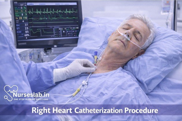
Understanding the anatomy of the venous system and right heart is critical. The catheter, commonly a balloon-tipped Swan-Ganz catheter, is usually inserted through a central vein and advanced sequentially through the superior vena cava (SVC), right atrium (RA), right ventricle (RV), and into the pulmonary artery (PA). The tip can be wedged in a small branch of the PA to measure pulmonary capillary wedge pressure (PCWP), an indirect estimate of left atrial pressure.
Technique and Procedural Steps
The procedure can be performed in a cardiac catheterization laboratory, intensive care unit, or operating room, depending on patient circumstances. The following outlines the typical steps:
- Patient Positioning: The patient is placed supine with the head turned away from the side of venous access.
- Sterile Preparation: The access site is prepped and draped in a sterile fashion.
- Local Anesthesia: Administered at the puncture site.
- Venous Access: Using ultrasound guidance, the chosen vein is cannulated with a needle. A guidewire is advanced, and a sheath is placed over the wire into the vein.
- Catheter Insertion: The Swan-Ganz catheter is advanced through the sheath. The balloon is inflated once the catheter is in the right atrium, facilitating smooth passage.
- Sequential Advancement: The catheter is advanced under continuous pressure waveform monitoring:
- Right Atrium: Characterized by low pressure waveforms (mean 2-6 mmHg).
- Right Ventricle: Higher systolic pressures (15-30 mmHg), low diastolic pressures (0-8 mmHg).
- Pulmonary Artery: Systolic pressures similar to RV, but diastolic pressures are higher (4-12 mmHg), with a characteristic dicrotic notch.
- Pulmonary Capillary Wedge: Balloon inflation in a distal PA branch occludes forward flow, allowing measurement of PCWP (6-12 mmHg).
- Pressure Measurements and Blood Sampling: At each station, pressures are recorded and blood samples may be drawn for oxygen saturation analysis.
- Cardiac Output Measurement: Thermodilution or Fick method can be used to determine cardiac output.
- Catheter Removal: Once all measurements are obtained, the catheter is withdrawn, and hemostasis is achieved at the access site.
Hemodynamic Measurements and Interpretation
A key advantage of RHC is the ability to obtain direct, real-time hemodynamic measurements. The primary parameters measured include:
- Right Atrial Pressure (RAP): Reflects right ventricular preload and central venous pressure.
- Right Ventricular Pressure (RVP): Assesses RV systolic function and afterload.
- Pulmonary Artery Pressure (PAP): Systolic, diastolic, and mean pressures indicate pulmonary vascular resistance and pressure load.
- Pulmonary Capillary Wedge Pressure (PCWP): Estimates left atrial pressure and left ventricular preload.
- Cardiac Output (CO): Quantifies blood flow from the heart, derived via thermodilution or Fick principle.
- Pulmonary Vascular Resistance (PVR): Calculated from PAP, PCWP, and CO, representing the resistance in pulmonary circulation.
- Systemic Vascular Resistance (SVR): Calculated from mean arterial pressure, RAP, and CO, reflecting systemic afterload.
Interpretation of these values, especially in the context of clinical presentation, can distinguish between different types of shock (cardiogenic, hypovolemic, distributive, obstructive), types of pulmonary hypertension, and the presence or absence of intracardiac shunts.
Sample Hemodynamic Data Table
| Parameter | Normal Range | Clinical Significance |
| Right Atrial Pressure (RAP) | 2-6 mmHg | Elevated in RV failure, fluid overload |
| Right Ventricular Pressure (RVP) | 15-30/0-8 mmHg | Elevated in pulmonary hypertension |
| Pulmonary Artery Pressure (PAP) | 15-30/4-12 mmHg | Elevated in pulmonary hypertension, left heart disease |
| Pulmonary Capillary Wedge Pressure (PCWP) | 6-12 mmHg | Elevated in left-sided heart failure, mitral disease |
| Cardiac Output (CO) | 4-8 L/min | Low in heart failure, shock |
| Pulmonary Vascular Resistance (PVR) | 20-130 dynes·sec/cm<sup>5</sup> | Elevated in pulmonary arterial hypertension |
| Systemic Vascular Resistance (SVR) | 800-1200 dynes·sec/cm<sup>5</sup> | Low in distributive shock; high in vasoconstriction |
Clinical Applications and Utility
Right heart catheterization remains invaluable in several clinical contexts:
- Pulmonary Hypertension: RHC is the gold standard for diagnosing and classifying pulmonary hypertension, distinguishing between pre-capillary and post-capillary causes, and assessing response to vasodilator challenge.
- Heart Failure: It aids in differentiating between right and left-sided heart failure, evaluating the need for advanced therapies, and optimizing volume status.
- Shock States: In critically ill patients, RHC can determine the underlying type of shock and guide fluid management, vasopressor, and inotropic therapy.
- Assessment of Intracardiac Shunts: By measuring oxygen saturations at multiple cardiac sites, RHC can detect and quantify left-to-right or right-to-left shunts.
- Cardiac Transplantation and Advanced Therapies: Pre-transplant evaluation and monitoring of response to therapies such as mechanical circulatory support devices rely on detailed hemodynamic assessment.
Complications of Right Heart Catheterization
Although generally safe, RHC carries risks, particularly in critically ill patients or those with coagulopathies. Complications include:
- Vascular injury (hematoma, bleeding, arteriovenous fistula, pseudoaneurysm)
- Infection at the access site
- Arrhythmias (premature ventricular contractions, atrial fibrillation, rarely ventricular tachycardia)
- Pulmonary artery rupture (rare but potentially fatal)
- Balloon rupture or catheter knotting
- Thromboembolism or air embolism
- Cardiac perforation and tamponade (extremely rare)
The overall complication rate is low, especially when the procedure is performed by experienced operators using modern techniques and imaging guidance.
Interpretation of Abnormal Findings
Abnormal hemodynamic measurements obtained during RHC can elucidate the etiology of a patient’s symptoms and guide management. For example:
- Elevated RAP with normal PCWP: Suggests isolated right ventricular dysfunction or pulmonary hypertension.
- Elevated PCWP with elevated PAP: Indicates left-sided heart failure or mitral valve disease.
- Low CO with high SVR: Consistent with cardiogenic shock.
- Low CO with low SVR: Suggests distributive (septic) shock.
- Step-up in oxygen saturation between RA and RV: Indicates left-to-right shunt (e.g., atrial septal defect).
Nursing Care for Patients Undergoing Right Heart Catheterization
Nurses play a pivotal role in ensuring the safety, comfort, and optimal outcomes of patients undergoing this procedure.
Pre-Procedure Nursing Care
Patient Assessment
Thorough patient assessment is the cornerstone of safe and effective right heart catheterization. Key aspects include:
- Medical History Review: Assess for underlying cardiac, pulmonary, renal, and hepatic conditions. Document allergies, especially to contrast agents, latex, and medications.
- Medication Reconciliation: Identify anticoagulants, antiplatelet agents, and other drugs that may affect bleeding or procedural risk. Confirm withholding or continuation as per protocol.
- Physical Examination: Evaluate vital signs, baseline oxygen saturation, peripheral pulses, and signs of heart failure or respiratory compromise.
- Laboratory Investigations: Ensure recent results for complete blood count, renal function, coagulation profile, and electrolytes are available and within acceptable ranges.
Preparation and Consent
- Patient Identification: Verify patient identity using at least two identifiers.
- Informed Consent: Confirm that the patient (or legal guardian) understands the procedure, risks, benefits, and alternatives. Witness and document consent as per institutional policy.
- Fasting and Pre-Procedural Instructions: Ensure the patient has fasted according to guidelines, typically 6–8 hours for elective procedures.
- Venous Access: Establish appropriate IV access for medication administration and fluid management.
- Skin Preparation: Prepare the insertion site (often the internal jugular, femoral, or subclavian vein) using antiseptic solution and ensure hair removal if necessary.
- Equipment Preparation: Confirm availability and functionality of required equipment, including monitoring devices, sterile trays, and emergency supplies.
Patient Education
- Procedure Explanation: Describe the purpose, steps, sensations (e.g., pressure, mild discomfort), and expected duration of the catheterization.
- Pre-Procedural Instructions: Instruct patients to report symptoms such as chest pain, palpitations, or shortness of breath.
- Addressing Anxiety: Provide reassurance, answer questions, and offer emotional support to alleviate fear and anxiety.
Intra-Procedure Nursing Responsibilities
Monitoring and Support
- Vital Signs: Continuously monitor heart rate, blood pressure, respiratory rate, oxygen saturation, and ECG for arrhythmias or ischemic changes.
- Hemodynamic Monitoring: Assist in recording real-time pressure measurements from the right atrium, right ventricle, pulmonary artery, and pulmonary capillary wedge position.
- Patient Observation: Assess for signs of distress, pain, dyspnea, or allergic reactions.
- Conscious Sedation: Monitor sedation levels, airway patency, and response to medications if sedation is used.
Assisting the Physician
- Instrument Handling: Provide sterile instruments, assist with catheter advancement, and manage equipment as directed.
- Sterile Technique: Maintain a sterile field throughout the procedure to minimize infection risk.
- Documentation: Record procedural events, medications administered, patient responses, and any deviations from protocol.
Ensuring Patient Comfort
- Positioning: Ensure the patient is comfortably positioned, typically supine, and provide additional support as needed.
- Pain Management: Administer analgesics as prescribed and monitor for effectiveness and adverse reactions.
- Communication: Offer verbal reassurance and inform the patient about each step to reduce anxiety and foster cooperation.
Post-Procedure Nursing Care
Monitoring for Complications
- Vital Signs and Hemodynamics: Continue close monitoring for at least 2–4 hours post-procedure or as per protocol. Watch for hypotension, tachycardia, arrhythmias, or hypoxemia.
- Insertion Site Assessment: Inspect for bleeding, hematoma, infection, or swelling at the catheter entry point.
- Peripheral Pulses: Evaluate distal pulses and limb perfusion, especially if femoral access was used.
- Respiratory Status: Monitor for signs of pulmonary embolism, pneumothorax, or respiratory distress.
Wound Care and Patient Positioning
- Dressing Management: Maintain a clean, dry, and intact dressing over the insertion site. Change as per protocol and observe for signs of infection.
- Immobilization: Advise the patient to remain flat and limit movement of the affected limb for a specified period to minimize bleeding risk.
- Pain Assessment: Evaluate and manage pain using appropriate analgesics and comfort measures.
Ongoing Assessment
- Neurological Status: Monitor for confusion, dizziness, or altered consciousness, which may indicate complications.
- Fluid Balance: Assess intake and output, monitor for signs of fluid overload or dehydration.
- Early Detection of Complications: Promptly report abnormal findings to the medical team for immediate intervention.
Patient Education
- Recovery Instructions: Advise on activity restrictions, wound care, and the importance of keeping the insertion site clean and dry.
- Signs of Complications: Educate on symptoms requiring immediate medical attention, such as excessive bleeding, swelling, fever, chest pain, shortness of breath, or palpitations.
- Medication Adherence: Review prescribed medications, their purposes, and potential side effects.
- Follow-Up Care: Emphasize the need for scheduled appointments, repeat testing, and ongoing communication with healthcare providers.
- Lifestyle Modification: Offer guidance on smoking cessation, diet, exercise, and management of underlying conditions as appropriate.
Complications and Management
Common Risks
- Bleeding and Hematoma: Most frequently at the insertion site; managed by pressure application and monitoring.
- Arrhythmias: May occur during catheter manipulation; monitor ECG and provide antiarrhythmic therapy if needed.
- Infection: Prevent with strict aseptic technique and monitor for fever, redness, or purulent discharge.
- Pulmonary Embolism: Rare but serious; monitor for sudden respiratory distress, chest pain, and provide supportive care.
- Pneumothorax: Particularly with subclavian access; assess for sudden dyspnea, decreased breath sounds, and initiate emergency management.
- Vascular Injury: Promptly identify signs of arterial puncture, limb ischemia, or expanding hematoma.
Early Detection and Nursing Interventions
- Routine Monitoring: Vigilant assessment enables rapid recognition and intervention for complications.
- Emergency Response: Activate protocols for cardiac arrest, severe bleeding, or respiratory compromise.
- Documentation: Accurately record all findings, interventions, and patient responses for continuity of care.
REFERENCES
- Chokkalingam Mani B, Chaudhari SS. Right Heart Cardiac Catheterization. [Updated 2022 May 9]. In: StatPearls [Internet]. Treasure Island (FL): StatPearls Publishing; 2022 Jan-.
- Government of Alberta. Cardiac Catheterization https://myhealth.alberta.ca/Health/pages/conditions.aspx?hwid=hw204075).
- Haleem SM, Sharma T, Chaudhari SS. Right Heart Cardiac Catheterization. [Updated 2025 Jul 5]. In: StatPearls [Internet]. Treasure Island (FL): StatPearls Publishing; 2025 Jan-. Available from: https://www.ncbi.nlm.nih.gov/sites/books/NBK557404/
- What is cardiac catheterization. National Heart, Lung, and Blood Institute. https://www.nhlbi.nih.gov/health/cardiac-catheterization.
- Cardiac catheterization. American Heart Association. https://www.heart.org/en/health-topics/heart-attack/diagnosing-a-heart-attack/cardiac-catheterization.
- Loscalzo J, et al., eds. Diagnostic cardiac catheterization and coronary angiography. In: Harrison’s Principles of Internal Medicine. 21st ed. McGraw Hill; 2022. https://accessmedicine.mhmedical.com.
- Health Education & Content Services. Preparing for your cardiac catheterization or heart rhythm procedure. Mayo Clinic; 2021.
Stories are the threads that bind us; through them, we understand each other, grow, and heal.
JOHN NOORD
Connect with “Nurses Lab Editorial Team”
I hope you found this information helpful. Do you have any questions or comments? Kindly write in comments section. Subscribe the Blog with your email so you can stay updated on upcoming events and the latest articles.


