


Last Updated on November 7, 2026 by Nurseslab.in Editorial Team
Learn about Coats disease, a rare retinal disorder. Explore its symptoms, causes, stages, diagnosis, treatment options and potential effects on vision.
Introduction
Coats disease is an idiopathic, non-hereditary retinal vascular disorder characterized by retinal telangiectasia and aneurysmal dilatation of retinal vessels, with progressive leakage producing massive intraretinal and subretinal lipid exudation and, if unchecked, exudative retinal detachment.
It was described by the Scottish ophthalmologist George Coats in 1908, and the original description still holds: abnormal vessels that leak, and lipid-rich exudate that accumulates where gravity takes it.
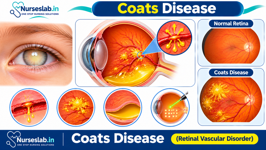
The essential mechanism is worth fixing clearly, because everything else follows from it:
Abnormal, incompetent retinal vessels leak → lipid-rich exudate accumulates under and within the retina → the retina separates and photoreceptors fail → irreversible vision loss, then neovascular glaucoma, then a blind painful eye.
Treatment at every stage is aimed at one thing: stopping the leak before the exudate reaches the fovea.
Epidemiology
- Laterality: Unilateral in approximately 95% of cases. Bilateral disease is uncommon and should prompt a search for an alternative diagnosis or an associated syndrome.
- Sex: Marked male predominance — at least 3:1, with many series reporting a considerably higher ratio.
- Age: Most often diagnosed in children under 8 years, though it occurs in infants and in adults. Adult-onset presentations tend to be milder and more localized.
- Incidence: Rare; reliable population figures are limited by the condition’s rarity and historical misclassification.
A clinically important pattern from the published series: patients presenting at an advanced stage are substantially younger. A 2025 global analysis found a mean age of 7.1 years in advanced-stage disease versus 17.4 years in early-stage disease, with advanced cases far more likely to present with strabismus or leukocoria rather than with a visual complaint.
Younger children present later and worse, because they cannot report what they cannot compare.
Pathophysiology
The underlying cause remains unknown. What is established is the sequence:
- Primary vascular abnormality. Telangiectatic and aneurysmal retinal vessels develop, predominantly in the temporal mid-periphery and periphery. The classic angiographic appearance is the “light-bulb” aneurysm — a saccular, bulbous dilatation.
- Breakdown of the blood–retinal barrier. The abnormal endothelium is incompetent, allowing plasma and lipid to escape.
- Exudation. Lipid-rich fluid accumulates intraretinally and subretinally. Because it is heavy, exudate gravitates to the inferior pole and can track to the posterior pole.
- Macular involvement. Exudate infiltrating the fovea causes macular oedema, subretinal fluid, and the formation of a foveal nodule — a fibrotic subretinal lesion that is one of the strongest predictors of poor visual outcome.
- Exudative retinal detachment. Progressive fluid accumulation separates the retina. In children, detachment may become bullous and immobile, extending forward to touch the lens capsule.
- End-stage complications. Peripheral capillary non-perfusion drives ischaemia, which drives neovascularization — of the retina, iris, and angle — producing neovascular glaucoma. The final common endpoint is a blind, painful eye, with phthisis bulbi in the most advanced cases.
Other late findings include discoid glial scarring, subretinal neovascularization, and intraretinal haemorrhage.
Genetics and associations
Coats disease itself is sporadic and non-hereditary. No causative gene has been confirmed, and families should be reassured on this point — it is one of the most common questions parents ask.
That said, several lines of investigation exist:
- Chromosomal instability involving chromosomes 3 and 13 has been proposed.
- Genes implicated in individual reports include the Norrie disease (NDP) gene, CRB1, and PANK2.
- A somatic (non-inherited) mechanism within the retina is consistent with the unilateral, sporadic pattern.
“Coats-like” exudative responses occur in a number of systemic and ocular conditions, and recognizing them matters because they change management:
- Turner syndrome
- Senior–Løken syndrome
- Retinitis pigmentosa
- Facioscapulohumeral dystrophy (FSHD)
- Linear scleroderma / Parry–Romberg syndrome
Clinical rule: in a female patient, or in bilateral disease, isolated Coats disease becomes much less likely. Bilateral involvement in a girl should specifically prompt evaluation for FSHD and consideration of the syndromic alternatives above.
Coats plus syndrome — a different disease
This is a frequent source of confusion, and the distinction is clean.
Coats plus syndrome (also called cerebroretinal microangiopathy with calcifications and cysts, CRMCC) is a separate, inherited, multisystem disorder that includes Coats-like retinal vasculopathy as one feature.
| Coats disease | Coats plus syndrome | |
|---|---|---|
| Inheritance | Sporadic, non-hereditary | Autosomal recessive |
| Gene | None confirmed | CTC1 (CST complex, telomere maintenance) |
| Extent | Eye only | Multisystem |
| Laterality | Usually unilateral | Typically bilateral |
| Prognosis | Ocular morbidity | Life-limiting |
Systemic features of Coats plus:
- Brain: intracranial calcifications, fluid-filled cysts, and leukodystrophy — progressive, causing movement disorders, seizures, and cognitive decline
- Skeletal: osteopenia with fragile bones
- Haematological and GI: anaemia, abnormal gastrointestinal bleeding, portal hypertension, and liver failure — potentially life-threatening
- Ectodermal: sparse, prematurely grey hair, nail malformations, café-au-lait pigmentation
For nursing practice: a child with bilateral Coats-like retinal findings plus growth failure, neurological signs, unexplained anaemia, or GI bleeding is not a routine Coats disease referral. That child needs genetics, neurology, haematology, and gastroenterology involvement.
Clinical presentation
Symptoms
Published series give a consistent picture. In a large 150-patient series, the commonest presenting complaints were:
- Decreased vision — 34%
- Strabismus — 23%
- Leukocoria
- Less commonly: pain, heterochromia, nystagmus, or an eye noticed to be “different”
Many children are asymptomatic from their own perspective. Unilateral disease in a young child is functionally silent until the parent notices a white reflex or a turning eye, or until school vision screening picks it up.
Signs
- Leukocoria — a white or abnormal pupillary reflex. In Coats disease it is often described as grey to pink or yellowish, in contrast to the chalky white of retinoblastoma
- Strabismus — typically a sensory deviation secondary to poor vision
- Retinal telangiectasia — involving the mid-periphery or periphery in nearly all eyes, most often temporal
- Yellowish subretinal and intraretinal exudate, gravitating inferiorly. In one large series, exudation involved all 12 clock hours in 55% of eyes
- Exudative retinal detachment in advanced disease
- Normal anterior segment in approximately 90% of eyes at presentation — a useful discriminator, since advanced retinoblastoma more often shows anterior segment involvement
- Late: iris and angle neovascularization, neovascular glaucoma, cataract, band keratopathy, phthisis bulbi
Visual acuity at presentation
The figure worth remembering, because it explains why early detection matters so much: in the large published series, 76% of eyes presented with vision between 20/200 and light perception or worse.
Three quarters of these children arrive with the eye already functionally lost or close to it.
Shields classification — the staging system to know
This is the standard classification and the one that appears in examinations and in every management discussion.
| Stage | Findings |
|---|---|
| Stage 1 | Retinal telangiectasia only |
| Stage 2 | Telangiectasia + exudation |
| — 2A | Extrafoveal exudation |
| — 2B | Foveal exudation |
| Stage 3 | Exudative retinal detachment |
| — 3A | Subtotal detachment |
| — 3A1 | Extrafoveal |
| — 3A2 | Foveal |
| — 3B | Total retinal detachment |
| Stage 4 | Total retinal detachment + glaucoma |
| Stage 5 | Advanced end-stage disease (blind, often painful eye; phthisis) |
The logic of the system: stage reflects progression from vessel abnormality alone → leakage → leakage reaching the fovea → retina separating → secondary glaucoma → end-stage eye. Foveal involvement is the hinge — it is what separates a retained reading eye from a lost one, which is why substages distinguish extrafoveal from foveal at every level.
Older systems — Gomez-Morales and Sigelman — follow a similar progression and occasionally appear in older literature.
Diagnostic evaluation
Fundoscopy — with full dilation and peripheral examination — is the primary diagnostic tool. In young children this generally requires examination under anaesthesia (EUA).
Fluorescein angiography (FA)
- Telangiectatic vessels and light-bulb aneurysms
- Early and progressive perivascular leakage
- Peripheral capillary non-perfusion — important, because it guides where scatter laser is needed
- Contrast with retinoblastoma, which shows a “double circulation” pattern
B-scan ultrasonography
- Retinal detachment without a solid mass and without calcification
- This is the single most useful bedside test for separating Coats disease from retinoblastoma
Optical coherence tomography (OCT)
- Intraretinal and subretinal fluid, macular oedema, and the foveal nodule
- Increasingly used to track response to treatment
CT / MRI
- CT demonstrates calcification, which is characteristic of retinoblastoma and not a feature of Coats disease
- MRI is generally preferred in children to avoid radiation exposure, particularly where a genetic predisposition to malignancy is possible
Differential diagnosis
Coats disease versus retinoblastoma — the comparison that matters most
Both cause leukocoria. Misdiagnosis in either direction is serious: a missed retinoblastoma is lethal, and historically many Coats eyes were enucleated for suspected retinoblastoma.
| Feature | Coats disease | Retinoblastoma |
|---|---|---|
| Age | Older child | Younger child |
| Laterality | Unilateral (~95%) | May be bilateral |
| Sex | Male predominance | No strong predominance |
| Leukocoria colour | Grey to pink / yellowish | Chalky white |
| Vessels | Traceable telangiectasia | Vessels dip into the tumour |
| Intraocular mass | Absent | Present (endophytic cases) |
| Calcification (CT / US) | Absent | Present |
| Vitreous seeding | Absent | May be present |
| Fluorescein angiography | Leakage from telangiectasia | “Double circulation” |
| Exudate | Massive yellow lipid exudate | Subretinal seeds |
Other differentials
- Familial exudative vitreoretinopathy (FEVR) — bilateral, hereditary, peripheral avascularity with dragged disc
- Retinopathy of prematurity (ROP) — history of prematurity, bilateral
- Persistent fetal vasculature (PFV) — microphthalmos, retrolental membrane
- Ocular toxocariasis — granuloma, inflammatory signs, serology
- Norrie disease — X-linked, bilateral, with deafness and cognitive involvement
- Incontinentia pigmenti — female, skin lesions
- Eales disease, pars planitis, retinal cavernous haemangioma
- Retinal vasoproliferative tumour — feeder and draining vessels, vascular mass, preretinal fibrosis, peripheral
- Leukaemic retinopathy and retinal metastases
Management
Treatment is stage-dependent and aims to ablate the leaking vessels and resolve the exudate before the fovea is destroyed.
| Modality | Indication | Notes |
|---|---|---|
| Observation | Minimal telangiectasia with little or no exudation and no threat to vision; also painless sightless eyes | Requires reliable follow-up |
| Laser photocoagulation | Telangiectasia with exudation and no or minimal subretinal fluid | Applied at low power over long duration; scatter laser to areas of peripheral non-perfusion. Mainstay of early disease |
| Cryotherapy | Exudation where subretinal fluid is too thick for laser to reach the retina | Often requires repeated sessions; more inflammation and exudative response |
| Anti-VEGF injection | Adjunct to reduce exudation and neovascularization | Promising but paediatric safety is not established; risk of fibrosis and tractional detachment |
| Intravitreal triamcinolone | Refractory exudation and macular oedema | Raises risk of glaucoma and cataract |
| Vitreoretinal surgery | Extensive detachment not amenable to laser or cryotherapy | Scleral buckle, vitrectomy, subretinal fluid drainage |
| Enucleation | Blind painful eye, or retinoblastoma cannot be excluded | The second indication is the historically important one |
In practice, early stages (1–2) are managed primarily with laser, and a 2025 global analysis found laser monotherapy used in 44.7% of early-stage eyes. Advanced stages increasingly receive combination therapy, which that analysis associated with improved odds of anatomical success.
Where the evidence is genuinely unsettled
Honesty matters here, because this is a rare disease with no randomized trial base.
A 2025 international consensus exercise found substantive disagreement among experts on the management of stages 3 and 4, and on the role of newer anti-VEGF agents such as faricimab. There is no settled protocol for advanced disease.
The 2025 global study of 656 eyes reported the most useful prognostic findings currently available:
- A foveal nodule was a key predictor of poor visual outcome
- Worse presenting visual acuity and advanced stage were the strongest predictors of both functional and anatomical failure
- The role of anti-VEGF in early disease remains unclear
Prognosis
- Better prognosis: milder disease, older age at presentation, extrafoveal exudation, good presenting acuity
- Worse prognosis: presentation under 3 years of age, advanced stage, total detachment, foveal nodule, poor presenting vision
The realistic framing for families: anatomical success — a reattached, comfortable, preserved eye — is achievable in many cases. Restoration of good central vision in an eye that presented with foveal exudate or detachment is often not. Those two goals must be discussed separately, because conflating them produces expectations that the disease cannot meet.
Nursing Care of Patient with Coats Disease
This is where nursing genuinely changes outcomes, and the leverage is heavily front-loaded toward detection.
Detection — the highest-value nursing contribution
Red reflex testing at every well-child visit is a nursing and primary-care task, and it is the main mechanism by which these children are found early.
- Perform the test in a darkened room, with the ophthalmoscope at arm’s length, examining both eyes simultaneously (Brückner test) to compare
- Any asymmetry, any white or dulled reflex, is abnormal and requires urgent referral
- Take parental photographic observations seriously. Flash photography frequently reveals leukocoria before any clinician does, and parents commonly raise it tentatively
- Assess for strabismus — new or intermittent deviation in a young child warrants referral, not reassurance
- Remember that a child with one good eye will not complain, and will pass informal “can you see this?” testing
The referral rule: leukocoria is an ophthalmic emergency until retinoblastoma has been excluded. Not a routine referral. Not a watch-and-wait.
Supporting the diagnostic period
The interval between “there is something in your child’s eye” and “it is not cancer” is acutely distressing, and nursing care during it is almost entirely communicative.
- Explain why retinoblastoma must be excluded first, without inviting the family to assume the worst
- Prepare the family for EUA, imaging, and possible repeat examinations
- Do not speculate on the diagnosis ahead of the ophthalmologist
- Provide a named contact and a realistic timeline
Examination under anaesthesia (EUA) — nursing care
Children with Coats disease undergo repeated EUAs over years, both for diagnosis and for monitoring and treatment.
Pre-procedure
- Confirm fasting status per paediatric protocol and verify consent, including consent for treatment at the same sitting if indicated
- Document baseline weight, vital signs, allergies, and any airway or anaesthetic history
- Administer dilating drops as prescribed, on schedule — inadequate dilation can mean a cancelled or incomplete examination
- Prepare the child developmentally appropriately; involve play specialists where available
- Allow a parent to accompany to induction where policy permits
Post-procedure
- Standard paediatric post-anaesthetic recovery: airway, consciousness, vital signs, pain, nausea
- Expect photophobia and blurred vision from dilation — darken the room, provide sunglasses, and warn the family it may last hours
- Assess for pain disproportionate to the procedure, and escalate
- Confirm understanding of the findings and the next appointment before discharge
After laser or cryotherapy
- Expect lid oedema, conjunctival injection, and discomfort — more pronounced after cryotherapy
- Administer and teach topical steroids, antibiotics, and cycloplegics as prescribed, with correct instillation technique demonstrated and teach-back obtained
- Teach cold compresses for lid swelling where permitted
- Red flags to report: worsening pain, increasing redness, purulent discharge, sudden vision loss, or a new floater shower
- Advise against rubbing the eye; consider a protective shield at night in younger children
After intravitreal injection
- Strict aseptic technique; confirm correct eye, drug, and dose with the second checker
- Monitor for raised intraocular pressure post-injection per unit protocol
- Teach the family the signs of endophthalmitis — increasing pain, worsening redness, decreasing vision, discharge — and that these require same-day assessment
- Reassure that subconjunctival haemorrhage and floaters are common and benign
Protecting the better eye — a lifelong priority
In unilateral disease, the fellow eye carries the child’s entire visual future. This is arguably the most important ongoing teaching point.
- Polycarbonate protective eyewear for sport and hazardous activity, and full-time spectacle protection where advised
- Prompt assessment of any injury or symptom in the good eye
- Up-to-date refraction and amblyopia management
- Teach the family explicitly: monocular status changes risk permanently, and protective eyewear is not optional
Vision rehabilitation and development
- Support amblyopia therapy — occlusion or atropine — where some potential remains in the affected eye; adherence is the main determinant of success, and it is poor without support
- Refer to low vision services and, where appropriate, services for visually impaired children
- Liaise with school — preferential seating, large print, extra time, and documented accommodations
- Monitor developmental and educational progress; unilateral vision loss affects depth perception, sport, and some practical tasks
- Address monocular depth-perception adjustment practically: stairs, ball sports, pouring, cycling
Psychosocial care
- Explore parental guilt — “we should have noticed sooner.” Framing matters: a silent unilateral disease in a preschool child is genuinely difficult to detect, and most parents detect it before any clinician does
- Reassure that Coats disease is sporadic and not inherited, and that siblings are not at identifiable increased risk
- Address the child’s own concerns about appearance, strabismus, glasses, or patching, age-appropriately
- Prepare for the possibility of enucleation gently and only when clinically relevant, and provide ocularist and prosthesis information when it is
- Connect families to rare disease support networks — isolation is a real feature of rare paediatric conditions
If enucleation is required
- Pre-operative preparation of child and family, including realistic description of the conformer and later prosthesis
- Post-operative socket care, dressing management, and infection surveillance
- Teach prosthesis insertion, removal, and cleaning once fitted, with teach-back
- Ongoing ocularist follow-up — the prosthesis requires periodic refitting as the child grows
- Continued emphasis on protection of the remaining eye
Follow-up adherence
Coats disease requires monitoring over years, with recurrence and reactivation both possible.
- Ensure the family understands that resolution of exudate is not cure
- Help navigate the practical barriers — transport, time off work, repeated anaesthetics, cost
- Flag missed appointments actively rather than passively
Family education checklist
Understanding the condition
- Coats disease affects blood vessels in the retina; they leak, and the leaked fluid damages the retina
- It is not cancer, not inherited, and not caused by anything the parents did
- It usually affects one eye only
Treatment expectations
- Treatment aims to stop the leakage and prevent further damage
- Repeated treatments and repeated examinations under anaesthesia are normal, not a sign of failure
- Preserving the eye and preserving good vision are different goals, and both will be discussed honestly
Daily care
- Eye drop schedule and correct instillation technique
- Patching or atropine regimen, if prescribed
- Protective eyewear for the good eye — every day it is needed
When to seek help urgently
- Increasing eye pain
- Worsening redness or discharge
- Sudden decrease in vision, or a shower of new floaters
- Any injury to either eye
- A new white reflex, or a newly turning eye
Follow-up
- Next appointment date and who to contact with questions
- Importance of attending even when the child seems well
REFERENCE
- Almutairi F, Esmaeil A, Al-Farsi N, AlKandari A, AlAli A. Coats disease: A narrative review of clinical spectrum, imaging features, and contemporary management. Sci Prog. 2026 Jul-Sep;109(3):368504261484841. doi: 10.1177/00368504261484841. Epub 2026 Aug 28. PMID: 42664491; PMCID: PMC13525312.
- American Academy of Ophthalmology. Coats Disease https://eyewiki.aao.org/Coats_Disease. Last updated 2/13/2024.
- American Association for Pediatric Ophthalmology and Strabismus. Coats’ Disease (https://www.aapos.org/glossary/coats-disease). Last updated 3/2021
- Gupta A, Paulbuddhe VS, Shukla UV, et al. Exudative Retinitis (Coats Disease) (https://www.ncbi.nlm.nih.gov/books/NBK560682/). 2023 Aug 25. In: StatPearls [Internet]. Treasure Island (FL): StatPearls Publishing; 2024 Jan.
- National Organization for Rare Disorders (U.S.). Coats Disease (https://rarediseases.org/rare-diseases/coats-disease/). Last updated 07/2015.
Stories are the threads that bind us; through them, we understand each other, grow, and heal.
JOHN NOORD
Connect with “Nurses Lab Editorial Team”
I hope you found this information helpful. Do you have any questions or comments? Kindly write in comments section. Subscribe the Blog with your email so you can stay updated on upcoming events and the latest articles.


