


Bone marrow examination is a vital diagnostic test that evaluates blood cell production and detects conditions such as anemia, leukemia, and other hematologic disorders. It guides treatment decisions, supports evidence-based practice, and enhances nursing and medical education.
Introduction
Bone marrow examination is a cornerstone in the evaluation and diagnosis of a wide spectrum of haematological and non-haematological disorders. As the primary site of blood cell production, the bone marrow reflects the dynamic processes of haematopoiesis and is frequently involved in systemic diseases. A thorough understanding of the pathology of bone marrow examination enables clinicians and pathologists to diagnose, classify, and guide the management of various conditions, ranging from anaemias to malignancies.
Anatomy and Physiology of Bone Marrow
Structure and Distribution
Bone marrow is a soft, vascular tissue located within the medullary cavities of bones. In adults, active (red) marrow is predominantly found in the axial skeleton—vertebrae, ribs, sternum, pelvis, and proximal ends of long bones—while yellow marrow (composed mainly of fat) occupies peripheral sites. The marrow microenvironment consists of a complex network of stromal cells, adipocytes, blood vessels, and extracellular matrix supporting haematopoietic cells.
Cellular Components and Function
Bone marrow is responsible for the continuous production of erythrocytes, leukocytes, and platelets through the process of haematopoiesis. The cellular constituents include:
- Haematopoietic stem cells (HSCs): Pluripotent cells capable of self-renewal and differentiation into all blood cell lineages.
- Progenitor and precursor cells: Intermediate stages in cell maturation.
- Stromal cells: Provide structural support and secrete growth factors.
- Adipocytes, macrophages, endothelial cells: Contribute to the regulation of haematopoiesis.
The balance between these elements ensures normal blood cell counts and immune function.
Indications for Bone Marrow Examination
Bone marrow examination is indicated in various clinical scenarios, primarily when peripheral blood findings are inconclusive or suggest an underlying marrow pathology. Common indications include:
- Unexplained cytopenias: Anaemia, leucopenia, thrombocytopenia, or pancytopenia.
- Leukaemias and lymphomas: Diagnosis, classification, staging, and monitoring.
- Myelodysplastic syndromes (MDS) and myeloproliferative neoplasms (MPN): Evaluation and subtyping of the syndrome and symptoms.
- Aplastic anaemia or marrow failure syndromes: Assessment of marrow cellularity.
- Fever of unknown origin: Detection of infections, granulomas, or malignancies.
- Staging of solid tumours: Identification of marrow infiltration in cancers such as neuroblastoma or breast carcinoma.
- Investigation of storage disorders, metabolic diseases, or unexplained splenomegaly.
Techniques and Procedures
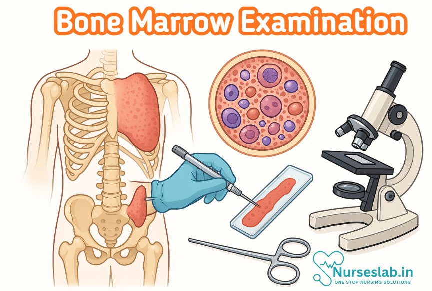
Bone Marrow Aspiration
Aspiration involves the removal of liquid marrow for cytological evaluation. The preferred sites are the posterior iliac crest in adults and the tibia or iliac spine in children. After local anaesthesia, a specialised needle is inserted into the marrow cavity, and a small volume (0.2–0.5 mL) of marrow is aspirated. Smears are prepared immediately to avoid clotting.
Bone Marrow Biopsy
A trephine biopsy provides a core of marrow tissue, allowing assessment of cellularity, architecture, and infiltration. It is especially valuable when aspiration yields a “dry tap” (due to fibrosis or packed marrow). The specimen is fixed in formalin and processed for histopathological examination.
Sample Handling and Staining
Proper handling is crucial to preserve cellular morphology. Aspirate smears are stained with Romanowsky stains (e.g., Leishman, Wright-Giemsa), while biopsy sections are stained with haematoxylin and eosin (H&E). Special stains (e.g., reticulin, Prussian blue) may be used to assess fibrosis or iron stores.
Additional Techniques
Flow cytometry, immunohistochemistry, cytogenetic, and molecular studies are often performed on aspirate or biopsy specimens for further characterisation and diagnosis of specific disorders.
Morphological Assessment
Normal Bone Marrow
A healthy adult bone marrow is cellular (30–70%, age-dependent) with normal representation of erythroid, myeloid, and megakaryocytic lineages. The myeloid:erythroid ratio is typically 2:1 to 4:1. Megakaryocytes are scattered and easily identified. The marrow architecture is orderly, with distinct maturation stages.
Abnormal Findings
Abnormalities may involve:
- Cellularity: Hypocellular (as in aplastic anaemia) or hypercellular (as in leukaemias).
- Maturation: Dysplastic changes, maturation arrest, or left-shifted maturation.
- Lineage distribution: Increased or decreased specific cell lines.
- Infiltrates: Malignant cells, granulomas, infectious organisms, or metastatic deposits.
- Stromal changes: Fibrosis, necrosis, or abnormal iron stores.
Morphological examination is the foundation for diagnosing most marrow pathologies.
Common Pathological Findings
Leukaemias
Acute leukaemias are characterised by increased blasts (>20% of nucleated cells), with suppression of normal haematopoiesis. Chronic leukaemias show increased mature cells of a particular lineage (e.g., granulocytes in chronic myeloid leukaemia). Morphology, cytochemistry, and immunophenotyping are essential for accurate classification.
Lymphomas
Marrow involvement by lymphomas (especially non-Hodgkin’s lymphoma) manifests as interstitial, nodular, or diffuse infiltrates of atypical lymphoid cells. Immunohistochemistry is vital for subtyping.
Myelodysplastic Syndromes (MDS)
MDS is marked by hypercellular marrow with dysplastic changes in one or more lineages, ineffective haematopoiesis, and increased risk of transformation to acute leukaemia. Ring sideroblasts and abnormal megakaryocytes are characteristic features.
Aplastic Anaemia
This condition presents as hypocellular or fatty marrow with marked reduction of all cell lines. It is distinguished from hypocellular MDS by the absence of significant dysplasia or abnormal cells.
Infections
Marrow may show granulomas (e.g., tuberculosis), fungal organisms, or haemophagocytosis in viral infections. Special stains and cultures aid in identification.
Metastatic Disease
Solid tumours, such as breast, prostate, or neuroblastoma, may infiltrate the marrow, often presenting as focal or diffuse replacement of normal elements by malignant cells. Immunohistochemistry assists in identifying the tumour origin.
Interpretation and Reporting
The interpretation of bone marrow findings requires correlation with clinical history, peripheral blood counts, and ancillary studies. Key elements of reporting include:
- Cellularity and lineage distribution.
- Morphological abnormalities and presence of infiltrates.
- Assessment of iron stores and fibrosis.
- Integration of immunophenotypic, cytogenetic, and molecular data.
- Clear, concise diagnostic summary and differential diagnoses.
Pitfalls such as sampling errors, artefacts, or inadequate specimens must be recognised and addressed.
Ancillary Studies
Immunohistochemistry (IHC)
IHC uses antibodies to detect cell surface or cytoplasmic markers, aiding in the identification and classification of neoplastic and non-neoplastic cells. It is indispensable in distinguishing lymphoid neoplasms and assessing metastatic disease.
Flow Cytometry
This technique analyses cell populations based on surface and cytoplasmic markers, enabling precise classification of leukaemias and lymphomas. It is also useful for detecting minimal residual disease.
Cytogenetics
Conventional karyotyping and fluorescent in situ hybridisation (FISH) detect chromosomal abnormalities significant for diagnosis, prognosis, and therapy selection, especially in leukaemias and MDS.
Molecular Testing
Polymerase chain reaction (PCR) and next-generation sequencing (NGS) identify gene mutations, translocations, and clonal rearrangements. These tests are increasingly used for risk stratification and targeted therapies.
Complications and Limitations
Bone marrow examination is generally safe but not without risks. Complications include:
- Pain and bleeding at the puncture site.
- Infection, especially in immunocompromised patients.
- Rarely, injury to nearby structures (nerves, vessels).
Contraindications encompass severe bleeding disorders and local infection at the intended site. Limitations arise from sampling error, patchy involvement, and interpretative challenges in cases of poor specimen quality or overlapping features.
Clinical Significance
Bone marrow examination has a profound impact on patient management. It guides diagnosis, informs prognosis, and influences treatment strategies, such as the need for chemotherapy, stem cell transplantation, or supportive care. Serial marrow assessments monitor disease progression or remission and detect minimal residual disease. Integration of morphological, immunophenotypic, cytogenetic, and molecular findings ensures precision medicine in haematology and oncology.
REFERENCES
- Ramadas Nayak, Textbook of Pathology and Genetics for Nurses, 2nd Edition,2024, Jaypee Publishers, ISBN: 978-93-5270-031-8.
- Suresh Sharma, Textbook of Pharmacology, Pathology & Genetics for Nurses II, 2nd Edition, 31 August 2022, Jaypee Publishers, ISBN: 978-9354655692.
- Kumar, V., Abbas, A.K., & Aster, J.C. (2020). Robbins and Cotran Pathologic Basis of Disease. 10th Edition. Elsevier.
- McCance, K.L., & Huether, S.E. (2018). Pathophysiology: The Biologic Basis for Disease in Adults and Children. 8th Edition. Elsevier.
- King O, West E, Lee S, Glenister K, Quilliam C, Wong Shee A, Beks H. Research education and training for nurses and allied health professionals: a systematic scoping review. BMC Med Educ. 2022 May 19;22(1):385. https://pmc.ncbi.nlm.nih.gov/articles/PMC9121620/
- Barría P RM. Use of Research in the Nursing Practice: from Statistical Significance to Clinical Significance. Invest Educ Enferm. 2023 Nov;41(3):e12. doi: 10.17533/udea.iee.v41n3e12. PMID: 38589312; PMCID: PMC10990586.
Stories are the threads that bind us; through them, we understand each other, grow, and heal.
JOHN NOORD
Connect with “Nurses Lab Editorial Team”
I hope you found this information helpful. Do you have any questions or comments? Kindly write in comments section. Subscribe the Blog with your email so you can stay updated on upcoming events and the latest articles.


