


Last Updated on May 27, 2026 by Nurseslab.in Editorial Team
The human stomach is a vital organ within the digestive system, playing a central role in the mechanical and chemical breakdown of food.
Introduction:
Nursing professionals routinely encounter patients with gastrointestinal issues, making knowledge of the stomach’s anatomy indispensable. From administering medications to recognising signs of distress, nurses must be able to integrate anatomical understanding into assessment, intervention, and patient education.
Overview of the Stomach
Location, Shape, and General Function
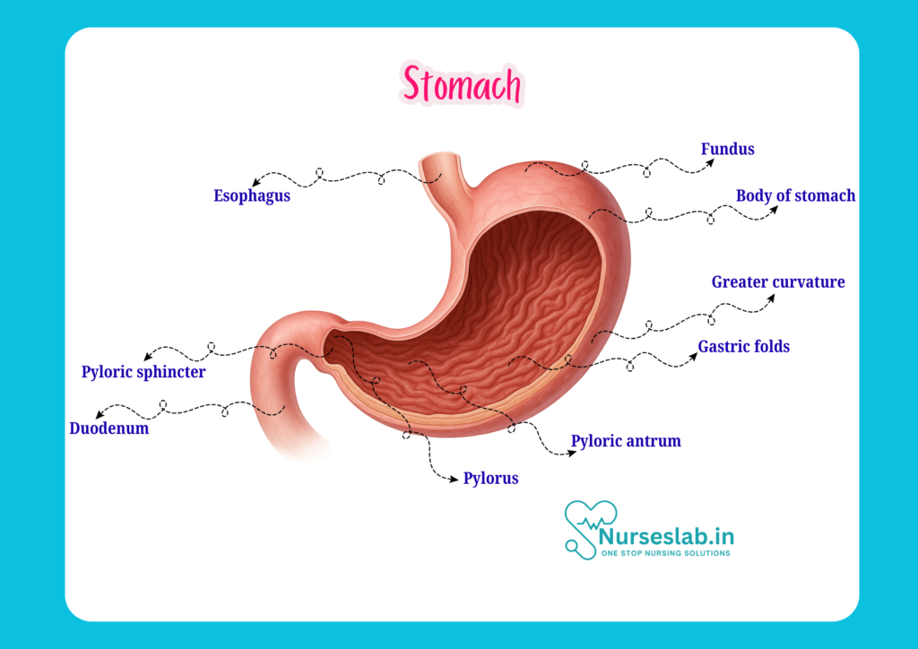
The stomach is a muscular, hollow organ situated in the upper left quadrant of the abdomen, just below the diaphragm. It lies between the oesophagus and the small intestine (duodenum), serving as a reservoir for ingested food. The stomach’s shape can vary depending on its contents and the individual’s body habitus, but it is generally described as J-shaped or curved.
Functionally, the stomach acts as a mixing chamber, blending food with gastric secretions to form chyme. It initiates protein digestion, regulates the passage of food into the small intestine, and absorbs certain substances such as alcohol and some medications. Its ability to expand and contract allows it to accommodate varying volumes of food and liquid.
Gross Anatomy
Regions of the Stomach
The stomach is divided into four main anatomical regions, each with specific features and functions:
- Cardia: This is the region where the oesophagus connects to the stomach. The cardia contains the lower oesophageal sphincter, which prevents reflux of gastric contents into the oesophagus.
- Fundus: The fundus is the dome-shaped area above the level of the cardia. It acts as a storage site for undigested food and gases produced during digestion.
- Body (Corpus): The body is the largest region, responsible for mixing and churning food with gastric juices. It contains the majority of gastric glands.
- Pylorus: The pylorus is the distal region, which narrows as it approaches the small intestine. It contains the pyloric sphincter, regulating the release of chyme into the duodenum.
Anatomical Relations
Understanding the anatomical relations of the stomach is crucial for nursing practice, especially during assessment and surgical procedures. The anterior surface of the stomach is related to the left lobe of the liver, diaphragm, and anterior abdominal wall. Posteriorly, it is adjacent to the pancreas, spleen, left kidney, left adrenal gland, and transverse colon. These relationships have clinical significance, as disorders of neighbouring organs can affect the stomach and vice versa.
Histological Layers
Mucosa, Submucosa, Muscularis Externa, and Serosa
The stomach wall is composed of four distinct layers, each contributing to its structure and function:
- Mucosa: The innermost layer, consisting of:
- Epithelium: Made up of simple columnar cells that secrete mucus, protecting the lining from acid and enzymes.
- Lamina propria: Contains blood vessels, lymphatics, and immune cells.
- Muscularis mucosae: A thin layer of smooth muscle aiding in local movements.
The mucosa houses gastric glands, which contain several cell types:
- Parietal cells: Secrete hydrochloric acid and intrinsic factor.
- Chief cells: Produce pepsinogen, an enzyme precursor.
- Mucous cells: Release protective mucus.
- Enteroendocrine cells: Secrete hormones such as gastrin.
- Submucosa: A layer of connective tissue containing blood vessels, lymphatics, and nerves. It supports the mucosa and allows it to move independently of the muscular layer.
- Muscularis externa: This layer consists of three layers of smooth muscle (unlike the rest of the gastrointestinal tract, which has two):
- Inner oblique: Unique to the stomach, assists in churning food.
- Middle circular: Responsible for mixing and propelling contents.
- Outer longitudinal: Helps with peristalsis and movement of chyme.
- Serosa: The outermost layer, consisting of a thin membrane (visceral peritoneum) that covers the stomach and reduces friction with surrounding organs.
Cell Types and Their Functions
Each cell type within the gastric glands plays a specific role:
- Parietal cells: Essential for creating an acidic environment, aiding digestion and absorption of vitamin B12.
- Chief cells: Initiate protein breakdown through pepsinogen activation.
- Mucous cells: Protect the stomach lining from self-digestion and injury.
- Enteroendocrine cells: Regulate digestive processes via hormone secretion.
Blood Supply and Lymphatics
Arterial Supply
The stomach receives a rich arterial supply primarily from branches of the coeliac trunk:
- Left gastric artery: Supplies the lesser curvature.
- Right gastric artery: Also supplies the lesser curvature, arising from the hepatic artery.
- Right and left gastroepiploic arteries: Supply the greater curvature.
- Short gastric arteries: Supply the fundus.
Venous Drainage
Venous blood from the stomach drains into the portal vein system, which transports blood to the liver for detoxification. The main veins involved are the left and right gastric veins, left and right gastroepiploic veins, and short gastric veins.
Lymphatic Drainage
Lymph from the stomach drains into several groups of lymph nodes, mainly along the lesser and greater curvatures. These nodes ultimately drain into the coeliac lymph nodes. Lymphatic drainage is clinically significant, especially in the spread of gastric cancer.
Nerve Supply
Sympathetic and Parasympathetic Innervation
The stomach’s nerve supply is complex, involving both autonomic and enteric nervous systems:
- Parasympathetic supply: Provided by the vagus nerve (cranial nerve X). It stimulates gastric secretions, increases motility, and relaxes sphincters.
- Sympathetic supply: Originates from the thoracic spinal cord via the coeliac plexus. It inhibits gastric activity, constricts blood vessels, and contracts sphincters.
Enteric Nervous System
The enteric nervous system is a network of neurons embedded within the stomach wall, regulating local reflexes, motility, and secretions independently of the central nervous system. This system allows the stomach to coordinate digestion effectively.
Functions of the Stomach
Mechanical and Chemical Digestion
The stomach performs both mechanical and chemical digestion:
- Mechanical digestion: Muscular contractions churn and mix food with gastric juices, breaking it down into smaller particles.
- Chemical digestion: Gastric glands secrete hydrochloric acid and enzymes (primarily pepsin), which begin the breakdown of proteins.
Secretion
The stomach secretes several substances essential for digestion:
- Hydrochloric acid: Creates an acidic environment for enzyme activation and pathogen destruction.
- Pepsinogen: Converted to pepsin, which digests proteins.
- Intrinsic factor: Necessary for vitamin B12 absorption.
- Mucus: Protects the stomach lining from acid and mechanical injury.
Absorption
While most absorption occurs in the small intestine, the stomach can absorb certain substances, such as water, alcohol, and some medications. This has implications for drug administration in nursing practice.
Clinical Relevance
Common Stomach Disorders
Nurses frequently encounter patients with stomach disorders that may require specialised care and monitoring:
- Peptic Ulcers: Breaks in the stomach lining due to acid and pepsin. Symptoms include pain, bleeding, and risk of perforation.
- Gastritis: Inflammation of the stomach lining, often caused by infection (e.g., Helicobacter pylori), alcohol, or medications.
- Stomach Cancer: Malignant growths in the stomach, often detected late due to subtle symptoms. Early recognition is vital for effective treatment.
- Gastroesophageal Reflux Disease (GERD): Reflux of gastric contents into the oesophagus, leading to heartburn and potential mucosal injury.
- Gastroparesis: Delayed stomach emptying due to nerve or muscle dysfunction, common in diabetic patients.
Implications for Nursing Care
Nurses must be vigilant in assessing patients for signs and symptoms of stomach disorders, understanding risk factors, and implementing appropriate interventions. This includes monitoring for bleeding, pain, changes in appetite, and complications such as perforation or obstruction. Patient education regarding lifestyle modifications, medication adherence, and symptom management is fundamental.
Nursing Considerations
Assessment
Comprehensive assessment is key to identifying stomach-related issues. Nurses should:
- Obtain a detailed history, including dietary habits, medication use, and family history.
- Perform physical examination, focusing on abdominal inspection, palpation, and auscultation.
- Monitor vital signs and look for signs of acute distress, such as guarding, rigidity, or rebound tenderness.
Monitoring
Ongoing monitoring is essential for patients with stomach disorders:
- Track fluid and electrolyte balance, especially in cases of vomiting or bleeding.
- Observe for changes in stool colour or consistency, which may indicate bleeding or malabsorption.
- Monitor laboratory values, such as haemoglobin, haematocrit, and markers of infection.
Patient Education
Nurses play a crucial role in educating patients about stomach health:
- Encourage balanced nutrition and avoidance of irritants (e.g., spicy foods, alcohol, NSAIDs).
- Explain the importance of medication adherence, especially for acid suppression therapy.
- Provide guidance on recognising warning signs and seeking timely medical attention.
Interventions
Nursing interventions may include:
- Administering prescribed medications, such as proton pump inhibitors, antacids, or antibiotics.
- Providing comfort measures, such as positioning and pain management.
- Coordinating care with the multidisciplinary team, including dieticians, physicians, and pharmacists.
- Preparing patients for diagnostic procedures, such as endoscopy or imaging.
REFERENCES
- Ross and Wilson, Anatomy and Physiology in Health and Illness, Fourteenth Edition, 1 July 2022, ISBN-13: 978-0323834612.
- Roger Watson, Anatomy and Physiology for Nurses, 14th Edition, 12-06-2018, ISBN: 9780702077418
- P.R Asha Latha, Text Book of Applied Anatomy & Physiology for Nurses, 7th Edition,3 January 2024, ISBN-13: 978-9356968622.
- Bryan H. Derikson, Tortora’s Principles of Anatomy and Physiology, 16th Edition, August 2023, ISBN: 978- 1119400066.
- Anatomy.co.uk, Reproductive System, Last updated on April 24, 2025, https://anatomy.co.uk/reproductive-system
Stories are the threads that bind us; through them, we understand each other, grow, and heal.
JOHN NOORD
Connect with “Nurses Lab Editorial Team”
I hope you found this information helpful. Do you have any questions or comments? Kindly write in comments section. Subscribe the Blog with your email so you can stay updated on upcoming events and the latest articles.


