


Last Updated on May 27, 2026 by Nurseslab.in Editorial Team
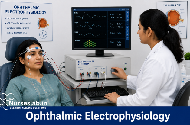
Ophthalmic electrophysiology uses tests such as ERG, VEP, and EOG to evaluate retinal activity and visual pathway function. It helps diagnose inherited retinal diseases, optic nerve disorders, and unexplained vision loss, supporting accurate clinical assessment.
Introduction
Ophthalmic electrophysiology encompasses a set of diagnostic procedures that evaluate the electrical activity generated by the visual system in response to visual stimuli. These procedures provide objective, quantifiable insights into the function of the retina, optic nerve, and visual pathways, complementing structural assessments such as fundus examination and imaging. As ophthalmic diseases often involve subtle or early functional changes preceding anatomical alterations, electrophysiological diagnostics have become indispensable in both clinical practice and research.
Principles of Ophthalmic Electrophysiology
Definition and Basic Science
Ophthalmic electrophysiology refers to the measurement of electrical potentials produced by the retina, optic nerve, and visual cortex in response to light or visual patterns. These bioelectrical signals arise from the activity of neurons and supporting cells within the visual system. By capturing and analyzing these responses, clinicians can assess the integrity and function of different visual pathway components.
Physiological Basis
The retina, as a highly organized neural tissue, transduces light into electrical signals through phototransduction. Photoreceptors (rods and cones) initiate this process, transmitting signals to bipolar, amacrine, and ganglion cells. These signals converge and are relayed via the optic nerve to the visual cortex. Electrophysiological tests exploit this cascade, applying controlled stimuli and recording the resultant electrical activity using surface electrodes.
Electrical Responses in the Visual System
The primary responses measured in ophthalmic electrophysiology include:
- Retinal responses: Generated by photoreceptors, bipolar, and ganglion cells (e.g., ERG components).
- Oculomotor responses: Resulting from retinal pigment epithelium (RPE) and eye movement (e.g., EOG).
- Cortical responses: Originating from visual cortex activation (e.g., VEP).
Each test isolates and quantifies specific aspects of these responses, enabling targeted functional assessment.
Major Diagnostic Procedures
Several electrophysiological tests are routinely employed in ophthalmic diagnostics, each with unique applications and methodologies.
Electroretinogram (ERG)
The ERG measures global retinal electrical activity in response to light stimuli. Electrodes are placed on the cornea or periocular skin, and flashes or pattern stimuli are presented. The resulting waveform comprises distinct components:
- a-wave: Negative deflection representing photoreceptor activity.
- b-wave: Positive deflection reflecting bipolar and Müller cell activity.
- Oscillatory potentials: High-frequency wavelets associated with inner retinal processing.
ERG protocols include full-field (flash) ERG, multifocal ERG, and pattern ERG, each targeting different retinal regions or cell types.
Electrooculogram (EOG)
The EOG assesses the function of the retinal pigment epithelium (RPE) and photoreceptors by measuring changes in the standing potential of the eye during alternating periods of light and dark adaptation. Skin electrodes are placed near the canthi, and eye movements between fixed targets generate voltage changes. The key parameter is the Arden ratio (light peak/dark trough), which reflects RPE integrity.
Visual Evoked Potential (VEP)
VEP evaluates the functional integrity of the visual pathways from the retina to the occipital cortex. Scalp electrodes over the visual cortex record potentials evoked by pattern-reversal or flash stimuli. The latency and amplitude of the P100 wave are critical indicators of optic nerve and cortical function.
Multifocal ERG (mfERG)
mfERG provides topographical mapping of retinal function, particularly in the macula. Multiple focal stimuli are presented simultaneously, and responses are mathematically separated to generate a functional map. This is invaluable for detecting localized retinal dysfunction.
Pattern ERG (PERG)
PERG isolates ganglion cell and macular function using patterned (checkerboard) stimuli. It is sensitive to diseases affecting the inner retina, such as glaucoma and optic neuropathies.
Clinical Applications
Indications for Electrophysiological Testing
Ophthalmic electrophysiology is indicated in a wide spectrum of clinical scenarios, including:
- Unexplained visual loss with normal fundus findings
- Inherited retinal dystrophies (e.g., retinitis pigmentosa, cone-rod dystrophy)
- Acquired retinal disorders (e.g., drug toxicity, central serous chorioretinopathy)
- Optic nerve disorders (e.g., optic neuritis, multiple sclerosis)
- Maculopathies and unexplained macular dysfunction
- Assessment of visual function in infants and non-verbal patients
- Preoperative evaluation for retinal surgery or gene therapy
Diseases Diagnosed and Case Examples
Electrophysiological procedures are pivotal in diagnosing and differentiating:
- Retinitis Pigmentosa: Marked reduction or absence of ERG responses.
- Congenital Stationary Night Blindness: Abnormal scotopic ERG with preserved photopic responses.
- Best Disease: Characteristic EOG Arden ratio reduction with relatively preserved ERG.
- Glaucoma: PERG abnormalities preceding visual field loss.
- Optic Neuritis: Delayed VEP P100 latency indicating optic nerve dysfunction.
- Drug Toxicity (e.g., hydroxychloroquine): mfERG reveals localized macular dysfunction before fundus changes.
Patient Preparation and Procedure
Pre-Test Instructions
Proper patient preparation is crucial for reliable results:
- Explain the procedure and its non-invasive nature to reduce anxiety.
- Avoid topical medications that may affect pupil size or retinal function unless required by the protocol.
- Ensure refractive correction is in place for pattern-based tests.
- Advise patients to avoid caffeine or stimulants on the day of testing.
Special considerations apply for children, elderly, and patients with disabilities, including the use of sedation or specialized electrodes.
Equipment and Environment
Testing requires standardized equipment and controlled conditions:
- Dark-adapted and light-adapted environments for ERG/EOG protocols.
- Calibrated stimulators (LED, CRT, or LCD screens) for stimulus delivery.
- Gold foil, DTL fiber, or skin electrodes for recording signals.
- Shielded cables and low-noise amplifiers to minimize electrical interference.
Ambient lighting, patient comfort, and minimization of movement are critical for artifact-free recordings.
Step-by-Step Process
A typical workflow for an ERG or VEP includes:
- Obtain informed consent and review medical history.
- Prepare skin or ocular surface, and apply electrodes according to standardized positions.
- Verify electrode impedance and signal quality.
- Dark-adapt or light-adapt the patient as per protocol requirements.
- Present appropriate visual stimuli (flashes, patterns) while recording responses.
- Monitor patient cooperation and repeat trials if necessary.
- Remove electrodes, clean skin/ocular surface, and debrief the patient.
Interpretation of Results
Normal vs Abnormal Findings
Interpretation requires comparison to age-matched normative data and consideration of clinical context. Key interpretative points include:
- Amplitude: Reduced amplitudes suggest loss of function or cell death.
- Latency: Prolonged latencies indicate slowed conduction or synaptic dysfunction.
- Waveform morphology: Altered shapes may pinpoint the affected retinal layer or pathway.
For example, a flat ERG suggests widespread retinal dysfunction, while a selective reduction in the b-wave with preserved a-wave points to inner retinal disease.
Clinical Significance
Electrophysiological findings must be integrated with clinical examination, imaging, and genetic testing. For instance, an abnormal mfERG in a patient with subtle macular changes may prompt early intervention or guide further investigations. The Arden ratio in EOG can confirm or rule out Best disease in ambiguous cases.
Limitations and Challenges
Despite their utility, ophthalmic electrophysiological tests have certain limitations:
- Technical: Signal noise, electrode placement errors, and equipment variability can affect results.
- Patient-related: Poor cooperation, movement, or media opacities (e.g., cataract) may impede testing.
- Interpretative: Overlap in findings among different diseases may complicate diagnosis; normative data must be population-specific.
Additionally, some conditions—such as early glaucoma—may produce subtle changes that are challenging to detect without advanced analysis.
Nursing Care in Ophthalmic Electrophysiology Diagnostic Procedures
As these procedures often require specialised preparation and monitoring, nurses are integral to ensuring patient safety, comfort, and diagnostic accuracy.
Pre-Procedure Nursing Care
1. Patient Assessment
Thorough patient assessment is the foundation of safe and effective care. Prior to the procedure, nurses should:
- Review the patient’s medical history, with particular attention to ocular, neurological, and systemic conditions that may affect the procedure or results.
- Assess for allergies, particularly to medications, topical anaesthetics, or contact lens materials used during testing.
- Evaluate the patient’s current medications, as certain drugs (e.g., antiepileptics, sedatives) may influence electrophysiological measurements.
- Identify any history of seizures or photosensitivity, as some tests involve exposure to flashing lights.
- Determine the patient’s level of understanding and address any language or communication barriers.
2. Informed Consent
Obtaining informed consent is essential. Nurses should ensure that the patient (or guardian) understands the nature, purpose, risks, and benefits of the procedure. This involves:
- Providing clear, jargon-free explanations of the test process and its significance.
- Answering any questions and alleviating concerns.
- Verifying that written consent is obtained and documented appropriately.
3. Patient Preparation
Preparation varies depending on the specific test, but generally includes:
- Ensuring the patient has refrained from wearing contact lenses for the recommended period prior to the test (usually 24 hours for ERG/EOG).
- Advising on medication restrictions if required (e.g., withholding certain eye drops).
- Instructing the patient to avoid caffeine or sedatives on the day of the procedure, unless medically indicated.
- Confirming that the patient has eaten a light meal to prevent hypoglycaemia during prolonged testing.
- For paediatric or anxious patients, discussing the option of sedation and ensuring appropriate fasting if sedation is planned.
4. Patient Education
Patient education is vital for cooperation and reducing anxiety. Nurses should:
- Explain the step-by-step process of the procedure, including what sensations the patient may experience (e.g., flashing lights, application of electrodes or contact lens).
- Discuss the importance of remaining still and following instructions during the test to ensure accurate results.
- Reassure the patient that the procedures are generally painless and of short duration.
- Provide written instructions and contact details for further queries.
Intra-Procedure Nursing Responsibilities
1. Monitoring and Patient Comfort
During the procedure, nurses play a critical role in monitoring the patient and ensuring their comfort:
- Assist with positioning the patient comfortably, ensuring proper head and body alignment.
- Apply electrodes or contact lens electrodes as per protocol, ensuring skin is clean and free of oils or makeup.
- Monitor the patient’s vital signs, especially in those with a history of seizures or cardiovascular conditions.
- Observe for signs of anxiety, discomfort, or distress, and intervene as appropriate.
- Provide reassurance and clear instructions throughout, particularly during periods of light stimulation.
2. Assisting the Ophthalmologist
Nurses support the ophthalmologist or technician by:
- Ensuring all necessary equipment is functioning and properly calibrated.
- Handing over materials, adjusting lighting, or assisting with electrode placement as needed.
- Documenting procedural details, patient responses, and any adverse events.
3. Infection Control
Strict adherence to infection control protocols is essential:
- Use single-use or properly sterilised electrodes and contact lenses.
- Clean and disinfect surfaces and equipment between patients.
- Utilise appropriate personal protective equipment (PPE) as indicated.
Post-Procedure Nursing Care
1. Observation and Monitoring
After the procedure, nurses should:
- Monitor the patient for immediate complications, such as allergic reactions to contact materials or topical anaesthetics.
- Check for signs of ocular irritation, redness, or discharge.
- Assess for systemic symptoms (e.g., headache, dizziness, nausea) which may result from prolonged light stimulation.
2. Patient Comfort and Recovery
Support the patient in their recovery by:
- Offering tissues or eye drops to relieve mild ocular discomfort if necessary.
- Ensuring the patient is stable before allowing them to leave, especially if sedation was used.
- Providing a comfortable environment for recovery, with minimal sensory stimulation if the patient reports light sensitivity.
3. Documentation
Accurate documentation is critical for continuity of care and audit purposes. Nurses should record:
- Details of the procedure performed, including type of test, duration, and any deviations from protocol.
- Patient responses, including cooperation and any adverse events.
- Post-procedure instructions given and follow-up arrangements.
4. Discharge Instructions
Before discharge, nurses should:
- Advise the patient to report any persistent discomfort, visual disturbances, or signs of infection.
- Provide contact information for urgent concerns or complications.
- Remind the patient of any follow-up appointments and when to expect test results.
Patient Education
Patient education should be an ongoing process, tailored to the individual’s needs and comprehension level:
- Provide clear information about the purpose and process of the test.
- Explain any sensations or side effects that might be experienced and their expected duration.
- Discuss the importance of aftercare, including avoiding eye rubbing and applying prescribed eye drops if indicated.
- Emphasise the need for follow-up, especially if results may impact ongoing treatment.
- Offer written information and resources for further reading or support.
Potential Complications and Nursing Interventions
While ophthalmic electrophysiology procedures are generally safe, nurses must be vigilant for potential complications and prepared to intervene:
- Ocular Irritation or Allergic Reactions: Monitor for redness, itching, swelling, or discharge. Administer prescribed anti-allergic medications or lubricating drops as needed. Notify the ophthalmologist if symptoms persist.
- Corneal Abrasion: Rarely, improper electrode placement or contact lens use can cause corneal injury. Observe for pain, photophobia, or excessive tearing. Provide prompt ophthalmic assessment and care.
- Seizures or Neurological Symptoms: Patients with known photosensitivity or epilepsy may be at risk during tests with flashing lights. Ensure emergency protocols are in place, monitor closely, and cease testing at the first sign of symptoms.
- Vasovagal Reactions: Some patients may experience faintness or syncope due to anxiety or prolonged immobility. Position the patient safely, monitor vital signs, and provide reassurance.
- Infection: Adhere strictly to infection control measures and monitor for signs of conjunctivitis or other infections post-procedure.
REFERENCES
- Minzhong Yu, Donnell Creel, Alessandro Iannaccone, Handbook of Clinical Electrophysiology of Vision, Published: 27 December 2019, https://doi.org/10.1007/978-3-030-30417-1
- Yu M, Kurup SK. Application of Ophthalmic Electrophysiology in Inflammatory Disorders of Retina and Optic Nerve. J Clin Med. 2024 Jun 29;13(13):3829. doi: 10.3390/jcm13133829.
- Etienne M. Schönbach, Marwan Abdulaal, Lalita Gupta, Lily Kim and Shree K. Kurup, Graefe’s Archive for Clinical and Experimental Ophthalmology, Vol. 258, 2020
- Sara Lee, Electrophysiology in Ophthalmic Research, June 15, 2025, https://www.numberanalytics.com/blog/electrophysiology-ophthalmic-research-methods
- Hamilton R. Clinical electrophysiology of vision-commentary on current status and future prospects. Eye (Lond). 2021 Sep;35(9):2341-2343. doi: 10.1038/s41433-021-01592-0. Epub 2021 May 27. PMID: 34045684; PMCID: PMC8376889.
Stories are the threads that bind us; through them, we understand each other, grow, and heal.
JOHN NOORD
Connect with “Nurses Lab Editorial Team”
I hope you found this information helpful. Do you have any questions or comments? Kindly write in comments section. Subscribe the Blog with your email so you can stay updated on upcoming events and the latest articles.


