


Last Updated on August 28, 2026 by Nurseslab.in Editorial Team
A meningioma is a primary tumour of the central nervous system that develops from cells associated with the meninges, the protective membranes surrounding the brain and spinal cord.
Overview
It usually grows from arachnoid cap cells and is commonly attached to the dura mater, the tough outer meningeal layer. Because it arises outside the brain tissue itself, it is described as an extra-axial tumour. Nevertheless, it can compress the brain, cranial nerves, blood vessels, or spinal cord and produce significant neurological disability.
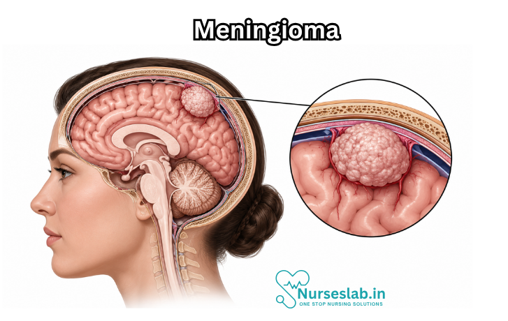
Meningiomas are the most common primary intracranial tumours in adults. Most are slow-growing World Health Organization grade 1 tumours, but “benign” does not necessarily mean harmless. A benign tumour in a confined space may still threaten vision, movement, speech, cognition, breathing, or spinal cord function. Grade 2 tumours recur more often, while grade 3 tumours grow more aggressively and may invade nearby tissue. Some meningiomas are discovered incidentally during imaging for an unrelated problem and never cause symptoms.
The Meninges and How a Meningioma Causes Disease
The meninges have three layers. The dura mater is the strong outer covering; the arachnoid mater is the web-like middle layer; and the pia mater lies closely over the surface of the brain and spinal cord. Cerebrospinal fluid circulates within the spaces between these membranes. A meningioma generally grows inward from the meningeal surface. It may displace the brain without initially invading it, involve a venous sinus, surround an artery or cranial nerve, extend through the skull base, or cause adjacent bone to thicken.
Symptoms are created mainly by location, pressure, surrounding oedema, interference with cerebrospinal fluid pathways, vascular involvement, and irritation of the cerebral cortex. A small tumour near an optic nerve can be more clinically important than a larger tumour over a relatively tolerant brain surface. Slow growth may allow the nervous system to compensate for years, so the first recognised symptom can appear only after the tumour becomes sizeable.
Who Develops Meningioma?
Meningiomas occur most often in middle-aged and older adults and are more frequently diagnosed in women, especially among lower-grade tumours. They are uncommon in children. The reason for the sex difference is not fully understood, although many tumours express progesterone receptors and hormonal biology is an area of continuing study. A person may have a single tumour or, less often, multiple meningiomas.
Causes and Risk Factors
For most patients, no single cause can be identified. Meningioma develops after genetic changes allow meningeal cells to grow abnormally. These changes are usually acquired in the tumour rather than inherited. Alteration of the NF2 gene on chromosome 22 is common in a substantial group of meningiomas, while other tumours may contain changes involving AKT1, TRAF7, KLF4, SMO, PIK3CA, or related pathways. Molecular patterns can correlate with tumour location and behaviour, but testing is interpreted by specialist neuropathology teams.
Established or recognised risk factors include previous ionising radiation to the head, particularly therapeutic radiation received during childhood, and neurofibromatosis type 2, an inherited tumour-predisposition syndrome. Increasing age is also associated with greater incidence. The relationship between reproductive hormones, pregnancy, and external hormone exposure is complex. Some tumours enlarge during pregnancy, possibly because of hormonal and vascular changes, but an individual should not start or stop hormonal medication without discussing personal risks with the treating clinician.
Classification and WHO Grade
A scan can strongly suggest meningioma, but definitive classification usually requires tissue examined by a neuropathologist. The current World Health Organization framework integrates microscopic features with selected molecular findings. Grade predicts biological behaviour and recurrence risk, although it is not a perfect forecast for an individual.
- WHO grade 1: Usually slow-growing and the most common category. Subtypes include meningothelial, fibrous, transitional, psammomatous, angiomatous, microcystic, secretory, and others. Complete removal may be curative, but recurrence remains possible.
- WHO grade 2: Includes atypical tumours and certain subtypes such as clear cell and chordoid meningioma. Increased mitotic activity, brain invasion, or a combination of specific microscopic features can support this grade. Recurrence risk is higher than in grade 1.
- WHO grade 3: An aggressive category with high recurrence risk. Marked mitotic activity, overtly malignant morphology, a TERT promoter mutation, or homozygous deletion of CDKN2A/B may establish grade 3 under current criteria.
Pathology reports may also describe subtype, mitotic count, brain invasion, necrosis, proliferation index, receptor staining, and molecular findings. Increasingly, chromosome patterns and DNA methylation profiles are used in specialist centres to refine recurrence risk. Grade, molecular biology, tumour location, and extent of resection are considered together rather than in isolation.
Common Locations
Meningiomas can arise anywhere meninges are present. Common intracranial sites include the cerebral convexity, parasagittal region, falx, sphenoid wing, olfactory groove, planum sphenoidale, tuberculum sellae, cavernous sinus, petroclival region, tentorium, posterior fossa, cerebellopontine angle, and foramen magnum. They can also develop around the spinal cord, most often in the thoracic region. Location strongly influences both symptoms and treatment risk.
Symptoms and Clinical Presentation
Many meningiomas cause no symptoms and are found incidentally. When symptoms occur, they usually develop gradually, although bleeding, acute swelling, seizure, or sudden obstruction of cerebrospinal fluid can produce an abrupt presentation. General symptoms may include progressive headache, seizures, nausea, vomiting, drowsiness, cognitive slowing, personality or behavioural change, weakness, altered sensation, problems with balance, and speech difficulty. Headache alone is common in the general population and does not by itself indicate a brain tumour.
Symptoms by location may include:
- Frontal or olfactory groove: reduced smell, impaired judgement, apathy, disinhibition, memory difficulty, or personality change.
- Convexity or parasagittal: seizure, one-sided weakness or numbness, or difficulty planning movement.
- Sphenoid wing, orbital, or optic pathway region: visual loss, double vision, protrusion of an eye, facial sensory change, or eye-movement weakness.
- Cavernous sinus: double vision, drooping eyelid, facial numbness, or pain caused by cranial nerve involvement.
- Petroclival or cerebellopontine angle: hearing difficulty, imbalance, facial numbness, swallowing problems, or brainstem compression.
- Posterior fossa or foramen magnum: unsteadiness, limb weakness, swallowing difficulty, or lower cranial nerve symptoms.
- Spinal: local or radiating pain, weakness, stiffness, altered sensation, walking difficulty, and—in advanced compression—bladder or bowel dysfunction.
Diagnostic Evaluation
Evaluation begins with a detailed history and neurological examination. The clinician assesses cognition, speech, vision, eye movements, hearing, cranial nerve function, strength, sensation, reflexes, coordination, gait, and seizure history. The pattern helps identify which structures may be affected and whether another disorder is more likely.
Contrast-enhanced magnetic resonance imaging is the preferred imaging study for most intracranial and spinal meningiomas. Typical features include a well-defined extra-axial mass with strong contrast enhancement and sometimes a “dural tail.” MRI shows compression, oedema, venous sinus involvement, and relationships to nerves and vessels. Computed tomography is particularly useful for calcification, bone thickening, bone destruction, and surgical planning. Vascular imaging or catheter angiography may occasionally be required when major vessels or a highly vascular tumour are involved.
Imaging cannot always distinguish meningioma from metastasis, solitary fibrous tumour, schwannoma, lymphoma, inflammatory lesions, or other dural masses. When surgery or biopsy is performed, histology confirms the diagnosis and grade. Selected centres may use somatostatin-receptor positron emission tomography, particularly to clarify tumour extent, recurrence, or radiotherapy targets. Baseline cognitive, visual, endocrine, hearing, or seizure evaluations may be added according to location.
Treatment Planning
Treatment is individualised by a multidisciplinary team. Important considerations include symptoms, age and general health, tumour size and location, growth on serial imaging, oedema, grade, molecular findings, surgical accessibility, involvement of critical nerves or vessels, previous treatment, and the patient’s goals. The principal options are active surveillance, surgery, focused radiosurgery, fractionated radiotherapy, and—in selected recurrent cases—clinical trials or systemic therapy.
Active Surveillance
Observation with scheduled clinical review and MRI is often appropriate for a small, asymptomatic, radiologically typical meningioma, especially when it is incidental or when treatment risk exceeds likely benefit. Surveillance is an active management strategy, not neglect. The team compares serial scans for growth and watches for symptoms. The interval between scans is tailored to tumour features, prior growth, age, and local guidance. Demonstrated growth, new symptoms, increasing oedema, or threat to a critical structure may trigger treatment.
Surgery
Surgery is generally considered for symptomatic, enlarging, large, or diagnostically uncertain tumours and for tumours causing substantial mass effect. Goals include obtaining tissue, relieving pressure, preserving or improving neurological function, and removing as much tumour as is safely possible. Gross total resection may cure many grade 1 meningiomas. However, radical removal can be unsafe when a tumour encases arteries, invades a venous sinus, or adheres to cranial nerves or the brainstem. In such cases, intentional subtotal resection followed by observation or radiotherapy may provide a better functional outcome.
Operative techniques may include image guidance, neurophysiological monitoring, specialised skull-base approaches, and reconstruction of involved dura or bone. Preoperative embolisation is occasionally used to reduce blood supply in selected tumours, but it has specific risks and is not routine. Surgical risks depend on location and may include bleeding, infection, stroke, seizures, cerebrospinal fluid leak, blood clots, swelling, and new deficits involving movement, sensation, speech, vision, hearing, swallowing, memory, or cranial nerves.
Radiation Therapy
Radiation may be used for residual, recurrent, inoperable, or higher-grade meningioma and sometimes as the primary treatment for a small but growing tumour in a difficult location. Stereotactic radiosurgery delivers a highly focused dose, usually in one or a few sessions, and is suitable only when tumour size and proximity to sensitive structures permit. Fractionated radiotherapy divides treatment into smaller doses over multiple sessions, which may be safer for larger lesions or tumours near the optic apparatus, brainstem, or other critical tissue.
Radiation usually aims to control further growth rather than make the tumour disappear immediately. Side effects vary with treatment volume and location. Early effects may include fatigue, scalp irritation, headache, nausea, and temporary swelling. Delayed effects can include cognitive change, endocrine dysfunction, hearing or vision injury, tissue necrosis, vascular effects, or a small long-term risk of a second tumour. Modern planning seeks to minimise exposure to healthy tissue.
Drug Therapy and Clinical Trials
There is no universally effective standard systemic drug treatment for meningioma. Medicines such as corticosteroids may reduce tumour-associated oedema temporarily, and antiseizure medication is used when clinically indicated. Hormonal therapy has not established a routine role. For progressive tumours that cannot be controlled with further surgery or radiation, neuro-oncology teams may consider clinical trials or selected targeted, anti-angiogenic, immune, or somatostatin-receptor-directed approaches. Evidence is evolving, and benefit is generally less predictable than with local treatment.
Recovery and Supportive Care
Recovery depends on pre-treatment function, tumour location, procedure type, complications, and general health. After surgery, patients are monitored for neurological change, bleeding, swelling, seizures, infection, electrolyte imbalance, and cerebrospinal fluid leakage. Pain control, early mobilisation, clot prevention, wound care, nutrition, and medication review are important. Steroids are usually tapered under medical supervision rather than stopped suddenly.
Rehabilitation may involve physiotherapy, occupational therapy, speech and language therapy, neuropsychology, visual rehabilitation, hearing support, or vocational counselling. Fatigue can persist even when scans are reassuring. Anxiety, low mood, altered cognition, and fear of recurrence deserve attention. Family members may notice subtle changes before the patient does, but the patient’s autonomy and privacy should remain central.
Prognosis and Recurrence
Many people with a completely removed grade 1 meningioma have excellent long-term tumour control. Prognosis becomes less favourable with higher grade, incomplete resection, aggressive molecular features, rapid growth, brain invasion, difficult location, or repeated recurrence. Functional outcome also depends on whether critical neurological structures were compressed or injured before treatment. Survival statistics describe groups and cannot precisely predict an individual’s course.
Recurrence can occur years after apparently successful treatment, including after complete removal. Grade 2 and grade 3 tumours require closer and often longer surveillance. Follow-up generally combines neurological review and contrast-enhanced MRI; frequency is individualised. A new or enlarging lesion may be managed with repeat surgery, radiosurgery, fractionated radiotherapy, a combined approach, or a clinical trial.
Potential Complications of the Disease
Untreated or progressive meningioma may cause seizures, persistent neurological deficits, hydrocephalus, visual or hearing loss, cranial nerve palsy, venous obstruction, spinal cord compression, and cognitive or behavioural change. Severe brain swelling or brainstem compression can be life-threatening. Higher-grade tumours can invade adjacent brain and bone; extracranial spread is rare but more likely in aggressive grade 3 disease.
Living With an Incidental Meningioma
An incidental diagnosis can be psychologically difficult even when treatment is unnecessary. It helps to ask whether the imaging appearance is typical, where the tumour is located, whether oedema is present, what previous scans show, which symptoms would be relevant, and when the next scan is planned. Keeping copies of imaging reports and an up-to-date medication list can support continuity. New symptoms should be reported rather than automatically attributed to the tumour, because common problems such as migraine, eye disease, medication effects, and vascular disease may have other causes.
When to Seek Urgent Help
Seek emergency medical assistance for a first seizure, a seizure lasting several minutes or repeated seizures without recovery, sudden weakness or numbness, new speech difficulty, sudden loss of vision, severe confusion, loss of consciousness, a sudden extremely severe headache, repeated vomiting with drowsiness, or rapidly worsening balance. After surgery, urgent review is also needed for increasing drowsiness, new neurological deficit, fever with wound redness or discharge, clear fluid leakage, uncontrolled pain, chest pain, shortness of breath, or a swollen painful leg.
Questions to Ask the Healthcare Team
- Is the diagnosis definite from imaging, or is tissue required?
- What are the tumour’s size, location, growth rate, and effects on nearby structures?
- What is the WHO grade and which pathological or molecular features matter?
- What are the benefits and risks of surveillance, surgery, and radiation in this case?
- If surgery is advised, is complete removal safe and what neurological functions are at risk?
- Would planned subtotal removal plus radiation better preserve function?
- How often will MRI follow-up be needed, and for how many years?
- Which symptoms require urgent review?
- Could the condition or treatment affect driving, work, pregnancy planning, or medication choices?
- Is review at a specialist skull-base, spine, neuro-oncology, or multidisciplinary centre appropriate?
Key Points
Meningioma is usually a slow-growing tumour arising from the coverings of the brain or spinal cord. Most tumours are grade 1, but impact depends as much on location as on pathology. MRI is the principal imaging tool, while tissue establishes definitive classification when obtained. Small asymptomatic tumours may be monitored; symptomatic or growing tumours may require surgery, radiosurgery, or fractionated radiotherapy. Higher-grade and incompletely removed tumours have greater recurrence risk and need closer follow-up. The best treatment plan balances tumour control with preservation of neurological function and quality of life.
Sources for Further Reading
- National Cancer Institute: Meningioma—Diagnosis and Treatment.
- European Association of Neuro-Oncology: Guideline on the Diagnosis and Management of Meningiomas.
- World Health Organization Classification of Tumours of the Central Nervous System, fifth edition.
- National Health Service guidance on non-cancerous brain tumours.
Nursing Care of a Patient with Meningioma
Nursing care focuses on neurological monitoring, seizure safety, symptom control, preoperative and postoperative support, radiation-care education, rehabilitation, psychosocial support, and long-term surveillance.
Assessment
- Assess neurological status, including level of consciousness, orientation, memory, behaviour, speech, pupils, cranial nerve function, motor strength, sensation, coordination, balance, gait, and seizure activity.
- Assess symptoms related to tumour location, such as headache, visual changes, hearing loss, facial numbness, limb weakness, personality changes, cognitive decline, dizziness, nausea, vomiting, or back pain if spinal involvement is present.
- Monitor for signs of increased intracranial pressure, including worsening headache, repeated vomiting, declining consciousness, unequal pupils, bradycardia with hypertension, abnormal posturing, or new focal deficits.
- Review imaging findings, tumour location, size, growth pattern, oedema, mass effect, WHO grade if known, biopsy or pathology results, and treatment plan such as observation, surgery, stereotactic radiosurgery, radiation therapy, or systemic therapy in selected cases.
- Assess seizure history, medication adherence, triggers, aura, frequency, duration, recovery, injuries, and need for anti-seizure medication education.
- Assess functional ability, falls risk, swallowing safety, nutrition, pain, sleep, fatigue, bowel and bladder function, skin integrity, and ability to perform activities of daily living.
- Assess emotional response, anxiety, fear of surgery or recurrence, family support, health literacy, work or driving concerns, advance care planning needs, and access to follow-up care.
Priority Nursing Diagnoses
- Risk for ineffective cerebral tissue perfusion related to tumour mass effect, cerebral oedema, increased intracranial pressure, or postoperative swelling.
- Risk for injury related to seizures, visual changes, weakness, impaired balance, confusion, or postoperative neurological deficits.
- Acute or chronic pain related to headache, tumour pressure, surgery, radiation effects, or musculoskeletal strain.
- Disturbed sensory perception related to tumour compression of visual, auditory, sensory, or cranial nerve pathways.
- Impaired physical mobility related to weakness, balance impairment, fatigue, neurological deficit, or postoperative restrictions.
- Anxiety related to brain tumour diagnosis, treatment uncertainty, surgery, recurrence risk, or changes in function.
- Deficient knowledge related to diagnosis, observation plan, surgery, radiation therapy, medications, seizure precautions, warning signs, and follow-up imaging.
Nursing Interventions
- Perform frequent neurological observations and report any decrease in consciousness, new weakness, speech changes, vision changes, seizures, unequal pupils, persistent vomiting, or worsening headache immediately.
- Maintain seizure precautions when indicated, including safe environment, suction and oxygen availability if ordered, padded side rails according to policy, and documentation of seizure features and recovery.
- Administer prescribed medications such as corticosteroids for oedema, anti-seizure medicines, analgesics, antiemetics, proton-pump inhibitors, anticoagulation prophylaxis, or other therapies, and monitor effectiveness and side effects.
- For patients under observation, reinforce the importance of scheduled MRI or CT imaging and reporting new or worsening neurological symptoms promptly.
- Prepare the patient for surgery when planned by supporting preoperative teaching, medication review, fasting instructions, consent process, baseline neurological assessment, and emotional support.
- After surgery, monitor airway, vital signs, neurological status, wound dressing, drainage, pain, nausea, fluid balance, glucose if receiving corticosteroids, infection signs, and complications such as bleeding, cerebrospinal fluid leak, seizures, or neurological deterioration.
- Support radiation therapy or stereotactic radiosurgery care by monitoring fatigue, scalp or skin irritation, headache, nausea, alopecia in the treatment area, and delayed neurological symptoms.
- Promote safety with fall precautions, assistive devices, adequate lighting, supervised mobility, driving restrictions until cleared, and referral to physiotherapy or occupational therapy when deficits affect function.
- Support nutrition, sleep, bowel function, skin integrity, and fatigue management, especially during recovery from surgery or radiation.
- Coordinate multidisciplinary care with neurosurgery, neurology, oncology, radiation oncology, rehabilitation, speech therapy, dietetics, pharmacy, psychology, social work, and community nursing.
Patient and Family Education
- Explain that many meningiomas grow slowly, but symptoms can occur when the tumour presses on brain, spinal cord, nerves, or blood vessels.
- Teach the patient to seek urgent care for new seizure, worsening headache, repeated vomiting, confusion, fainting, weakness, speech difficulty, vision loss, severe drowsiness, fever after surgery, wound drainage, or clear fluid leaking from the nose or incision.
- Review the treatment plan in understandable terms, including observation, surgery, radiation therapy, stereotactic radiosurgery, medications, rehabilitation, and follow-up imaging.
- Teach seizure safety if relevant: take anti-seizure medicines as prescribed, avoid missed doses, follow driving and work restrictions, avoid swimming alone or unsafe heights, and inform family what to do during a seizure.
- Review postoperative wound care, activity restrictions, infection signs, medication schedule, steroid taper instructions if prescribed, and when to contact the neurosurgical team.
- Encourage rest, gradual activity, balanced nutrition, hydration, bowel management, pain control, and participation in rehabilitation therapies if needed.
- Provide emotional support and encourage discussion of work, family roles, memory or personality changes, body image, financial concerns, advance care planning, and community support resources.
Expected Outcomes
- The patient maintains stable neurological status or shows improvement after treatment.
- The patient remains free from preventable seizure injury, falls, aspiration, wound infection, pressure injury, and avoidable neurological deterioration.
- Pain, headache, nausea, fatigue, anxiety, and treatment-related side effects are controlled as much as possible.
- The patient and family explain the diagnosis, treatment plan, medication use, seizure precautions, urgent warning signs, and follow-up imaging needs.
- The patient participates safely in mobility, self-care, rehabilitation, and daily activities according to the care plan.
- The patient receives coordinated neurosurgical, neurological, oncology, rehabilitation, psychosocial, and community follow-up.
Evaluation
Evaluate nursing care by reviewing neurological observations, seizure control, headache and pain level, vision or hearing changes, mobility, cognition, wound healing, medication response, steroid and anti-seizure side effects, radiation effects, functional independence, emotional adjustment, family understanding, and follow-up imaging attendance. Revise the care plan if neurological status worsens, seizures occur, intracranial pressure signs develop, wound complications appear, treatment toxicity occurs, tumour growth is identified, or the patient needs additional rehabilitation, oncology, neurosurgical, palliative, or psychosocial support.
REFERENCES
- Meningioma diagnosis and treatment. National Cancer Institute. https://www.cancer.gov/rare-brain-spine-tumor/tumors/meningioma.
- Alruwaili AA, De Jesus O. Meningioma (https://www.ncbi.nlm.nih.gov/books/NBK560538/). 2023 Aug 23. In: StatPearls [Internet]. Treasure Island (FL): StatPearls Publishing; 2025 Jan-. .
- American Association of Neurological Surgeons. Meningiomas (https://www.aans.org/patients/conditions-treatments/meningiomas/). Last reviewed 4/8/2024.
- Meningiomas. American Association of Neurological Surgeons. https://www.aans.org/en/Patients/Neurosurgical-Conditions-and-Treatments/Meningiomas.
- National Cancer Institute (U.S.). Meningioma: Diagnosis and Treatment (https://www.cancer.gov/rare-brain-spine-tumor/tumors/meningioma). Last reviewed 11/20/2024.
- Apra C, et al. Current treatment options for meningioma. Expert Review of Neurotherapeutics. 2018; doi:10.1080/14737175.2018.1429920.
- Ogasawara C, Philbrick BD, Adamson DC. Meningioma: A Review of Epidemiology, Pathology, Diagnosis, Treatment, and Future Directions (https://pmc.ncbi.nlm.nih.gov/articles/PMC8004084/). Biomedicines. 2021 Mar 21;9(3):319.
Stories are the threads that bind us; through them, we understand each other, grow, and heal.
JOHN NOORD
Connect with “Nurses Lab Editorial Team”
I hope you found this information helpful. Do you have any questions or comments? Kindly write in comments section. Subscribe the Blog with your email so you can stay updated on upcoming events and the latest articles.


