


Understanding the respiratory system’s structure is fundamental for nursing professionals. Among the essential components are the bronchi and bronchioles, which play a critical role in air conduction, filtration, and overall respiratory health. This educational article provides an in-depth exploration of the anatomy, histology, functions, clinical relevance, and anatomical variations of the bronchi and bronchioles, tailored specifically for nursing students and educators.
Introduction
The respiratory system is indispensable for life, facilitating the exchange of oxygen and carbon dioxide essential for cellular processes. For nurses, a thorough grasp of respiratory anatomy, especially the bronchi and bronchioles, is vital for effective patient assessment, intervention, and education.
Overview of the Respiratory System
The respiratory system is divided into the upper and lower tracts. The upper respiratory tract consists of the nose, nasal cavity, pharynx, and larynx, primarily involved in filtering, warming, and humidifying inspired air. The lower respiratory tract, which includes the trachea, bronchi, bronchioles, and alveoli, is directly responsible for conducting air to the lungs and facilitating gas exchange.
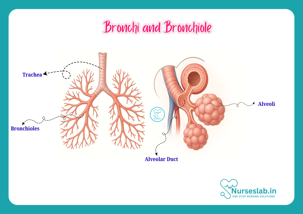
The bronchi and bronchioles are pivotal components of the lower respiratory tract. While the bronchi serve as major airways conducting air into each lung, the bronchioles represent finer passages that ultimately lead to the alveolar sacs, where gas exchange occurs. Their anatomical and functional integrity is crucial for adequate ventilation and oxygenation.
Anatomy of the Bronchi
Definition and Hierarchy
The bronchi are large, tubular airways that branch from the lower end of the trachea. The division of the trachea at the level of the fifth thoracic vertebra forms two primary (main) bronchi—right and left. These bronchi further branch into secondary (lobar) bronchi, each supplying a lobe of the lung, and then into tertiary (segmental) bronchi, which supply bronchopulmonary segments.
Primary Bronchi
The right main bronchus is shorter, wider, and more vertical compared to the left, making it more susceptible to aspirated foreign bodies. The left main bronchus is longer, narrower, and more horizontal, passing beneath the aortic arch and anterior to the oesophagus and thoracic aorta.
Secondary and Tertiary Bronchi
The right lung has three secondary bronchi (for its three lobes), while the left lung has two. These further subdivide into tertiary bronchi, each ventilating a bronchopulmonary segment—ten on the right and eight to ten on the left, depending on anatomical variations.
Structural Features
Bronchi are characterised by the presence of cartilaginous rings or plates in their walls, which maintain airway patency and prevent collapse during respiration. The wall of the bronchi comprises several layers:
- Mucosa: Lined by pseudostratified ciliated columnar epithelium with goblet cells for mucus secretion.
- Submucosa: Contains seromucous glands that further contribute to mucus production.
- Muscularis: Contains smooth muscle fibres arranged in a spiral pattern.
- Cartilage: C-shaped rings in larger bronchi, becoming irregular plates in smaller bronchi.
- Adventitia: Outermost connective tissue layer anchoring the bronchi to adjacent structures.
Cartilaginous Support
The cartilaginous framework provides structural rigidity, which is essential for maintaining open airways, especially during forced exhalation or coughing. As the bronchi branch and decrease in diameter, the cartilage transitions from C-shaped rings to irregular plates, eventually disappearing at the bronchiolar level.
Mucosal Lining
The mucosal lining is rich in ciliated epithelial cells and goblet cells, which together form the mucociliary escalator. This system traps inhaled particles and pathogens, moving them upwards towards the pharynx for expulsion or swallowing—an important defence mechanism for the lower respiratory tract.
Blood Supply
The bronchi receive arterial blood from the bronchial arteries, which arise from the thoracic aorta. Venous drainage is through the bronchial veins, which ultimately drain into the azygos and hemiazygos systems.
Nerve Supply
The bronchi are innervated by the autonomic nervous system:
- Parasympathetic fibres (from the vagus nerve) cause bronchoconstriction and increased mucus secretion.
- Sympathetic fibres (from the thoracic sympathetic chain) cause bronchodilation and decreased mucus secretion.
This innervation is clinically significant in conditions like asthma, where bronchial tone is altered.
Anatomy of the Bronchioles
Definition and Types
Bronchioles are the smallest conducting airways, arising from the terminal branches of the tertiary bronchi. Unlike the bronchi, bronchioles lack cartilage and glands in their walls. They are classified into:
- Terminal bronchioles: The final part of the conducting zone, conducting air to the respiratory bronchioles.
- Respiratory bronchioles: The beginning of the respiratory zone, containing occasional alveoli in their walls, and participating in gas exchange.
Structural Differences from Bronchi
Bronchioles are distinguished from bronchi by their smaller size (less than 1 mm in diameter), absence of cartilage, and absence of seromucous glands. Their walls are composed primarily of smooth muscle and are lined by ciliated simple columnar or cuboidal epithelium.
Smooth Muscle and Mucosal Lining
The smooth muscle layer in bronchioles is relatively thick compared to their diameter, allowing significant regulation of airway calibre. The mucosal lining becomes progressively thinner, with fewer goblet cells and a transition from columnar to cuboidal ciliated epithelium as the airway branches.
Blood and Nerve Supply
Bronchioles receive their blood supply from the pulmonary circulation. Like the bronchi, they are innervated by both sympathetic and parasympathetic fibres, enabling dynamic control of airway resistance.
Histology
A microscopic examination of the bronchi and bronchioles reveals distinct histological features:
Bronchi
- Epithelium: Pseudostratified ciliated columnar epithelium with abundant goblet cells.
- Lamina propria: Contains elastic fibres and lymphoid tissue.
- Submucosa: Houses seromucous glands and blood vessels.
- Cartilage: Hyaline cartilage in the form of rings or plates.
- Adventitia: Connective tissue with nerves, blood vessels, and lymphatics.
Bronchioles
- Epithelium: Transitions from ciliated columnar to ciliated cuboidal, with club (Clara) cells replacing goblet cells in terminal bronchioles. Club cells secrete protective proteins and surfactant-like substances.
- Muscularis: Prominent smooth muscle layer, especially in terminal bronchioles.
- Cartilage and Glands: Absent, distinguishing bronchioles from bronchi.
- Adventitia: Thin connective tissue layer.
Goblet Cells, Cilia, and Smooth Muscle
Goblet cells secrete mucus, which traps dust and microbes. Cilia beat rhythmically to move mucus towards the pharynx. The smooth muscle layer allows for constriction and dilation, regulating airflow and airway resistance—a key factor in diseases like asthma.
Functions
The bronchi and bronchioles serve several essential functions:
- Air Conduction: Serve as passageways for air to move from the trachea to the alveoli.
- Filtration and Defence: The mucociliary escalator traps and removes inhaled particles and pathogens.
- Humidification and Warming: Air is humidified and warmed as it passes through the mucosal lining.
- Regulation of Airway Resistance: Bronchiolar smooth muscle controls airway diameter, influencing airflow resistance and distribution.
- Participation in Gas Exchange: Although primarily a function of alveoli, respiratory bronchioles contribute due to their alveolar outpouchings.
Clinical Relevance
Knowledge of the anatomy and function of the bronchi and bronchioles is pivotal in recognising, assessing, and managing respiratory disorders. Some common clinical conditions include:
Bronchitis
Bronchitis is the inflammation of the bronchi, often caused by infection or irritants. It leads to increased mucus production, cough, and airway narrowing. Chronic bronchitis, a form of chronic obstructive pulmonary disease (COPD), is especially significant in smokers and those exposed to pollutants.
Asthma
Asthma is characterised by hyperresponsiveness of the bronchioles, leading to bronchoconstriction, inflammation, and increased mucus secretion. Symptoms include wheezing, breathlessness, and cough, often triggered by allergens, exercise, or infection. Understanding the anatomy and innervation of the bronchioles helps explain the effectiveness of bronchodilators and anti-inflammatory medications.
Bronchiolitis
Bronchiolitis is the inflammation of the bronchioles, most commonly affecting infants and young children. It is typically viral in origin (e.g., respiratory syncytial virus) and results in airway obstruction due to swelling and increased mucus.
Nursing Considerations and Assessment Techniques
- Assessment: Auscultation for wheezing, crackles, or diminished breath sounds; observation of respiratory rate, effort, and oxygen saturation.
- Intervention: Administering bronchodilators, ensuring airway clearance, monitoring for signs of respiratory distress, and educating patients on inhaler use.
- Prevention: Encouraging smoking cessation, vaccination, and avoidance of respiratory irritants.
Nurses play a central role in early detection, intervention, and education for patients with bronchial and bronchiolar diseases.
Anatomical Variations and Development
Embryological Development
The lower respiratory tract develops from the foregut during embryogenesis. The lung bud forms around the fourth week of gestation, branching into primary, secondary, and tertiary bronchi, followed by the development of bronchioles and alveoli. Disruptions in this process can lead to congenital anomalies such as tracheoesophageal fistula or bronchial atresia.
Normal Anatomical Variations
Anatomical variations can occur in the number and branching patterns of bronchi and bronchioles. For instance, the right lung typically has three lobes and ten bronchopulmonary segments, while the left has two lobes and eight to ten segments. Variations in bronchial branching may impact surgical planning and bronchoscopic procedures.
Implications for Nursing Practice
Awareness of developmental anomalies and anatomical variations is essential for accurate assessment and intervention. Nurses should recognise that variations may influence the presentation of disease and response to treatment, especially during procedures such as intubation, suctioning, and bronchoscopy.
Summary and Key Points
- The bronchi and bronchioles are critical components of the lower respiratory tract, responsible for air conduction, filtration, humidification, and regulation of airway resistance.
- Bronchi are supported by cartilage and lined by ciliated epithelium with goblet cells, while bronchioles lack cartilage and glands but have a prominent smooth muscle layer.
- Histological features, such as the presence of cilia and mucus-secreting cells, are vital for airway defence mechanisms.
- Common diseases like bronchitis, asthma, and bronchiolitis impact the bronchi and bronchioles, requiring specific nursing assessments and interventions.
- Anatomical variations and developmental anomalies can affect clinical presentation and management, highlighting the importance of thorough anatomical knowledge for nurses.
REFERENCES
- Ross and Wilson, Anatomy and Physiology in Health and Illness, Fourteenth Edition, 1 July 2022, ISBN-13: 978-0323834612.
- Roger Watson, Anatomy and Physiology for Nurses, 14th Edition, 12-06-2018, ISBN: 9780702077418
- P.R Asha Latha, Text Book of Applied Anatomy & Physiology for Nurses, 7th Edition, 3 January 2024, ISBN-13: 978-9356968622.
- Bryan H. Derikson, Tortora’s Principles of Anatomy and Physiology, 16th Edition, August 2023, ISBN: 978- 1119400066.
- Anatomy.co.uk, Reproductive System, Last updated on April 24, 2025, https://anatomy.co.uk/reproductive-system
Stories are the threads that bind us; through them, we understand each other, grow, and heal.
JOHN NOORD
Connect with “Nurses Lab Editorial Team”
I hope you found this information helpful. Do you have any questions or comments? Kindly write in comments section. Subscribe the Blog with your email so you can stay updated on upcoming events and the latest articles.


