


Last Updated on October 21, 2026 by Nurseslab.in Editorial Team
Pelvic ultrasound uses sound waves to examine pelvic organs. Learn about its purpose, preparation, procedure, types, results, and what to expect.
Introduction
Pelvic ultrasound is a non-invasive diagnostic imaging technique that utilises high-frequency sound waves to produce detailed images of the organs and structures within the pelvis. This procedure plays a pivotal role in modern medicine, offering clinicians valuable insights into gynaecological, urological, and obstetric conditions. Its broad scope encompasses assessment of reproductive organs, urinary tract, and surrounding tissues, making it a cornerstone in the evaluation of pelvic symptoms and pathologies. The importance of pelvic ultrasound lies in its safety, versatility, and ability to provide real-time visualisation without the risks associated with ionising radiation.
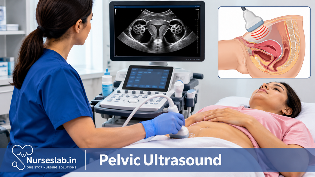
Definition and Overview
A pelvic ultrasound is an imaging study designed to visualise the pelvic organs, including the uterus, ovaries, fallopian tubes, bladder, prostate (in males), and adjacent structures. By transmitting sound waves into the body and interpreting the echoes that return, the ultrasound machine constructs images that help in the diagnosis of various medical conditions. The procedure can be performed using two primary approaches: transabdominal (through the lower abdomen) and transvaginal (through the vaginal canal), each with specific applications and advantages.
Indications: Common Uses and Clinical Applications
A Pelvic ultrasound is indicated for a range of clinical scenarios, serving both diagnostic and monitoring purposes. Common reasons for ordering a pelvic ultrasound include:
- Pelvic pain: To investigate causes such as ovarian cysts, fibroids, or pelvic inflammatory disease.
- Abnormal uterine bleeding: Assessment of endometrial thickness, polyps, or tumours.
- Infertility evaluation: Monitoring follicular development, uterine anatomy, and tubal patency.
- Pregnancy assessment: Confirmation of intrauterine pregnancy, gestational age estimation, evaluation for ectopic pregnancy, and foetal wellbeing.
- Suspected masses or tumours: Identification and characterisation of ovarian, uterine, or bladder masses.
- Urinary symptoms: Investigation of bladder abnormalities, post-void residual volume, or ureteric dilation.
- Prostate evaluation (in males): Assessment of prostate size and pathology.
In addition to these, pelvic ultrasound may be employed to guide certain procedures, such as oocyte retrieval in assisted reproduction or aspiration of cysts.
Preparation Steps
Proper preparation is crucial for obtaining optimal images and accurate results. Preparation steps vary depending on the type of pelvic ultrasound—transabdominal or transvaginal.
Transabdominal Ultrasound Preparation
- Hydration: Patients are typically instructed to drink 1–1.5 litres of water (about 4–6 glasses) one hour before the examination to ensure a full bladder. A distended bladder acts as an acoustic window, improving the visibility of pelvic organs.
- Fasting: Fasting is generally not required unless specified for combined abdominal and pelvic scans.
- Clothing: Comfortable, loose-fitting clothing is recommended. Patients may be asked to change into a hospital gown.
- Medical history: Patients should inform the sonographer of any recent pelvic procedures, ongoing symptoms, or pregnancy status.
Transvaginal Ultrasound Preparation
- Bladder status: Patients are usually asked to empty the bladder just before the procedure, as a full bladder is not required.
- Consent: Informed consent is obtained, as this is an internal examination.
- Attire: Patients may be provided with a gown and privacy to change.
- Menstrual cycle: The timing of the scan may be tailored to the patient’s menstrual cycle, especially for fertility assessments.
For both types, patients should bring previous imaging reports and relevant medical records for comparison and context.
Procedure Overview
The pelvic ultrasound procedure is performed by a trained sonographer, radiologist, or gynaecologist using specialised equipment. The choice between transabdominal and transvaginal approaches depends on the clinical indication, patient preference, and anatomical considerations.
Equipment
- Ultrasound machine: Equipped with high-frequency transducers and real-time display capabilities.
- Transducers: Curvilinear probe for transabdominal scans; high-frequency endocavitary probe for transvaginal scans.
- Gel: Water-based gel applied to the skin or probe to facilitate sound wave transmission and improve contact.
Transabdominal Technique
- The patient lies supine on the examination table, with the lower abdomen exposed.
- Ultrasound gel is applied to the skin over the pelvis.
- The sonographer moves the transducer gently across the lower abdomen, angling and repositioning as needed to obtain images of the uterus, ovaries, bladder, and surrounding tissues.
- Images are captured in multiple planes—longitudinal, transverse, and oblique—for comprehensive evaluation.
- The procedure typically lasts 15–30 minutes, depending on the complexity and findings.
Transvaginal Technique
- After obtaining consent, the patient lies in the lithotomy position (on the back, knees bent and feet supported).
- A protective cover is placed over the transvaginal probe, and gel is applied.
- The probe is gently inserted into the vaginal canal, allowing close proximity to the pelvic organs for higher-resolution images.
- The sonographer adjusts the probe’s position to visualise different structures, including the uterus, endometrium, ovaries, and adnexa.
- This approach is especially useful for early pregnancy evaluation, infertility assessments, and detailed gynaecological imaging.
- The procedure usually takes 15–20 minutes.
Throughout the procedure, the sonographer may ask the patient to adjust breathing, change position, or perform a gentle Valsalva manoeuvre to improve visualisation.
Interpretation of Results
The interpretation of pelvic ultrasound findings requires expertise and consideration of clinical context. Results are categorised as normal or abnormal based on the appearance, size, shape, and echogenicity of pelvic structures.
Normal Findings
- Uterus: Homogeneous myometrial texture; regular endometrial lining thickness appropriate for menstrual phase.
- Ovaries: Normal size, presence of follicles (varying with cycle); absence of masses or cysts.
- Bladder: Smooth wall; no intravesical lesions.
- Prostate (in males): Uniform echotexture; normal size and contour.
Abnormal Findings
- Ovarian cysts: Simple (thin-walled, anechoic) or complex (septations, solid components).
- Uterine fibroids: Well-defined, hypoechoic masses within the uterine wall.
- Endometrial abnormalities: Thickening, polyps, or irregularity suggestive of hyperplasia or malignancy.
- Ectopic pregnancy: Absence of intrauterine gestational sac; presence of adnexal mass.
- Pelvic inflammatory disease: Enlarged, tender adnexa; fluid collections.
- Bladder or prostate lesions: Masses, wall thickening, or irregularities.
The report typically includes measurements, descriptions, and recommendations for further investigations or management, tailored to the clinical scenario.
Risks and Limitations
Pelvic ultrasound is widely regarded as a safe procedure with minimal risks. However, certain limitations and potential discomforts should be acknowledged:
- Safety profile: No exposure to ionising radiation; suitable for repeated use, including during pregnancy.
- Discomfort: Some individuals may experience mild discomfort during transvaginal scans or from bladder distension in transabdominal scans.
- Limitations: Image quality may be affected by obesity, excessive bowel gas, or deep-seated lesions. Small or early-stage pathologies may be missed.
- Operator dependency: Results depend on the skill and experience of the sonographer and interpreter.
- Restricted scope: Ultrasound cannot assess all pelvic conditions; further imaging (CT, MRI) or invasive procedures may be required for comprehensive evaluation.
Benefits
The pelvic ultrasound diagnostic procedure offers several advantages:
- Non-invasiveness: No surgical incisions or internal instrumentation (except for the transvaginal probe, which is minimally invasive).
- Real-time imaging: Enables dynamic assessment of organ movement, blood flow (with Doppler), and immediate evaluation of findings.
- Diagnostic accuracy: Effective for detecting common pelvic pathologies, monitoring treatment response, and guiding interventions.
- Accessibility: Widely available in hospitals, clinics, and diagnostic centres across India and globally.
- Cost-effectiveness: More affordable compared to CT or MRI scans.
- Patient safety: Suitable for children, pregnant women, and individuals with contraindications to other imaging modalities.
Aftercare and Follow-up
Post-procedure care for pelvic ultrasound is minimal, reflecting the non-invasive nature of the test. However, certain steps and considerations are important:
- Immediate aftercare: Patients can resume normal activities immediately. No recovery period is needed.
- Hygiene: After transvaginal scans, patients may wish to freshen up; disposable covers and gel are removed by the staff.
- Results delivery: Preliminary findings may be communicated at the time of the scan. Detailed reports are typically available within 24–48 hours, depending on institutional protocols.
- Follow-up: Based on results, further tests, specialist referrals, or treatment may be recommended. Patients should discuss the findings with their referring clinician to understand implications and next steps.
- Repeat scans: In certain cases (e.g., monitoring cysts, assessing treatment response), repeat ultrasounds may be scheduled as advised by the healthcare provider.
Comprehensive Nursing Care for Patients Undergoing Pelvic Ultrasound
Pre-Procedure Nursing Responsibilities
Effective nursing care begins well before the patient enters the ultrasound suite. The pre-procedure phase is critical for ensuring patient safety, comfort, and readiness for the examination. This stage encompasses patient identification, consent, thorough assessment, preparation, and targeted patient education.
Patient Identification and Consent
Accurate patient identification is the foundation of safe practice. Nurses must verify the patient’s identity using at least two identifiers, such as full name and date of birth, ensuring alignment with the scheduled procedure. Obtaining informed consent is equally crucial; the nurse must confirm that the patient understands the purpose, process, benefits, and potential risks of the pelvic ultrasound. If the patient has any questions or hesitations, these should be addressed comprehensively before proceeding. Consent should be documented as per institutional protocols.
Comprehensive Patient Assessment
A focused assessment should be conducted prior to the examination. This includes reviewing the patient’s medical history, allergies (especially latex or contrast agents, if applicable), current medications, and any previous reactions to similar procedures. Nurses should also assess for factors that might affect the ultrasound, such as pregnancy status, recent pelvic surgery, or active infections. Baseline vital signs may be recorded to monitor physiological changes during the procedure.
Patient Preparation
Preparation varies based on the type of pelvic ultrasound. For transabdominal scans, patients are typically instructed to arrive with a full bladder, as this provides a clearer acoustic window for imaging pelvic organs. Nurses should explain the rationale for this requirement and provide specific guidance, such as the volume of water to consume and the timing relative to the appointment. In some cases, fasting may be required, particularly if additional abdominal imaging is planned; clear instructions should be given in advance.
For transvaginal ultrasounds, patients may be asked to empty their bladder beforehand. Nurses should offer privacy and support, especially as the prospect of an intimate procedure may cause anxiety or embarrassment. Ensuring the patient is appropriately gowned and covered with drapes helps maintain dignity.
Addressing Anxiety and Providing Patient Education
Pelvic ultrasound can be a source of anxiety, particularly for those unfamiliar with the procedure or concerned about potential findings. The nurse’s role includes offering empathetic reassurance, explaining the steps in clear, non-technical language, and addressing any misconceptions. Patients should be informed about what sensations to expect, the typical duration of the procedure, and the importance of remaining still. Open communication fosters trust and helps reduce procedural stress.
Intra-Procedure Nursing Care
Assisting with Positioning and Ensuring Privacy
Proper positioning is vital for optimal imaging and patient comfort. For transabdominal scans, the patient usually lies supine with the lower abdomen exposed. For transvaginal examinations, the patient is positioned in a lithotomy or semi-recumbent posture, with appropriate draping to maintain privacy. Nurses should explain each step before touching or moving the patient, always seeking consent and ensuring comfort.
Maintaining Dignity and Respect
The nurse acts as the patient’s advocate throughout the procedure, ensuring that their dignity is upheld. This includes minimising exposure, using chaperones when appropriate, and being attentive to verbal and non-verbal cues of discomfort or distress. Cultural sensitivity is paramount, especially when caring for patients from backgrounds where pelvic examinations may be particularly sensitive.
Assisting the Sonographer and Monitoring the Patient
Nurses play a collaborative role with the sonographer, assisting with equipment preparation, gel application, and probe handling, if required. They remain vigilant for signs of pain, discomfort, or anxiety, intervening promptly if the patient experiences distress. Continuous monitoring of vital signs may be warranted in patients with underlying medical conditions or heightened anxiety.
Infection Control Practices
Strict adherence to infection control protocols is non-negotiable. This includes hand hygiene before and after patient contact, use of gloves, and ensuring that probes and equipment are cleaned and disinfected according to manufacturer and institutional guidelines. For transvaginal procedures, probe covers must be used, and single-use lubricating gel is recommended to minimise infection risk.
Post-Procedure Nursing Care
Assisting with Clean-Up and Patient Hygiene
After the ultrasound, the nurse assists the patient in cleaning off any residual gel and ensures they have adequate privacy to dress. Used linens, probe covers, and other disposables should be appropriately discarded, and the examination area cleaned in preparation for subsequent patients.
Monitoring for Adverse Reactions
Although pelvic ultrasound is generally safe, nurses should observe for any immediate adverse reactions, such as faintness, allergic response to the gel, pain, or bleeding (particularly after transvaginal scans). Prompt intervention and reporting are essential if any complications arise.
Providing Post-Procedure Instructions
The nurse should inform the patient about any necessary follow-up, such as when and how results will be communicated, and advise on any specific post-procedure care. For example, if a biopsy or other intervention was performed concurrently, tailored instructions regarding rest, signs of infection, or when to seek medical attention should be provided.
Ensuring Patient Comfort and Emotional Support
Before discharge, the nurse should verify that the patient feels physically comfortable and emotionally reassured. This includes addressing any lingering concerns, providing written information if appropriate, and offering contact details for further questions or support.
Patient Education and Communication
Explaining the Procedure and Addressing Questions
Clear communication is central to patient-centred care. Nurses should provide a concise explanation of the pelvic ultrasound, including its purpose, what to expect during the procedure, and any preparatory steps required. Visual aids or written leaflets can enhance understanding, particularly for patients with limited health literacy.
Active listening is equally important. Nurses must be prepared to answer questions, clarify doubts, and respond empathetically to emotional reactions. By fostering an open, non-judgemental environment, nurses empower patients to participate actively in their care.
Providing Reassurance and Emotional Support
Pelvic ultrasound may be associated with anxiety, embarrassment, or fear. Nurses should normalise these feelings while offering practical coping strategies, such as relaxation techniques or distraction. For patients awaiting results, reassurance about the process and timelines helps alleviate uncertainty.
Special Considerations
Paediatric Patients
When caring for children, nurses must adapt their communication to the child’s developmental level, involving parents or guardians as necessary. Comfort items, distraction techniques, and a gentle, reassuring manner are especially valuable. Consent and assent processes should be age-appropriate and child-friendly.
Elderly Patients
Elderly patients may have mobility limitations, sensory impairments, or cognitive challenges. Nurses should allow extra time for positioning, speak clearly, and ensure that instructions are understood. Assistance with walking, dressing, and toileting may be required, and additional support should be provided for those with dementia or confusion.
Pregnant Patients
Pregnant women may be especially anxious about the health of the foetus. Nurses should provide clear information about the safety of ultrasound, explain the rationale for the examination, and offer support throughout. Extra care should be taken with positioning to avoid supine hypotensive syndrome, and the nurse should be alert to signs of discomfort or distress.
Culturally Diverse Patients
Cultural beliefs and practices may influence attitudes towards pelvic examinations. Nurses should be sensitive to these issues, offer interpreters if language barriers exist, and respect requests for same-gender providers or chaperones. Culturally appropriate explanations and a respectful approach foster trust and cooperation.
Documentation and Reporting
Accurate Record-Keeping
Thorough documentation is an essential component of nursing care. The nurse must record all relevant details, including patient identification, consent, assessment findings, preparation steps, intra-procedure observations, and post-procedure outcomes. Any adverse events, interventions, or patient concerns should be documented promptly and accurately.
Reporting Findings or Concerns
If the nurse observes any abnormalities, patient distress, or procedural complications, these should be reported immediately to the appropriate medical personnel. Timely communication ensures that any issues are addressed promptly, safeguarding patient wellbeing and continuity of care.
Conclusion: The Impact of Nursing Care in Pelvic Ultrasound
Nurses are pivotal in delivering holistic, patient-centred care before, during, and after pelvic ultrasound examinations. Their responsibilities go beyond technical tasks, encompassing emotional support, effective communication, meticulous infection control, and vigilant monitoring. By fostering trust, ensuring safety, and upholding dignity, nurses not only facilitate accurate diagnostic outcomes but also enhance the overall patient experience. An ongoing commitment to education, cultural sensitivity, and best practice ensures that nursing professionals continue to play a vital role in the evolving landscape of diagnostic imaging.
REFERENCES
- John Hopkins, Pelvic Ultrasound, https://www.hopkinsmedicine.org/health/treatment-tests-and-therapies/pelvic-ultrasound
- American Academy of Family Physicians. Pelvic Ultrasound (https://familydoctor.org/pelvic-ultrasound/).
- Cohen HL, Narayanan M, Tafti D. Pelvic Ultrasound (https://www.ncbi.nlm.nih.gov/books/NBK470360/). In: StatPearls [Internet]. Treasure Island, Florida: StatPearls Publishing; 2020.
- Radiologyinfo.org. Ultrasound – Pelvis https://www.radiologyinfo.org/en/info.cfm?pg=pelvus.
Stories are the threads that bind us; through them, we understand each other, grow, and heal.
JOHN NOORD
Connect with “Nurses Lab Editorial Team”
I hope you found this information helpful. Do you have any questions or comments? Kindly write in comments section. Subscribe the Blog with your email so you can stay updated on upcoming events and the latest articles.


