


Understanding the anatomy of the pharynx and larynx is essential for nursing students and practising nurses. These structures play a crucial role in vital processes such as breathing, swallowing, and speech. A thorough knowledge of their anatomy, functions, and clinical relevance will enable nurses to assess, monitor, and manage patients with related disorders more effectively.
Introduction
The pharynx and larynx are integral components of the upper respiratory and digestive systems. Disorders affecting these structures can lead to life-threatening complications such as airway obstruction, aspiration, and voice loss. Nurses are often the first healthcare professionals to assess and respond to airway emergencies, making it imperative to possess a clear understanding of the relevant anatomy and functions. Moreover, effective communication with patients and interdisciplinary teams depends on accurate anatomical knowledge. This foundation supports safe practice, early recognition of pathology, and optimal patient outcomes.
Overview of the Pharynx
Definition, Location, and General Function
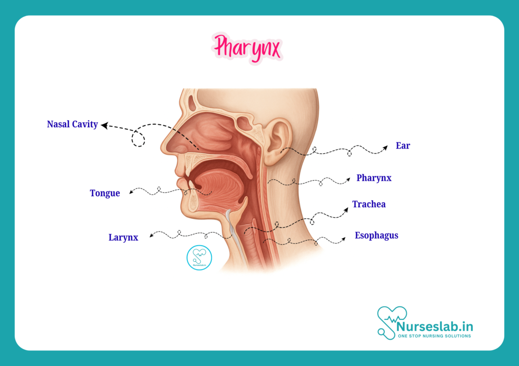
The pharynx is a muscular, funnel-shaped tube that forms the upper part of both the digestive and respiratory tracts. It extends from the base of the skull to the level of the sixth cervical vertebra, where it continues as the oesophagus. The pharynx serves as a common passageway for air (to the larynx and lungs) and food (to the oesophagus), playing a pivotal role in swallowing and respiration.
Detailed Anatomy of the Pharynx
Subdivisions of the Pharynx
The pharynx is anatomically divided into three regions:
- Nasopharynx: The uppermost part, located behind the nasal cavity and above the soft palate. It serves primarily as an airway.
- Oropharynx: The middle section, situated behind the oral cavity, extending from the soft palate to the upper border of the epiglottis. It serves as a passage for both air and food.
- Laryngopharynx (Hypopharynx): The lower part, lying behind the larynx and extending from the upper border of the epiglottis to the lower border of the cricoid cartilage, where it continues as the oesophagus.
Boundaries of the Pharynx
- Superior boundary: Base of the skull
- Inferior boundary: Lower border of the cricoid cartilage (C6 vertebra)
- Anterior boundaries: Posterior nasal apertures (choanae), oral cavity, laryngeal inlet
- Posterior boundary: Prevertebral fascia covering the cervical vertebrae
Walls and Layers of the Pharynx
The pharyngeal wall consists of four layers:
- Mucous membrane: The innermost lining, continuous with the nasal and oral mucosa.
- Submucosa: Contains connective tissue and pharyngeal glands.
- Pharyngobasilar fascia: A fibrous layer providing support, especially in the upper pharynx.
- Muscular layer: Composed of circular and longitudinal muscles that facilitate swallowing.
Muscles of the Pharynx
Pharyngeal muscles are classified as:
- Circular muscles (constrictors):
- Superior constrictor
- Middle constrictor
- Inferior constrictor
These muscles contract sequentially to propel the food bolus towards the oesophagus.
Longitudinal muscles:
- Stylopharyngeus
- Palatopharyngeus
- Salpingopharyngeus
These muscles elevate the pharynx during swallowing and speaking.
Blood Supply of the Pharynx
- Arterial supply: Branches from the external carotid artery, mainly the ascending pharyngeal, facial, maxillary, and lingual arteries.
- Venous drainage: Pharyngeal venous plexus drains into the internal jugular vein.
Nerve Supply of the Pharynx
- Motor supply: Mainly by the pharyngeal plexus, formed by branches of the glossopharyngeal (IX), vagus (X), and accessory (XI) nerves. The stylopharyngeus is supplied by the glossopharyngeal nerve.
- Sensory supply: Nasopharynx – maxillary nerve (V2); Oropharynx – glossopharyngeal nerve (IX); Laryngopharynx – vagus nerve (X).
Functions of the Pharynx
- Swallowing (Deglutition): The pharynx coordinates the passage of food from the mouth to the oesophagus through a series of muscular contractions, preventing aspiration into the airway.
- Respiration: Acts as a conduit for air from the nasal cavity to the larynx and lungs.
- Protection: The pharynx contains lymphoid tissue (e.g., tonsils) that forms part of the body’s immune defence, trapping pathogens and initiating immune responses.
Overview of the Larynx
Definition, Location, and General Function
The larynx, often called the “voice box,” is a cartilaginous structure in the anterior neck. It lies opposite the third to sixth cervical vertebrae and connects the laryngopharynx above to the trachea below. The larynx is responsible for voice production, airway protection during swallowing, and serving as a passage for air to the lower respiratory tract.
Detailed Anatomy of the Larynx
Cartilages of the Larynx
The larynx is composed of nine cartilages:
Three unpaired cartilages:
- Thyroid cartilage: The largest, forms the laryngeal prominence (“Adam’s apple”).
- Cricoid cartilage: A complete ring located below the thyroid cartilage.
- Epiglottis: A leaf-shaped cartilage that covers the laryngeal inlet during swallowing to prevent aspiration.
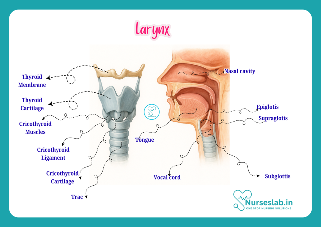
Three paired cartilages:
- Arytenoid cartilages: Pyramid-shaped, located on the posterior cricoid cartilage, essential for vocal cord movement.
- Corniculate cartilages: Small, cone-shaped, on the apex of arytenoids.
- Cuneiform cartilages: Rod-shaped, embedded in the aryepiglottic folds.
Muscles of the Larynx
- Intrinsic muscles: Responsible for controlling the tension and position of the vocal cords.
- Major intrinsic muscles include:
- Cricothyroid
- Posterior cricoarytenoid
- Lateral cricoarytenoid
- Transverse and oblique arytenoids
- Thyroarytenoid and vocalis
- Extrinsic muscles: Attach the larynx to surrounding structures and move it as a whole.
Examples include:
- Sternothyroid
- Thyrohyoid
- Inferior pharyngeal constrictor
Ligaments and Membranes
- Thyrohyoid membrane: Connects the thyroid cartilage to the hyoid bone.
- Cricothyroid ligament: Connects the cricoid and thyroid cartilages.
- Vocal ligaments: Form the core of the vocal cords.
- Quadrangular membrane: Forms the vestibular folds (false vocal cords).
Vocal Cords
The larynx contains two sets of folds:
- Vocal folds (true vocal cords): Responsible for sound production.
- Vestibular folds (false vocal cords): Play a minor role in voice but help protect the airway.
Blood Supply of the Larynx
- Arterial supply: Superior and inferior laryngeal arteries (branches of the superior thyroid and inferior thyroid arteries respectively).
- Venous drainage: Superior and inferior laryngeal veins drain into the corresponding thyroid veins.
Nerve Supply of the Larynx
- Motor and sensory supply: Mainly via branches of the vagus nerve (cranial nerve X).
- Superior laryngeal nerve: Internal branch (sensory to mucosa above vocal cords), external branch (motor to cricothyroid muscle).
- Recurrent laryngeal nerve: Motor to all other intrinsic muscles and sensory to mucosa below vocal cords.
Functions of the Larynx
- Voice Production (Phonation): Vibration of the vocal cords produces sound, modified by the tongue, lips, and palate to form speech.
- Airway Protection: During swallowing, the epiglottis closes over the laryngeal inlet to prevent food and liquids from entering the trachea.
- Breathing: The larynx maintains an open airway, allowing the free passage of air to the trachea and lungs.
Clinical Relevance
Common Disorders
- Pharyngitis: Inflammation of the pharynx, typically due to viral or bacterial infection, presenting with sore throat, fever, and dysphagia.
- Laryngitis: Inflammation of the larynx, often resulting in hoarseness or loss of voice. Common causes include infection, overuse, or irritants such as smoke.
- Airway Obstruction: Swelling, foreign bodies, trauma, or tumours can obstruct the pharynx or larynx, leading to life-threatening respiratory distress.
Assessment Techniques
- History taking: Enquire about symptoms such as sore throat, difficulty swallowing, hoarseness, cough, and breathing difficulty.
- Physical examination: Inspection of the oral cavity and pharynx using a torch and tongue depressor; palpation of the neck for tenderness or masses.
- Laryngoscopy: Direct or indirect visualization of the larynx to assess vocal cords and detect abnormalities.
- Swallowing assessment: Observing for signs of dysphagia, coughing, or choking during eating and drinking.
Emergency Situations
- Acute airway obstruction: Rapid recognition and intervention are required. Signs include stridor, cyanosis, difficulty breathing, and loss of consciousness. Emergency airway management (e.g., suction, airway adjuncts, or tracheostomy) may be necessary.
- Anaphylaxis: Severe allergic reactions can cause swelling of the pharynx and larynx. Immediate administration of adrenaline and airway support is critical.
Nursing Considerations
Patient Assessment
- Monitor for signs of airway compromise: stridor, hoarseness, use of accessory muscles, cyanosis.
- Assess for pain, swelling, or masses in the neck and throat region.
- Evaluate voice changes, swallowing difficulties, and presence of cough or aspiration.
Care during Airway Emergencies
- Ensure patency of airway using appropriate positioning (e.g., head-tilt, chin-lift, jaw thrust).
- Be prepared to initiate basic airway management (e.g., suctioning, insertion of oropharyngeal or nasopharyngeal airways).
- Assist with advanced procedures (e.g., endotracheal intubation, tracheostomy) as per protocol and training.
- Monitor oxygen saturation and provide supplemental oxygen as required.
Communication with Patients
- Use clear and simple language, especially for patients with voice or swallowing difficulties.
- Utilise alternative communication methods (e.g., writing, gestures) for patients who cannot speak.
- Provide reassurance and psychological support, as airway or voice disorders can cause significant distress.
Health Education
- Educate patients about maintaining throat and vocal health (e.g., avoiding irritants, staying hydrated, voice rest when needed).
- Instruct on signs that require urgent medical attention (e.g., sudden breathing difficulty, severe pain, blood in saliva).
- Promote adherence to prescribed treatments for infections or chronic conditions affecting the pharynx or larynx.
REFERENCES
- Ross and Wilson, Anatomy and Physiology in Health and Illness, Fourteenth Edition, 1 July 2022, ISBN-13: 978-0323834612.
- Roger Watson, Anatomy and Physiology for Nurses, 14th Edition, 12-06-2018, ISBN: 9780702077418
- P.R Asha Latha, Text Book of Applied Anatomy & Physiology for Nurses, 7th Edition,3 January 2024, ISBN-13: 978-9356968622.
- Bryan H. Derikson, Tortora’s Principles of Anatomy and Physiology, 16th Edition, August 2023, ISBN: 978- 1119400066.
- Anatomy.co.uk, Reproductive System, Last updated on April 24, 2025, https://anatomy.co.uk/reproductive-system
Stories are the threads that bind us; through them, we understand each other, grow, and heal.
JOHN NOORD
Connect with “Nurses Lab Editorial Team”
I hope you found this information helpful. Do you have any questions or comments? Kindly write in comments section. Subscribe the Blog with your email so you can stay updated on upcoming events and the latest articles.


