


Last Updated on May 28, 2026 by Nurseslab.in Editorial Team
Introduction
The human kidney is a remarkable organ, vital for maintaining homeostasis and overall health. For nurses, a thorough understanding of kidney anatomy is essential for assessing renal function, managing patient care, and recognising signs of kidney disease.
Overview of the Kidneys
Humans possess two kidneys, located retroperitoneally on either side of the vertebral column in the upper abdominal region. Shaped like beans, each kidney weighs around 120-150 grams in adults and measures approximately 10-12 cm in length, 5-7 cm in width, and 2-3 cm in thickness. The right kidney is generally positioned slightly lower than the left due to the presence of the liver. The kidneys are protected by the lower ribs, muscles of the back, and a layer of adipose tissue.
Location and Relations
- Vertebral Level: The kidneys extend from the T12 to L3 vertebrae.
- Right Kidney: Lies just below the liver, anterior to the 12th rib.
- Left Kidney: Slightly higher, anterior to the 11th and 12th ribs, near the spleen.
- Surrounding Structures: The kidneys are surrounded by renal fascia, perirenal fat, and pararenal fat, which provide protection and anchorage.
External Structure
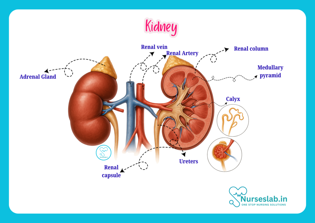
The kidney’s external surface is smooth and convex, with a medial indentation called the hilum. The hilum serves as the entry and exit point for the renal artery, renal vein, nerves, and ureter. The kidney is covered by three layers:
- Renal Capsule: A tough, fibrous outer layer that protects the kidney from trauma and infection.
- Perirenal Fat: A layer of adipose tissue cushioning the organ.
- Renal Fascia: Connective tissue anchoring the kidney to surrounding structures.
Internal Structure
On sectioning the kidney, two distinct regions are visible:
- Renal Cortex: The outer portion, granular in appearance, containing glomeruli and convoluted tubules.
- Renal Medulla: The inner region, composed of 8-18 cone-shaped renal pyramids. The apex of each pyramid, called the renal papilla, projects into minor calyces.
Between the pyramids are renal columns, extensions of cortical tissue that separate adjacent pyramids.
Renal Hilum and Sinus
The hilum leads into the renal sinus, a cavity containing the renal pelvis, calyces, blood vessels, nerves, and fat. The renal pelvis is a funnel-shaped structure that collects urine from the calyces and directs it into the ureter.
Microscopic Anatomy
Nephron: The Functional Unit
Each kidney contains approximately 1 to 1.5 million nephrons, the basic structural and functional units responsible for urine formation. A nephron consists of two main parts:
- Renal Corpuscle: Composed of the glomerulus and Bowman’s capsule.
- Renal Tubule: Consists of the proximal convoluted tubule (PCT), loop of Henle, distal convoluted tubule (DCT), and connecting tubule.
Renal Corpuscle
- Glomerulus: A network of capillaries where blood filtration begins.
- Bowman’s Capsule: A double-walled structure encasing the glomerulus and collecting the filtrate.
Renal Tubule
- Proximal Convoluted Tubule (PCT): Located in the cortex, responsible for reabsorption of water, ions, and nutrients.
- Loop of Henle: Descends into the medulla and returns to the cortex, playing a key role in concentrating urine.
- Distal Convoluted Tubule (DCT): Involved in further selective reabsorption and secretion.
- Collecting Duct: Receives urine from multiple nephrons and drains into the minor calyces.
Types of Nephrons
- Cortical Nephrons: Make up about 85% of nephrons, located primarily in the cortex, with short loops of Henle.
- Juxtamedullary Nephrons: Situated near the medulla, with long loops of Henle extending deep into the medulla, essential for producing concentrated urine.
Blood Supply
The kidneys receive about 20-25% of cardiac output. The renal artery, a branch of the abdominal aorta, enters each kidney at the hilum and divides into smaller arteries: segmental, interlobar, arcuate, and interlobular arteries. Blood is filtered in the glomerulus, and the filtered blood exits via the renal vein.
Venous drainage mirrors arterial supply, with blood flowing from interlobular to arcuate, interlobar, and finally into the renal vein, which drains into the inferior vena cava.
Nerve Supply
The renal plexus, a network of autonomic nerves, supplies the kidneys. Sympathetic stimulation regulates blood flow and influences renin secretion, while parasympathetic fibres are less prominent in humans.
Functions of the Kidney
The kidneys perform multiple vital functions that go beyond urine formation:
- Filtration: Removal of waste products, toxins, and excess substances from the blood.
- Reabsorption: Recovery of essential nutrients, ions, and water.
- Secretion: Active transport of certain substances into the tubular fluid.
- Excretion: Elimination of metabolic waste and drugs via urine.
- Regulation of Blood Pressure: Via the renin-angiotensin-aldosterone system (RAAS).
- Electrolyte Balance: Maintenance of sodium, potassium, calcium, and phosphate levels.
- Acid-Base Balance: Regulation of blood pH by excreting hydrogen ions and reabsorbing bicarbonate.
- Production of Hormones: Erythropoietin (stimulates red blood cell production), renin (regulates blood pressure), and activation of vitamin D (for calcium absorption).
Clinical Relevance for Nurses
Assessment of Kidney Function
Nurses are responsible for monitoring signs and symptoms of renal dysfunction. Common indicators include:
- Oedema (swelling due to fluid retention)
- Change in urine output and characteristics (colour, volume, odour)
- Hypertension
- Electrolyte imbalances (e.g., hyperkalaemia, hyponatraemia)
- Fatigue and anaemia (due to reduced erythropoietin)
Laboratory investigations such as blood urea, serum creatinine, glomerular filtration rate (GFR), and urinalysis are routinely used to assess renal function.
Common Renal Pathologies
- Acute Kidney Injury (AKI): Sudden loss of kidney function, identified by reduced urine output and rising creatinine levels.
- Chronic Kidney Disease (CKD): Gradual loss of kidney function, often associated with diabetes, hypertension, or glomerulonephritis.
- Glomerulonephritis: Inflammation of the glomeruli, presenting with proteinuria and haematuria.
- Nephrotic Syndrome: Characterised by heavy proteinuria, hypoalbuminaemia, oedema, and hyperlipidaemia.
- Renal Calculi (Kidney Stones): Hard deposits of minerals causing pain, obstruction, and haematuria.
- Urinary Tract Infections (UTI): May ascend to involve the kidneys, resulting in pyelonephritis.
Kidney Transplantation
For patients with end-stage renal disease, kidney transplantation offers a chance for improved quality of life. Nurses play a crucial role in preoperative assessment, postoperative care, and educating patients about immunosuppressive therapy and infection prevention.
Histological Features
On microscopic examination, the kidney reveals specialised cells and structures:
- Podocytes: Specialised cells in Bowman’s capsule aiding filtration.
- Juxtaglomerular Apparatus (JGA): Regulates blood pressure and GFR, composed of juxtaglomerular cells, macula densa, and extraglomerular mesangial cells.
- Mesangial Cells: Provide structural support and regulate filtration.
The arrangement of tubules and blood vessels in the cortex and medulla is essential for the countercurrent mechanism, enabling urine concentration.
Development and Embryology
The kidneys develop from the intermediate mesoderm during embryogenesis. Three successive structures form: pronephros (non-functional), mesonephros (transiently functional), and metanephros (permanent kidney). The metanephros begins functioning by the 8th week of gestation.
Applied Anatomy
Renal Angle
The renal angle is the area between the 12th rib and lateral border of the erector spinae muscle, clinically significant for assessing tenderness in cases of pyelonephritis or perinephric abscess.
Imaging Techniques
Ultrasonography, CT scan, and MRI are commonly used to visualise kidney structure and pathology. Intravenous urography (IVU) and renal biopsy may be performed as indicated.
Catheterisation and Nursing Care
Nurses are often involved in catheterisation procedures, monitoring for complications such as infection and obstruction. Proper technique and aseptic precautions are vital.
Indian Context: Kidney Health and Nursing
Kidney disease is a significant health concern in India, with increasing prevalence due to lifestyle factors, diabetes, and hypertension. Nurses play a pivotal role in early detection, patient education, and management. Culturally sensitive communication, dietary guidance, and community outreach programmes are essential for effective kidney care.
Traditional Indian diets, high in salt and spices, may influence kidney health. Nurses should encourage patients to adopt balanced diets, maintain adequate hydration, and seek regular health check-ups.
Summary and Key Points
- The kidneys are bean-shaped organs located retroperitoneally, essential for filtration, excretion, and homeostasis.
- Each kidney contains millions of nephrons, the functional units responsible for urine formation.
- Renal function is assessed clinically and through laboratory investigations.
- Common pathologies include AKI, CKD, glomerulonephritis, nephrotic syndrome, renal calculi, and UTI.
- Nurses are central to patient care, education, and management of kidney-related conditions.
- Understanding kidney anatomy is vital for safe and effective nursing practice.
REFERENCES
- Ross and Wilson, Anatomy and Physiology in Health and Illness, Fourteenth Edition, 1 July 2022, ISBN-13: 978-0323834612.
- Roger Watson, Anatomy and Physiology for Nurses, 14th Edition, 12-06-2018, ISBN: 9780702077418
- P.R Asha Latha, Text Book of Applied Anatomy & Physiology for Nurses, 7th Edition,3 January 2024, ISBN-13: 978-9356968622.
- Bryan H. Derikson, Tortora’s Principles of Anatomy and Physiology, 16th Edition, August 2023, ISBN: 978- 1119400066.
- Anatomy.co.uk, Reproductive System, Last updated on April 24, 2025, https://anatomy.co.uk/reproductive-system
Stories are the threads that bind us; through them, we understand each other, grow, and heal.
JOHN NOORD
Connect with “Nurses Lab Editorial Team”
I hope you found this information helpful. Do you have any questions or comments? Kindly write in comments section. Subscribe the Blog with your email so you can stay updated on upcoming events and the latest articles.


