


Last Updated on July 10, 2026 by Nurseslab.in Editorial Team
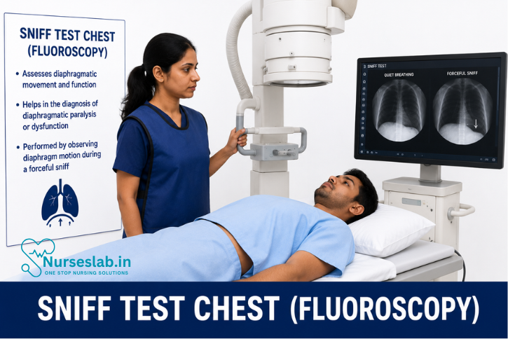
A sniff test using chest fluoroscopy evaluates diaphragm movement during breathing to diagnose diaphragmatic paralysis, weakness, or nerve dysfunction with real‑time imaging.
Introduction
The sniff test chest fluoroscopy is a specialised diagnostic procedure employed to evaluate diaphragmatic motion and function. Utilising real-time X-ray imaging, this test offers valuable insights into the mechanics of respiration, particularly the movement of the diaphragm during voluntary sniffing. It is a minimally invasive technique that has become a cornerstone in the assessment of diaphragmatic paralysis or dysfunction.
Clinical Indications
The sniff test chest fluoroscopy is primarily indicated in scenarios where diaphragmatic dysfunction is suspected. Common clinical indications include:
- Unexplained Dyspnoea: Patients presenting with shortness of breath without obvious cardiac or pulmonary causes.
- Suspected Diaphragmatic Paralysis or Paresis: Particularly in cases following thoracic or cervical surgery, trauma, or neurological disorders affecting the phrenic nerve.
- Evaluation of Neuromuscular Disorders: Conditions such as amyotrophic lateral sclerosis (ALS), myasthenia gravis, or muscular dystrophy, where diaphragmatic involvement may occur.
- Pre-operative and Post-operative Assessment: For thoracic surgeries, especially those involving the mediastinum, heart, or lungs.
- Assessment of Unilateral or Bilateral Diaphragmatic Elevation: Detected through chest radiography or clinical examination.
- Chronic Respiratory Insufficiency: When diaphragmatic weakness is suspected as a contributing factor.
The sniff test is also occasionally utilised to differentiate between true diaphragmatic paralysis and conditions mimicking diaphragmatic elevation, such as subphrenic abscess or hepatomegaly.
Patient Preparation
Proper preparation is crucial to ensure patient safety and the accuracy of the sniff test chest fluoroscopy. The steps include:
- Pre-procedure Assessment:
- Detailed clinical history, focusing on respiratory symptoms, previous surgeries, neurological disorders, and trauma.
- Review of prior imaging studies, such as chest X-rays and CT scans, to assess baseline diaphragmatic position.
- Contraindications:
- Absolute: Pregnancy (due to radiation risk), inability to cooperate with instructions, severe respiratory distress, or unstable cardiovascular status.
- Relative: Recent thoracic surgery (unless specifically indicated), active pulmonary infection, or allergy to contrast (if used).
- Instructions to Patients:
- Fasting is generally not required.
- Comfortable clothing is preferred; metallic objects around the chest should be removed.
- Explanation of the procedure, including the need to perform forceful sniffing on command.
- Reassurance regarding the minimal discomfort and low radiation exposure.
- Safety Measures:
- Lead shielding for the abdomen and pelvis, especially in women of childbearing age.
- Verification of patient identity and consent.
- Monitoring for any contraindications or risk factors.
Procedure Technique
The sniff test is performed under fluoroscopic guidance, providing real-time visualisation of diaphragmatic movement. The following steps outline the standard technique:
- Patient Positioning:
- The patient is typically positioned in the upright (standing or seated) posture, with the chest facing the fluoroscopy unit.
- Lateral views may be obtained if required.
- Baseline Imaging:
- Initial fluoroscopic images are obtained during quiet breathing to establish the baseline diaphragmatic excursion.
- Sniff Maneuver:
- The patient is instructed to perform a rapid, forceful sniff through the nose, mimicking the action of smelling.
- During the maneuver, fluoroscopic images are captured in real-time, focusing on the movement of both hemidiaphragms.
- Image Acquisition:
- Dynamic cine loops or video sequences may be recorded for subsequent analysis.
- Multiple sniffs may be performed to ensure reproducibility and accuracy.
- Post-procedure:
- No recovery period is generally required; patients can resume normal activities immediately.
- Images are reviewed by a radiologist or pulmonologist for interpretation.
The entire procedure typically lasts 10–15 minutes, and does not require sedation or contrast agents in most cases.
Interpretation of Results
Interpretation of the sniff test chest fluoroscopy centres on the movement pattern of the diaphragm during the sniff maneuver. Key points include:
Normal Findings:
- Both hemidiaphragms should move downwards (caudally) synchronously and briskly during a forceful sniff.
- The excursion is symmetrical, with no paradoxical movement.
Abnormal Findings:
- Paradoxical Movement: Instead of descending, the affected hemidiaphragm elevates during sniffing, indicating diaphragmatic paralysis or severe weakness.
- Reduced Excursion: Limited downward movement, suggestive of partial paresis or restrictive lung disease.
- Unilateral vs Bilateral Dysfunction: Unilateral paralysis shows paradoxical elevation of one hemidiaphragm, while bilateral involvement may present with minimal or absent movement of both.
False Positives/Negatives:
- Technical factors, such as poor patient effort or suboptimal positioning, can lead to misinterpretation.
- Subphrenic pathology (e.g., abscess, mass) may mimic diaphragmatic dysfunction.
- The findings must be correlated with clinical presentation and other investigations, such as pulmonary function tests, nerve conduction studies, or ultrasonography, for a definitive diagnosis.
Clinical Significance
The sniff test chest fluoroscopy holds substantial clinical value, primarily in diagnosing and managing conditions involving diaphragmatic dysfunction:
- Diaphragmatic Paralysis: Enables prompt identification, guiding further management such as surgical plication, rehabilitation, or ventilatory support.
- Neuromuscular Disease Assessment: Assists in evaluating the extent of respiratory muscle involvement, influencing treatment decisions.
- Pre-operative Evaluation: Determines suitability for thoracic or upper abdominal surgery, reducing perioperative complications.
- Post-operative Follow-up: Monitors recovery of diaphragmatic function after procedures involving the phrenic nerve or diaphragm.
- Differential Diagnosis: Differentiates between true diaphragmatic dysfunction and mimicking conditions, improving diagnostic accuracy.
The non-invasive nature and immediate results make the sniff test an invaluable tool in both acute and chronic settings, enhancing patient care and outcomes.
Risks and Limitations
While generally considered safe, the sniff test chest fluoroscopy is not without risks and limitations:
- Radiation Exposure: Although the dose is low, repeated procedures or cumulative exposure may pose risks, especially in children and pregnant women.
- Patient Cooperation: Accurate results depend on the patient’s ability to perform the sniff maneuver correctly; non-cooperation or poor effort can invalidate the test.
- Limited Sensitivity: Mild or early diaphragmatic weakness may not be detected, especially if compensation by accessory muscles occurs.
- Technical Limitations: Suboptimal imaging, operator inexperience, or equipment malfunction can affect diagnostic accuracy.
- Contraindications: Pregnant women and critically ill patients may not be suitable candidates due to safety concerns.
- Diagnostic Boundaries: The test does not provide information on the underlying cause of dysfunction; further investigations are often required.
The risks must be balanced against the diagnostic benefits, and alternative modalities such as ultrasonography may be considered in select cases.
Nursing Care of Patients Undergoing Sniff Test Chest (Fluoroscopy)
The procedure is performed in a radiology suite using specialised fluoroscopic equipment that emits ionising radiation. The nurse’s responsibilities include preparing the patient, providing emotional support, monitoring for adverse reactions, and ensuring adherence to safety protocols.
Patient Assessment: Pre-Procedure Evaluation
Thorough patient assessment is the foundation of safe and effective nursing care for individuals undergoing a Sniff Test Chest. The nurse should begin by reviewing the patient’s medical history, focusing on respiratory illnesses, neuromuscular disorders, recent chest trauma, or previous surgeries that may affect diaphragmatic function. It is essential to identify any co-existing conditions, such as chronic obstructive pulmonary disease (COPD), heart failure, or recent infections, which might impact the procedure or its interpretation.
Additionally, a detailed medication history must be obtained, especially noting drugs that may interfere with respiratory effort or consciousness, such as sedatives or muscle relaxants. Allergies, particularly to contrast agents or medications, should be documented. Baseline vital signs, oxygen saturation, and a focused respiratory examination—including inspection for use of accessory muscles, auscultation for abnormal breath sounds, and assessment of breathing patterns—should be conducted and recorded.
Psychosocial assessment is equally important. Patients may experience anxiety or fear related to the procedure or possible diagnoses. The nurse should explore the patient’s understanding, expectations, and concerns, providing reassurance and clarifying doubts as needed. Identifying language barriers, sensory impairments, or cognitive limitations will help tailor communication and support strategies.
Preparation: Patient Education, Consent, and Readiness
Preparation for the Sniff Test Chest involves a series of coordinated steps to ensure the patient’s physical and emotional readiness. Patient education is paramount; the nurse should explain the purpose of the procedure, the steps involved, and what the patient can expect during the test. Emphasise that the test is generally painless and non-invasive, and highlight the importance of following instructions, particularly regarding sniffing manoeuvres.
Obtain informed consent as per institutional policy, ensuring the patient (or legal guardian) understands the risks, benefits, and alternatives. The nurse should verify that the patient has adhered to any pre-procedure instructions, such as fasting requirements or withholding certain medications, if applicable.
Physical preparation includes ensuring the patient is appropriately attired in a hospital gown and has removed any jewellery or metallic items that may interfere with imaging. Assess the need for intravenous access if contrast administration is anticipated. Confirm that the patient has emptied their bladder to maximise comfort during the examination.
Emotional readiness is fostered through active listening and empathetic communication. Address any fears of radiation exposure by explaining the safety measures in place and the minimal risk involved. If the patient requires assistance with mobility or positioning, make arrangements for additional support.
Procedural Support: Monitoring and Safety
During the Sniff Test Chest, the nurse acts as a crucial liaison between the patient and the radiology team, ensuring both safety and comfort. Position the patient as instructed, typically in a standing or semi-recumbent posture, and assist in maintaining the required position throughout the procedure.
Continuous monitoring of vital signs is essential, particularly in patients with compromised respiratory status or anxiety. Observe for signs of distress, such as increased work of breathing, cyanosis, or agitation. The nurse should be prepared to intervene promptly should any acute symptoms arise.
Adherence to radiation safety protocols is mandatory. Ensure that lead aprons or shields are used as appropriate, and that only necessary personnel are present in the fluoroscopy suite. Maintain clear and open communication with the radiologist and technicians, relaying any concerns or changes in patient status without delay.
Patient privacy and dignity should be respected at all times. Provide reassurance and gentle guidance during the sniffing manoeuvres, encouraging the patient to perform to the best of their ability. For paediatric or cognitively impaired patients, creative communication strategies or the presence of a family member may facilitate cooperation.
Post-Procedure Care: Immediate Observations and Recovery
Following the Sniff Test Chest, the nurse is responsible for ensuring the patient’s safe recovery and monitoring for any immediate complications. Assess vital signs, oxygen saturation, and overall clinical status. Instruct the patient to report any new or unusual symptoms, such as chest pain, shortness of breath, or dizziness.
If contrast media was used, monitor for allergic reactions, including rash, itching, or difficulty breathing. Provide hydration as appropriate to facilitate renal elimination of contrast agents. For patients with pre-existing conditions or those who experienced distress during the procedure, a longer observation period may be warranted.
Document all post-procedure observations, interventions, and patient responses in the medical record. Ensure that the patient is stable, comfortable, and has recovered baseline function before discharge from the radiology suite. Provide clear instructions regarding mobility, activity restrictions (if any), and follow-up appointments.
Complication Management: Identification and Intervention
While the Sniff Test Chest is generally safe, nurses must remain vigilant for potential complications. Adverse reactions to contrast media, though rare, require immediate intervention—administer antihistamines or corticosteroids as prescribed and initiate emergency protocols if anaphylaxis is suspected. Respiratory distress, hypoxaemia, or syncope during or after the procedure should be managed according to advanced life support guidelines.
Other potential complications include vasovagal reactions, particularly in anxious or frail patients, and radiation exposure incidents. In the event of a vasovagal episode, position the patient supine, elevate the legs, and monitor vital signs closely. Report and document any incidents of excessive radiation exposure as per institutional policy.
Effective complication management relies on prompt recognition, rapid intervention, and thorough documentation. The nurse should communicate any concerns to the multidisciplinary team and participate in root cause analysis if adverse events occur, contributing to quality improvement initiatives.
Patient Education: Home Care, Follow-Up, and Symptom Reporting
Patient education extends beyond the procedure itself, empowering individuals to participate actively in their recovery and ongoing care. Before discharge, provide verbal and written instructions regarding expected post-procedure symptoms, such as mild fatigue or transient discomfort, and advise when to seek medical attention.
Educate patients about the signs and symptoms of complications, including persistent chest pain, breathlessness, fever, or allergic reactions. Emphasise the importance of adhering to follow-up appointments for review of test results and further management. For patients with underlying neuromuscular or respiratory conditions, reinforce disease-specific self-management strategies and connect them with relevant support services or rehabilitation programmes.
Encourage patients to maintain open communication with their healthcare team and to report any concerns promptly. Provide contact information for the hospital or clinic, and ensure that patients know how to access emergency services if needed.
REFERENCES
- Criner G, Marchetti N. Effects of Neuromuscular Diseases on Ventilation. In: Grippi MA, Antin-Ozerkis DE, Dela Cruz CS, Kotloff RM, Kotton C, Pack AI, eds. Fishman’s Pulmonary Diseases and Disorders. 6th ed. McGraw-Hill Education; 2023.
- Laghi FA Jr, Saad M, Shaikh H. Ultrasound and non-ultrasound imaging techniques in the assessment of diaphragmatic dysfunction (https://pubmed.ncbi.nlm.nih.gov/33722215/). BMC Pulm Med. 2021 Mar 15;21(1):85.
- Minami T, Manzoor K, McCool FD. Assessing Diaphragm Function in Chest Wall and Neuromuscular Diseases (https://pubmed.ncbi.nlm.nih.gov/29779593/). Clin Chest Med. 2018 Jun;39(2):335-344.
Stories are the threads that bind us; through them, we understand each other, grow, and heal.
JOHN NOORD
Connect with “Nurses Lab Editorial Team”
I hope you found this information helpful. Do you have any questions or comments? Kindly write in comments section. Subscribe the Blog with your email so you can stay updated on upcoming events and the latest articles.


