


Last Updated on February 12, 2026 by Nurseslab.in Editorial Team
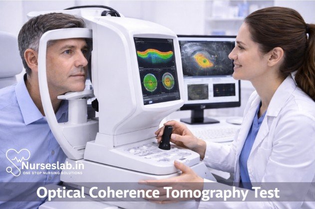
Optical Coherence Tomography (OCT) is a non‑invasive imaging technique that captures detailed cross‑sectional views of the retina. It is vital for diagnosing macular diseases, glaucoma, and retinal disorders, supporting accurate assessment and ongoing monitoring.
Introduction
Optical Coherence Tomography (OCT) is a non-invasive imaging technology that has revolutionised the way medical professionals diagnose and monitor a variety of conditions, particularly in ophthalmology, cardiology, and dermatology. By providing high-resolution, cross-sectional images of biological tissues, OCT enables clinicians to visualise microstructures at near-microscopic detail, facilitating early detection and precise management of diseases.
Since its inception in the early 1990s, OCT has undergone significant evolution, expanding its utility across multiple medical specialties. Its ability to deliver real-time imaging without physical contact or radiation exposure has made it an indispensable tool in contemporary clinical practice.
Principles of Optical Coherence Tomography
Basic Physics
OCT is based on the principle of low-coherence interferometry, which employs light waves to capture micrometre-resolution, two- and three-dimensional images from within optical scattering media, such as biological tissue. When a beam of light from a broadband source is split into reference and sample arms, the reflected light from both paths recombines to produce an interference pattern. The analysis of this pattern enables the reconstruction of tissue microarchitecture.
Types of OCT
- Time-domain OCT (TD-OCT): The original form of OCT, TD-OCT, relies on a moving reference mirror to measure the echo time delay of light reflected from tissue. While effective, its acquisition speed and resolution are limited compared to newer modalities.
- Spectral-domain OCT (SD-OCT): SD-OCT utilises a stationary reference mirror and a spectrometer to detect the interference spectrum. This approach offers higher speed and improved axial resolution, making it the preferred choice in many clinical settings.
- Swept-source OCT (SS-OCT): SS-OCT employs a tunable laser source that rapidly sweeps across a range of wavelengths. This technology allows deeper tissue penetration and faster acquisition, particularly advantageous in imaging larger structures such as the choroid in ophthalmology.
Technology and Equipment
Key Components
- Light Source: Typically a superluminescent diode or tunable laser, providing broadband illumination essential for high-resolution imaging.
- Interferometer: Splits and recombines the light beam to generate an interference signal, which contains depth information about the tissue.
- Detector: A photodiode array or line-scan camera detects the interference pattern and converts it into a digital signal for processing.
- Scanning System: Mechanisms (often galvanometric mirrors) control the lateral movement of the beam across the tissue, enabling the construction of two- or three-dimensional images.
- Computer and Software: Advanced algorithms process raw data, reconstruct images, and provide tools for measurement and analysis.
Imaging Modalities
OCT systems offer various imaging modalities, including cross-sectional (B-scan), en face (C-scan), and volumetric imaging. These modalities allow clinicians to visualise tissue layers, surface topology, and three-dimensional reconstructions.
Safety Considerations
OCT is considered exceptionally safe, as it uses non-ionising light and does not involve physical contact with the tissue. However, proper calibration, maintenance, and adherence to manufacturer guidelines are essential to ensure patient safety and optimal image quality.
Clinical Applications
Ophthalmology
OCT is most widely used in ophthalmology, where it enables high-resolution imaging of the retina, optic nerve head, and anterior segment. It is invaluable in diagnosing and monitoring conditions such as age-related macular degeneration, diabetic retinopathy, glaucoma, and macular oedema. OCT facilitates the visualisation of retinal layers, helping clinicians detect subtle pathological changes and guide treatment decisions.
Cardiology
In cardiology, intravascular OCT is employed to assess coronary arteries. This application allows the visualisation of arterial wall microstructure, identification of plaque composition, and evaluation of stent deployment. OCT provides superior resolution compared to intravascular ultrasound, aiding in the precise management of coronary artery disease.
Dermatology
OCT is increasingly utilised in dermatology for imaging skin architecture, aiding in the diagnosis of basal cell carcinoma, melanoma, and other skin pathologies. Its non-invasive nature allows repeated imaging without discomfort or risk to the patient.
Other Specialties
Beyond ophthalmology, cardiology, and dermatology, OCT has found applications in gastroenterology (imaging mucosal layers), dentistry (assessing periodontal tissues), and oncology (tumour margin evaluation), reflecting its versatility across medical disciplines.
Patient Preparation
Pre-Procedure Instructions
- Inform patients about the nature and purpose of the OCT procedure, including its non-invasive and painless characteristics.
- Reassure patients regarding the absence of ionising radiation and the safety of the procedure.
- Advise patients to remove contact lenses, spectacles, or any facial accessories prior to ophthalmic OCT.
- Instruct patients to avoid using eye drops or topical medications immediately before the procedure unless specifically indicated.
- For cardiovascular OCT, ensure fasting or specific medication adjustments as per clinical protocol.
Contraindications
- Patients with severe photophobia or inability to maintain steady fixation may not be suitable for ophthalmic OCT.
- Individuals with active ocular infection or inflammation should defer the procedure until resolution.
- Cardiovascular OCT may be contraindicated in patients with unstable haemodynamics or severe arrhythmia.
Patient Consent
Obtain informed consent prior to the procedure, outlining the benefits, potential risks (however minimal), and alternatives. Address patient queries and concerns to ensure cooperation and compliance during imaging.
Step-by-Step Diagnostic Procedure
Equipment Setup
- Calibrate the OCT device according to manufacturer specifications.
- Check the alignment of the scanning system and ensure the cleanliness of optical components.
- Prepare the computer and software for data acquisition and storage.
Patient Positioning
- For ophthalmic OCT, seat the patient comfortably with chin and forehead support to minimise movement.
- In cardiovascular OCT, position the patient as per interventional protocol, ensuring vascular access and monitoring equipment are in place.
- For dermatological OCT, expose the target area and ensure the patient remains still during scanning.
Image Acquisition
- Align the imaging beam with the target tissue, using fixation devices or markers as needed.
- Instruct the patient to maintain steady gaze or remain still during image capture.
- Initiate the scan, acquiring cross-sectional or volumetric images as per clinical requirement.
- Monitor image quality in real-time, adjusting focus and alignment as necessary.
- Repeat scans if required to ensure optimal visualisation of the area of interest.
Troubleshooting
- If image artefacts or poor quality are observed, check for patient movement, improper alignment, or equipment malfunction.
- Re-instruct the patient or reposition as necessary.
- Consult technical support or refer to the device manual for persistent issues.
Interpretation of Results
Normal vs Abnormal Findings
- Normal Findings: Well-delineated tissue layers, regular microarchitecture, absence of abnormal reflectivity or fluid accumulation.
- Abnormal Findings: Disruption of tissue layers, presence of cystic spaces, hyper- or hyporeflective lesions, abnormal thickness, or structural distortion.
Common Pathologies Detected
- Ophthalmology: Macular oedema, retinal detachment, drusen, glaucoma-related changes.
- Cardiology: Plaque characterisation, stent malapposition, thrombus detection.
- Dermatology: Tumour margins, subepidermal structures, inflammatory changes.
Reporting and Documentation
Interpretation should be performed by trained professionals, correlating OCT findings with clinical presentation and other diagnostic modalities. Documentation must include image quality assessment, anatomical landmarks, pathological findings, and recommendations for further management.
Advantages of OCT
- Non-Invasive: OCT does not require tissue biopsy or exposure to ionising radiation, minimising patient risk.
- High Resolution: Capable of resolving microstructures at the scale of 1–15 micrometres, far surpassing conventional imaging techniques.
- Rapid Acquisition: Real-time imaging allows immediate assessment and decision-making.
- Versatility: Applicable across multiple medical specialties and tissue types.
- Comparative Advantage: Compared to ultrasound, OCT offers superior resolution for superficial tissues, though ultrasound provides deeper penetration for certain applications.
Limitations and Challenges
- Technical Limitations: Limited penetration depth (typically 1–2 mm), making it less suitable for imaging deep structures.
- Patient-Related Factors: Movement, poor fixation, or anatomical variations can compromise image quality.
- Interpretative Challenges: Requires specialised training for accurate interpretation; artefacts may mimic pathology.
- Cost and Accessibility: High initial investment and maintenance costs may limit availability in resource-constrained settings.
- Limited Functional Information: OCT primarily provides structural rather than functional or metabolic data.
Nursing Care of Patients Undergoing Optical Coherence Tomography (OCT) Procedure
The procedure plays a crucial role in diagnosing and monitoring various retinal diseases, glaucoma, and macular conditions. As with any diagnostic procedure, the role of the nurse is vital in ensuring patient safety, comfort, education, and optimal outcomes throughout the peri-procedural period.
Role of the Nurse in OCT Procedures
Nursing responsibilities span the entire course of the OCT procedure, encompassing pre-procedural assessment and preparation, intra-procedural support, and post-procedural care and education. The nurse acts as a patient advocate, ensuring comfort, safety, and understanding while facilitating the smooth execution of the imaging process.
Pre-Procedural Nursing Care
1. Patient Assessment
A thorough pre-procedural assessment is essential to identify any contraindications, potential challenges, and individual patient needs. The nurse should:
- Review the patient’s medical history, focusing on ocular conditions, allergies (especially to mydriatic agents), and systemic diseases (e.g., diabetes, hypertension).
- Verify the indication for the OCT procedure and ensure appropriate documentation and consent are in place.
- Assess the patient’s level of understanding and anxiety regarding the procedure.
- Evaluate visual acuity and baseline ocular status as needed.
2. Patient Preparation and Education
Patient education is paramount in alleviating anxiety and ensuring cooperation during the procedure. The nurse should:
- Explain the purpose, process, and benefits of OCT in simple, non-technical language.
- Describe what the patient will experience: sitting at the OCT device, focusing on a target light, and the need to remain still for a few seconds.
- Discuss the non-invasive, painless nature of the test and reassure the patient about the absence of radiation or injections (except in specific angiography protocols).
- Inform the patient about the possible use of mydriatic (dilating) eye drops and potential temporary effects such as blurred vision and light sensitivity.
- Advise the patient to arrange for assistance with transportation if dilation is planned, as driving may be impaired for several hours.
- Address any questions or concerns and provide written information if available.
3. Physical Preparation
Nurses should ensure:
- Proper hand hygiene and infection control measures are observed.
- Availability of necessary equipment and supplies (OCT machine, mydriatic drops, tissues, gloves, etc.).
- Patient identification and confirmation of the correct eye(s) to be imaged.
- Administration of mydriatic drops if required, observing for adverse reactions (e.g., allergy, angle-closure glaucoma in susceptible individuals).
- Monitoring of vital signs if indicated, especially in patients with cardiovascular or systemic comorbidities.
- Assistance with removal of contact lenses or ocular prostheses, if necessary.
Intra-Procedural Nursing Care
1. Patient Positioning and Comfort
Proper positioning is crucial for obtaining high-quality OCT images. The nurse should:
- Guide the patient to sit comfortably at the OCT device, adjusting the chin rest and head support as needed.
- Encourage the patient to relax, keep both eyes open, and focus on the fixation target.
- Provide reassurance and clear instructions to remain still during image acquisition, as movement can result in artefacts and repeat scans.
- Support patients with physical disabilities or those requiring assistance to maintain position.
2. Observation and Support
During the procedure, the nurse should:
- Monitor the patient for discomfort, anxiety, or signs of distress.
- Offer verbal encouragement and maintain a calm, supportive presence.
- Assist the ophthalmic technician or physician as necessary, for example, by administering additional eye drops or helping with patient repositioning.
- Ensure strict adherence to infection control protocols, especially when the OCT device’s chin and forehead rests are used by multiple patients.
Post-Procedural Nursing Care
1. Immediate Post-Procedure Care
Immediately after the OCT procedure, the nurse should:
- Assist the patient in moving away from the machine and provide tissues to wipe any excess eye drops.
- Assess for any adverse reactions to medications (e.g., redness, irritation, allergic response).
- Instruct the patient to report any new or worsening symptoms such as pain, vision loss, or severe discomfort.
- Document the procedure, noting any difficulties encountered, medications administered, and patient responses.
2. Patient Education and Discharge Instructions
Comprehensive post-procedural education includes:
- Explaining that the results will be reviewed by the ophthalmologist and discussed at a follow-up visit.
- Advising on the expected duration of blurred vision and light sensitivity if dilation was used, and the importance of wearing sunglasses outdoors.
- Emphasising the need to avoid driving or operating machinery until vision returns to normal.
- Providing guidance on when to seek urgent medical attention (e.g., if experiencing severe pain, sudden vision changes, or allergic reactions).
- Reminding the patient of upcoming appointments and the importance of ongoing monitoring for chronic conditions.
Special Considerations in Nursing Care
1. Paediatric and Geriatric Patients
OCT may be challenging in very young, elderly, or cognitively impaired individuals. Nurses should:
- Use age-appropriate language and techniques to explain the procedure.
- Involve caregivers or family members for reassurance and assistance.
- Allow extra time and provide additional support for positioning and cooperation.
- Monitor more closely for medication side effects or distress.
2. Patients with Disabilities
Patients with physical or visual disabilities may need tailored support:
- Arrange for accessible facilities and equipment modifications if needed.
- Provide hands-on guidance and verbal cues for positioning.
- Assess for the need for additional assistance or adaptive devices.
3. Infection Prevention and Control
Strict infection control is essential as OCT devices are used by multiple patients. Nurses must:
- Clean and disinfect chin rests, forehead bands, and other contact surfaces between patients.
- Practice hand hygiene before and after patient contact.
- Use gloves when administering eye drops or handling ocular secretions.
Documentation and Communication
Accurate and timely documentation supports continuity of care and legal protection. Nurses should record:
- Date and time of the procedure
- Type of OCT performed (e.g., macular, optic nerve)
- Medications administered, including type, dose, and time
- Patient’s response and any adverse events
- Education provided and patient understanding
- Any special considerations or accommodations made
Effective communication with the multidisciplinary team—including ophthalmologists, optometrists, and technicians—is vital for optimal patient outcomes.
REFERENCES
- Popescu DP, Choo-Smith LP, Flueraru C, Mao Y, Chang S, Disano J, Sherif S, Sowa MG. Optical coherence tomography: fundamental principles, instrumental designs and biomedical applications. Biophys Rev. 2011 Sep;3(3):155. doi: 10.1007/s12551-011-0054-7. Epub 2011 Aug 6. PMID: 28510064; PMCID: PMC5418377.
- American Academy of Ophthalmology. What Is Optical Coherence Tomography? https://www.aao.org/eye-health/treatments/what-is-optical-coherence-tomography Reviewed 4/27/2023.
- Roy, M., Khan, R., Sala Uddin Pathan, A.Q.M., Raman, R. (2025). Fundamentals of Optical Coherence Tomography. In: Cengiz, I.F., Oliveira, J.M., Reis, R.L. (eds) Bioimaging Modalities in Bioengineering. Biomaterials, Bioengineering and Sustainability, vol 9. Springer, Cham. https://doi.org/10.1007/978-3-031-99410-4_3
- Aumann S, Donner S, Fischer J, et al. Optical Coherence Tomography (OCT): Principle and Technical Realization https://www.ncbi.nlm.nih.gov/books/NBK554044/. 2019 Aug 14. In: Bille JF, editor. High Resolution Imaging in Microscopy and Ophthalmology: New Frontiers in Biomedical Optics [Internet]. Cham (CH): Springer; 2019. Chapter 3.
- Optometrists Network. What Is an OCT Eye Exam? https://www.optometrists.org/general-practice-optometry/guide-to-eye-exams/eye-exams/what-is-an-oct-eye-exam
- Diagnostic performance of virtual fractional flow reserve derived from optical coherence tomography in coronary functional stenosis evaluation, November 2025, European Heart Journal 46(Supplement_1) https://www.researchgate.net/publication/400473398_Diagnostic_performance_of_virtual_fractional_flow_reserve_derived_from_optical_coherence_tomography_in_coronary_functional_stenosis_evaluation
Stories are the threads that bind us; through them, we understand each other, grow, and heal.
JOHN NOORD
Connect with “Nurses Lab Editorial Team”
I hope you found this information helpful. Do you have any questions or comments? Kindly write in comments section. Subscribe the Blog with your email so you can stay updated on upcoming events and the latest articles.


