


Last Updated on August 28, 2026 by Nurseslab.in Editorial Team
Overview
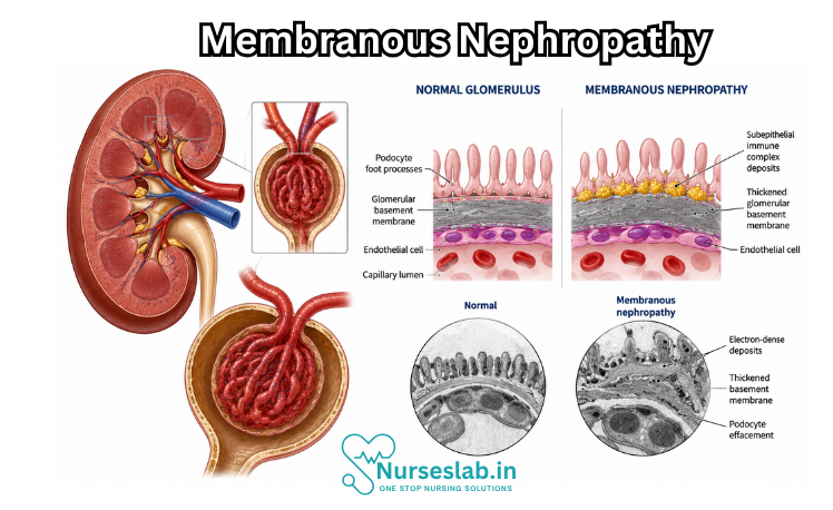
Membranous nephropathy, also called membranous glomerulopathy, is an immune-mediated disease of the kidney’s microscopic filters, the glomeruli. It is characterised by immune deposits along the outer surface of the glomerular basement membrane. These deposits injure podocytes—the specialised cells that help prevent valuable blood proteins from leaking into urine. The resulting protein loss may cause nephrotic syndrome, with heavy proteinuria, low serum albumin, oedema, and elevated blood lipids.
The disease most often affects adults and is an important cause of nephrotic syndrome in people without diabetes. Its course is variable. Some patients enter spontaneous remission, some continue to have proteinuria but stable kidney function, and others progressively lose kidney function. Modern care therefore combines accurate identification of the disease mechanism, investigation for associated conditions, intensive supportive therapy, and risk-based use of immunosuppressive treatment.
Normal Glomerular Filtration and What Goes Wrong
Each kidney contains many nephrons, and each nephron begins with a glomerulus. Blood passes through a network of capillaries whose filtration barrier has three major components: fenestrated endothelial cells, the glomerular basement membrane, and podocyte foot processes bridged by slit diaphragms. This barrier allows water and small solutes to be filtered while retaining blood cells and most proteins.
In membranous nephropathy, antibodies bind to antigens associated with podocytes or become involved in deposits beneath the podocytes. Complement activation and related immune injury disrupt podocyte structure and the slit diaphragm. The barrier becomes abnormally permeable to albumin and other proteins. The basement membrane forms new material around the deposits, producing the “membranous” thickening visible on biopsy. There is usually less inflammatory-cell proliferation than in many nephritic diseases, so urine sediment may be relatively bland.
Classification: Primary, Secondary, and Antigen-Defined Disease
Historically, membranous nephropathy was divided into primary disease, in which no external cause was identified, and secondary disease associated with another illness, infection, medication, or exposure. Discovery of specific target antigens has made classification more precise. The most common is M-type phospholipase A2 receptor, or PLA2R. Other antigens include THSD7A, NELL1, EXT1/EXT2, semaphorin 3B, and additional less common targets. Antigen identification may suggest a kidney-limited autoimmune process or prompt investigation for a particular association.
A positive circulating anti-PLA2R antibody in an appropriate clinical setting strongly supports PLA2R-associated membranous nephropathy. However, a negative blood test does not completely exclude it, because antibody levels may be low, disappear after an immunological response, or be detectable only in kidney tissue. Clinical context and biopsy findings remain important.
Causes and Associated Conditions
- Autoimmune kidney-limited disease: Autoantibodies are directed against podocyte antigens, most commonly PLA2R.
- Systemic autoimmune disease: Systemic lupus erythematosus is a major association; rheumatoid arthritis, thyroid autoimmunity, and other disorders may occasionally coexist.
- Infections: Hepatitis B, hepatitis C, syphilis, and certain other chronic infections can be associated with the membranous pattern.
- Medications and supplements: Non-steroidal anti-inflammatory drugs, older agents such as gold or penicillamine, and some supplements or traditional medicines can be implicated.
- Malignancy: Some cases occur in association with solid or haematological cancer. Evaluation is guided by age, symptoms, risk factors, antigen pattern, and usual screening recommendations.
- Other settings: Membranous nephropathy may rarely occur after stem-cell transplantation or in transplanted kidneys.
Finding an association does not automatically prove causation. The nephrology team considers timing, serology, biopsy staining, response after removing an exposure, and whether treatment of the associated condition improves proteinuria. This distinction matters because secondary disease is treated primarily by addressing the underlying cause, while kidney-directed immunosuppression may be unnecessary or unsafe in some cases.
Who Is Affected?
Membranous nephropathy can occur at any age but is diagnosed mainly in adults, with a peak in middle and later life. It is more common in men than women in many adult populations. The disease is less common in children; paediatric cases warrant especially careful evaluation for associated conditions and unusual antigens. It is usually acquired rather than inherited, although genetic background may influence susceptibility to autoimmune disease.
Signs and Symptoms
The onset is often gradual. Some patients are identified after protein is found during routine urine testing, while others present with nephrotic syndrome. Kidney filtration may initially remain normal even when protein loss is severe.
- Foamy or frothy urine: Often caused by excess urinary protein, although appearance alone is not diagnostic.
- Oedema: Puffiness around the eyes and swelling of the feet, ankles, legs, abdomen, or genital area.
- Rapid weight gain: Usually reflects fluid retention rather than increased body fat.
- Fatigue and reduced stamina: May relate to oedema, inflammation, treatment effects, or low albumin.
- Reduced appetite or abdominal fullness: Can occur with marked fluid accumulation.
- High blood pressure: May accompany sodium retention or declining kidney function.
- Breathlessness: Can result from severe fluid overload, pleural fluid, infection, pulmonary embolism, or another cardiopulmonary problem.
- Blood clot symptoms: One-sided leg swelling, sudden chest pain, shortness of breath, or abdominal/flank pain require urgent assessment.
Diagnostic Evaluation
Evaluation confirms the nephrotic pattern, identifies the antigen or biopsy pattern, estimates disease activity and progression risk, and searches for an associated cause.
- History and examination: Timing of swelling, urine changes, weight gain, blood pressure, recent infections, thrombotic symptoms, autoimmune features, medication and supplement exposure, cancer warning signs, family history, and pregnancy plans are reviewed. Examination assesses oedema, volume status, blood pressure, heart and lung findings, skin changes, and signs of thrombosis or systemic disease.
- Urinalysis and microscopy: Dipstick detects protein and blood. Microscopy often shows a relatively bland sediment, sometimes with microscopic haematuria, fatty casts, or oval fat bodies. Prominent red-cell casts suggest another or additional glomerular process.
- Quantification of protein loss: A urine protein-to-creatinine ratio from a spot sample is convenient; albumin-to-creatinine ratio or a 24-hour collection may also be used. Serial measurements are more informative than an isolated result.
- Kidney function and serum proteins: Creatinine and estimated glomerular filtration rate assess filtration. Serum albumin and total protein indicate the severity of protein loss. Electrolytes, urea, liver tests, and full blood count help evaluate complications and alternative causes.
- Lipid profile: Total cholesterol, LDL cholesterol, and triglycerides are often elevated in nephrotic syndrome.
- Anti-PLA2R testing: The antibody supports diagnosis, helps assess immunological activity, contributes to risk stratification, and can be followed during treatment. Falling antibodies usually precede improvement in proteinuria.
- Other antigen or antibody testing: Anti-THSD7A or tissue-based antigen testing may be considered where available, particularly if PLA2R studies are negative.
- Secondary-cause screen: Tests are selected according to clinical context and can include antinuclear and anti-double-stranded DNA antibodies, complement levels, hepatitis B and C studies, HIV testing, syphilis testing, and other infection investigations.
- Medication and exposure review: Prescription drugs, over-the-counter pain medicines, supplements, injections, and traditional remedies are checked carefully.
- Age-appropriate malignancy evaluation: Standard cancer screening should be up to date. Additional tests are guided by symptoms, examination, smoking history, laboratory abnormalities, biopsy antigen pattern, and clinician judgement rather than indiscriminate scanning.
- Kidney ultrasound: Assesses kidney size, structure, obstruction, and biopsy suitability; it does not itself diagnose membranous nephropathy.
- Kidney biopsy: Tissue is examined by light microscopy, immunofluorescence, and electron microscopy. Biopsy confirms the membranous pattern, identifies immune deposits and antigen staining, assesses chronic scarring, and checks for another kidney disease.
What the Kidney Biopsy Shows
- Light microscopy: Diffuse thickening of glomerular capillary walls with little cellular proliferation. Silver stains may reveal “spikes” of new basement membrane material between deposits.
- Immunofluorescence: Granular deposits of immunoglobulin G and complement along capillary loops. The immunoglobulin subclass pattern can provide clues about disease type.
- Electron microscopy: Electron-dense deposits in the subepithelial space, podocyte foot-process effacement, and newly formed basement membrane material.
- Antigen staining: Detection of PLA2R or another target in deposits helps classify disease and focus the search for associations.
- Chronicity: Interstitial fibrosis, tubular atrophy, and glomerular scarring help estimate irreversible damage and likely treatment benefit.
When a Biopsy May or May Not Be Needed
In an adult with nephrotic syndrome, preserved kidney function, and a convincingly positive anti-PLA2R test, a nephrologist may diagnose PLA2R-associated membranous nephropathy without biopsy. Biopsy can still add information when kidney function declines unexpectedly, the presentation is atypical, another kidney disease is possible, serology is negative, treatment decisions are uncertain, or chronic damage needs assessment. The risks and value of biopsy are discussed individually.
Differential Diagnosis
- Minimal change disease and focal segmental glomerulosclerosis.
- Diabetic kidney disease and amyloidosis.
- Lupus nephritis and other immune-complex glomerulonephritides.
- Infection-related glomerular disease.
- Monoclonal immunoglobulin-associated kidney disease.
- Proteinuria caused by severe hypertension, obesity-related glomerulopathy, or pre-eclampsia.
- Non-kidney causes of oedema such as heart failure, liver disease, venous insufficiency, or severe malnutrition.
Risk Stratification
Not every patient needs immediate immunosuppression. Clinicians estimate low, moderate, high, or very high risk of progressive kidney injury using a combination of findings rather than one threshold.
- Amount, duration, and trend of proteinuria.
- Serum albumin and severity of nephrotic complications.
- Estimated glomerular filtration rate and its rate of decline.
- Anti-PLA2R level and trajectory where applicable.
- Urinary markers of tubular injury or poor protein selectivity where available.
- Age, comorbidity, infection risk, fertility considerations, and treatment tolerance.
- Thromboembolic events, severe oedema, acute kidney injury, or other life-threatening complications.
Treatment Goals
- Reduce proteinuria and achieve partial or complete clinical remission.
- Suppress pathogenic antibody production and obtain immunological remission.
- Preserve kidney function and prevent kidney failure.
- Control oedema, blood pressure, and cardiovascular risk.
- Prevent thrombosis, infection, and treatment toxicity.
- Treat any associated disease or remove a causative exposure.
- Maintain quality of life and support informed treatment decisions.
Supportive Treatment for All Patients
- Sodium restriction: A reduced-salt diet helps control oedema and blood pressure. The exact target should reflect medical advice and nutritional needs.
- Diuretics: Loop diuretics, sometimes combined with another diuretic, remove excess fluid. Doses require supervision because over-diuresis can cause dehydration, low blood pressure, electrolyte imbalance, or acute kidney injury.
- Renin–angiotensin system blockade: An ACE inhibitor or angiotensin receptor blocker may reduce proteinuria and treat hypertension. Potassium and creatinine are monitored.
- Blood-pressure control: Targets are individualised according to age, proteinuria, kidney function, and tolerance.
- Lipid management: Diet, activity, smoking cessation, and often a statin are considered because nephrotic syndrome raises cholesterol and cardiovascular risk.
- Anticoagulation assessment: Severe hypoalbuminaemia increases venous and arterial thrombosis risk. Preventive anticoagulation may be offered after balancing albumin, other clotting risks, and bleeding risk; it is not automatic for every patient.
- Vaccination and infection prevention: Recommended vaccines should be reviewed before immunosuppression when possible. Live vaccines may be unsafe during certain therapies.
- Avoid kidney toxins: Non-steroidal anti-inflammatory drugs and unreviewed supplements may worsen kidney function or be associated with the disease.
- Nutrition: Adequate calories and a balanced protein intake are preferred. Very high-protein intake can increase proteinuria; severe restriction can worsen malnutrition.
- Monitoring: Weight, blood pressure, oedema, urine protein, albumin, creatinine, eGFR, electrolytes, and disease-specific antibodies are followed at intervals appropriate to risk.
When Immunosuppressive Therapy Is Considered
Immunosuppression is generally reserved for patients whose risk of progression or complications outweighs treatment harm. Low-risk disease may be observed with supportive care because spontaneous remission can occur. Treatment is considered sooner when kidney function is declining, nephrotic syndrome is persistent and severe, antibody levels remain high, or serious complications develop.
Immunosuppressive Options
- Rituximab: An anti-CD20 monoclonal antibody that depletes B cells and reduces antibody production. It is a common first-line option for patients at meaningful risk of progression. Before treatment, clinicians assess infection history, hepatitis B status, immunoglobulins, vaccinations, and infusion risks. Possible harms include infusion reactions, infections, low immunoglobulin levels, hepatitis B reactivation, and rare serious complications.
- Cyclophosphamide plus glucocorticoids: A cyclical regimen has strong evidence for high- or very-high-risk disease and may be preferred when kidney function is deteriorating rapidly or nephrotic syndrome is life-threatening. Risks include infection, low blood counts, infertility, bladder toxicity, malignancy risk, diabetes, bone loss, mood change, and other steroid effects. Cumulative exposure and fertility preservation require careful discussion.
- Calcineurin inhibitors: Tacrolimus or ciclosporin can reduce proteinuria relatively quickly and may be used alone or in selected combinations. Relapse after withdrawal is common, and treatment may cause hypertension, tremor, electrolyte disturbance, diabetes, or nephrotoxicity. Drug levels and kidney function must be monitored.
- Alternative or rescue approaches: Patients with resistant or repeatedly relapsing disease should be reviewed at an expert centre. Re-evaluation includes adherence, secondary causes, chronic scarring, ongoing antibody activity, and whether residual proteinuria reflects active membranous nephropathy or secondary focal segmental scarring. Clinical trials may offer newer B-cell, plasma-cell, complement, or antigen-directed strategies.
Treatment of Secondary Membranous Nephropathy
- Drug-associated disease: Stop the suspected medicine or supplement with clinician guidance and monitor for improvement.
- Infection-associated disease: Treat the infection using specialist-directed therapy; immunosuppression may be hazardous if infection is uncontrolled.
- Lupus-associated disease: Management follows lupus nephritis principles and the overall pattern of organ involvement.
- Malignancy-associated disease: Treating the cancer may improve the kidney disorder. Kidney-directed therapy is coordinated with oncology.
- Persistent nephrotic complications: Supportive kidney treatment continues even while the underlying cause is addressed.
Understanding Treatment Response
An immunological response and a clinical response occur on different timelines. Anti-PLA2R levels may fall or disappear months before proteinuria improves because damaged podocytes and the filtration barrier require time to recover. Persistent proteinuria immediately after antibody suppression does not necessarily mean treatment failure. Conversely, persistently high or rising antibody levels can indicate ongoing immune activity even before clinical worsening is obvious.
- Complete remission: Proteinuria falls to a very low level with stable kidney function, according to the criteria used by the treating team.
- Partial remission: Proteinuria falls substantially and below the nephrotic range, with stable kidney function.
- Immunological remission: Disease-associated circulating antibodies become undetectable or markedly decline.
- Relapse: Proteinuria returns after remission, ideally interpreted alongside albumin, kidney function, and antibody activity.
Complications
- Venous thromboembolism: Deep-vein thrombosis, pulmonary embolism, and renal-vein thrombosis are important nephrotic complications.
- Arterial thrombosis: Stroke, myocardial infarction, or limb ischaemia can occur, especially with additional cardiovascular risks.
- Infection: Urinary loss of immune proteins, oedema, and immunosuppressive treatment increase susceptibility.
- Acute kidney injury: May result from severe oedema, reduced circulating volume, diuretics, infection, thrombosis, nephrotoxic medicines, or rapidly progressive disease.
- Chronic kidney disease and kidney failure: Persistent active disease can lead to irreversible scarring and loss of filtration.
- Malnutrition and vitamin loss: Prolonged protein loss may reduce muscle mass and alter transport of hormones, vitamin D, and medications.
- Cardiovascular disease: Hypertension, dyslipidaemia, inflammation, and kidney dysfunction increase long-term risk.
- Treatment toxicity: Infection, cytopenias, infertility, malignancy, metabolic complications, and drug-specific organ injury require prevention and monitoring.
Special Situations
Pregnancy and Fertility
Pregnancy with active nephrotic syndrome carries risks including hypertension, pre-eclampsia, thrombosis, preterm birth, and fetal growth problems. Several kidney and immunosuppressive medicines are unsafe before conception or during pregnancy. People planning pregnancy should consult nephrology and maternal–fetal medicine early. Cyclophosphamide can impair fertility, so fertility preservation should be discussed before exposure.
Children
Membranous nephropathy is uncommon in children. Secondary causes, congenital infections, autoimmune disease, and paediatric-associated antigens require specialist evaluation. Treatment evidence is less extensive than in adults, so decisions should be made in a paediatric nephrology centre.
Kidney Transplantation
Membranous nephropathy can recur after kidney transplantation, particularly when disease-associated antibodies remain active. Anti-PLA2R measurement before and after transplant can help estimate and monitor risk. New proteinuria in a transplant recipient requires prompt evaluation because recurrent disease is only one possible cause. Rituximab is often considered for clinically significant recurrent PLA2R-associated disease.
Prognosis and Natural History
The natural history is heterogeneous. A meaningful proportion of patients experience spontaneous partial or complete remission, often over months or years. Others remain stable with persistent proteinuria, and a subgroup progresses to chronic kidney disease or kidney failure. Favourable features include declining proteinuria, stable eGFR, rising serum albumin, and falling anti-PLA2R levels. Persistent heavy proteinuria, high antibody activity, declining eGFR, severe complications, and substantial chronic damage indicate greater risk.
A remission does not eliminate the need for follow-up because relapse can occur. Long-term outlook also depends on blood-pressure control, cardiovascular risk, avoidance of kidney-toxic agents, and management of therapy complications. Prognostic statistics describe groups and cannot determine an individual outcome.
Living With Membranous Nephropathy
- Track daily weight during active oedema and report rapid changes according to the care plan.
- Measure blood pressure at home if advised and bring readings to appointments.
- Take medicines consistently; do not stop steroids, antihypertensives, anticoagulants, or immunosuppressants without guidance.
- Check before using pain medicines, herbal remedies, protein powders, or supplements.
- Stay physically active within tolerance and use strategies to reduce clot risk during prolonged immobility or travel.
- Follow food-safety and infection precautions during immunosuppression.
- Discuss vaccination, contraception, pregnancy, travel, work exposure, and dental procedures with the treating team when relevant.
- Seek support for fatigue, anxiety, financial burden, and uncertainty; chronic kidney disease can affect emotional as well as physical health.
Nursing Care of a Patient with Membranous Nephropathy
Nursing care focuses on fluid and oedema management, blood pressure control, proteinuria monitoring, thromboembolism and infection prevention, medication safety, nutrition education, and long-term nephrology follow-up.
Assessment
- Assess oedema location and severity, including periorbital swelling, leg or ankle swelling, ascites, weight gain, skin tightness, and response to diuretics.
- Monitor urine characteristics, including foamy urine, urine output, haematuria, and ordered urine protein-to-creatinine ratio or 24-hour urine protein results.
- Assess vital signs, especially blood pressure, heart rate, respiratory status, oxygen saturation, daily weight, intake and output, and signs of fluid overload or dehydration.
- Review laboratory results, including serum albumin, creatinine, estimated glomerular filtration rate, electrolytes, lipid profile, urine protein, complete blood count, coagulation tests when ordered, and anti-PLA2R antibody results if available.
- Assess for thromboembolism symptoms such as sudden shortness of breath, chest pain, calf pain, unilateral leg swelling, flank pain, haematuria, or sudden neurological symptoms.
- Assess infection risk and signs of infection, including fever, cough, urinary symptoms, skin infection, wound problems, and immunosuppressive medication effects.
- Review possible secondary causes, including hepatitis B or C, HIV, syphilis, systemic lupus erythematosus, thyroid disease, malignancy screening, NSAID use, penicillamine, gold therapy, and other relevant medicines or exposures.
Priority Nursing Diagnoses
- Excess fluid volume related to sodium and water retention, hypoalbuminaemia, and nephrotic syndrome.
- Risk for impaired renal function related to persistent proteinuria, hypertension, disease progression, or medication toxicity.
- Risk for thromboembolism related to nephrotic syndrome, hypoalbuminaemia, hypercoagulability, and reduced mobility.
- Risk for infection related to urinary protein loss, oedema, immunosuppressive therapy, or invasive procedures.
- Imbalanced nutrition related to altered protein needs, sodium restriction, oedema, hyperlipidaemia, or poor appetite.
- Deficient knowledge related to disease process, medication use, diet, home monitoring, warning signs, and follow-up needs.
- Fatigue related to oedema, anaemia, chronic illness, medication effects, or reduced activity tolerance.
Nursing Interventions
- Monitor daily weight, oedema, intake and output, urine output, blood pressure, respiratory status, and response to diuretics or fluid-management plan.
- Implement sodium restriction and fluid guidance as prescribed, and teach the patient how to identify hidden sodium in processed foods.
- Administer prescribed medications such as diuretics, ACE inhibitors or ARBs, SGLT2 inhibitors if ordered, lipid-lowering therapy, anticoagulation, immunosuppressive therapy, or corticosteroids, and monitor effectiveness and adverse effects.
- Monitor for complications of immunosuppression, including fever, infection, leukopenia, liver or kidney toxicity, hyperglycaemia, gastrointestinal symptoms, and vaccination needs.
- Promote skin integrity by protecting swollen skin, elevating oedematous limbs, repositioning, moisturizing dry skin, preventing pressure injury, and monitoring for cellulitis or wounds.
- Encourage safe mobility, leg exercises, hydration guidance as ordered, and early reporting of clot symptoms; follow prescribed anticoagulation precautions if therapy is used.
- Support nutrition with dietitian referral for individualized sodium, protein, lipid, weight, and kidney-friendly dietary planning.
- Prepare and support the patient for diagnostic tests such as kidney biopsy, serologic testing, malignancy screening, infection screening, or imaging when ordered.
- Teach home monitoring of weight, blood pressure, swelling, urine changes, medication adherence, and symptoms requiring urgent care.
- Coordinate care with nephrology, primary care, dietetics, pharmacy, social work, anticoagulation clinic, and specialists involved in secondary-cause evaluation.
Patient and Family Education
- Explain that membranous nephropathy causes the kidney filters to leak protein, which can lead to swelling, foamy urine, high cholesterol, blood-clot risk, and kidney-function changes.
- Teach the patient to measure weight regularly, monitor swelling, check blood pressure if advised, and report reduced urine output or sudden weight gain.
- Review medication purpose, timing, side effects, and safety precautions, especially for diuretics, blood-pressure medicines, anticoagulants, lipid medicines, corticosteroids, rituximab, cyclophosphamide, calcineurin inhibitors, or other immunosuppressants when prescribed.
- Teach sodium reduction, heart-healthy eating, safe protein intake according to nephrology advice, fluid guidance if prescribed, and avoidance of NSAIDs unless approved by the provider.
- Review urgent warning signs: sudden shortness of breath, chest pain, coughing blood, calf pain or swelling, severe headache, weakness on one side, severe flank pain, fever, spreading skin redness, very low urine output, or rapid worsening oedema.
- Encourage infection prevention, hand hygiene, vaccination review, avoiding sick contacts when immunosuppressed, and early reporting of fever or signs of infection.
- Encourage follow-up appointments, laboratory monitoring, urine protein checks, kidney-function tests, antibody monitoring when ordered, and evaluation for secondary causes.
Expected Outcomes
- The patient demonstrates reduced oedema, stable weight, and improved comfort.
- Blood pressure, urine protein, serum albumin, kidney function, electrolytes, and lipid levels improve or remain stable according to individualized goals.
- The patient remains free from thromboembolism, severe infection, skin breakdown, medication toxicity, and avoidable kidney-function decline.
- The patient follows the prescribed diet, medication, monitoring, and follow-up plan.
- The patient and family explain warning signs, clot prevention, infection prevention, medication safety, and when to seek urgent care.
- The patient receives timely nephrology follow-up and secondary-cause evaluation when indicated.
Evaluation
Evaluate nursing care by reviewing oedema, daily weight, blood pressure, urine output, urine protein trends, serum albumin, kidney function, electrolytes, lipid levels, infection signs, thromboembolism symptoms, medication response, side effects, diet adherence, skin integrity, fatigue, and follow-up attendance. Revise the care plan if proteinuria worsens, kidney function declines, oedema increases, thrombosis or infection is suspected, immunosuppressive toxicity occurs, blood pressure remains uncontrolled, or the patient has barriers to medication access, diet, monitoring, or nephrology follow-up.
REFERENCES
- Ronco, P., Beck, L., Debiec, H. et al. Membranous nephropathy. Nat Rev Dis Primers 7, 69 (2021). https://doi.org/10.1038/s41572-021-00303-z
- Alok A, Yadav A. Membranous Nephropathy (https://www.ncbi.nlm.nih.gov/books/NBK559169/). [Updated 2022 Jun 11]. In: StatPearls [Internet]. Treasure Island (FL): StatPearls Publishing; 2023 Jan-.
- Beck LH Jr, Salant DJ. Membranous nephropathy: from models to man (https://pubmed.ncbi.nlm.nih.gov/24892704/). J Clin Invest. 2014;124(6):2307-2314.
- Merck Manual. Membranous Nephropathy (Membranous Glomerulonephritis) https://www.merckmanuals.com/professional/genitourinary-disorders/glomerular-disorders/membranous-nephropathy.
- Dantas M, Silva LBB, Pontes BTM, Dos Reis MA, de Lima PSN, Moysés Neto M. Membranous nephropathy. J Bras Nefrol. 2023 Apr-Jun;45(2):229-243. doi: 10.1590/2175-8239-JBN-2023-0046en. PMID: 37527529; PMCID: PMC10627124.
- UK Kidney Association. Membranous Nephropathy https://ukkidney.org/rare-renal/patient/membranous-nephropathy.
Stories are the threads that bind us; through them, we understand each other, grow, and heal.
JOHN NOORD
Connect with “Nurses Lab Editorial Team”
I hope you found this information helpful. Do you have any questions or comments? Kindly write in comments section. Subscribe the Blog with your email so you can stay updated on upcoming events and the latest articles.


