


Last Updated on May 28, 2026 by Nurseslab.in Editorial Team
Introduction
Understanding the anatomy of the brain is essential for every nursing student and practising nurse. The brain is the command centre of the human body, controlling not only movement and sensation but also thought, emotion, and vital life functions.
Nurses are often at the frontline of patient care, managing individuals with neurological conditions, monitoring for changes in consciousness, and providing support during recovery from brain injuries or surgeries. A solid grasp of brain anatomy enhances the nurse’s ability to assess, recognise early signs of neurological issues, and deliver effective patient care.
Gross Anatomy of the Brain
Major Divisions
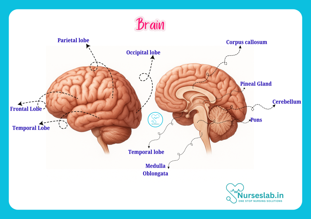
The human brain, weighing about 1.3 to 1.4 kilograms in adults, is divided into three main regions:
- Cerebrum: The largest part, responsible for higher cognitive functions, sensation, and voluntary movement.
- Cerebellum: Located beneath the cerebrum, it regulates motor control, balance, and coordination.
- Brainstem: Connects the brain to the spinal cord and controls vital functions such as breathing, heart rate, and consciousness.
External Features
Externally, the brain presents a complex surface marked by grooves (sulci) and ridges (gyri), which increase its surface area and allow for a greater number of neurons. The brain is divided into right and left hemispheres by the longitudinal fissure. These hemispheres are connected by the corpus callosum, a thick band of nerve fibres that enables communication between both sides.
Cerebrum
Lobes of the Cerebrum
The cerebrum is divided into four main lobes, each associated with specific functions:
- Frontal Lobe: Located at the front, involved in voluntary movement, reasoning, planning, problem-solving, and speech production (Broca’s area).
- Parietal Lobe: Situated behind the frontal lobe, responsible for processing sensory information such as touch, temperature, and pain.
- Temporal Lobe: Located on the sides of the brain, concerned with hearing, memory, and language comprehension (Wernicke’s area).
- Occipital Lobe: At the back of the brain, dedicated to visual processing.
Cortical Areas and Their Functions
The surface of the cerebrum, known as the cerebral cortex, is highly specialised. Key cortical areas include:
- Primary Motor Cortex (precentral gyrus): Controls voluntary muscle movements.
- Primary Somatosensory Cortex (postcentral gyrus): Receives and processes sensory information from the body.
- Primary Visual Cortex: Located in the occipital lobe, interprets visual signals.
- Primary Auditory Cortex: Found in the temporal lobe, processes sound information.
- Association Areas: Integrate information from various sensory and motor areas, involved in learning, memory, and complex thought processes.
Clinical Relevance
Damage to specific lobes or cortical areas can lead to distinct neurological deficits. For example, injury to the frontal lobe may cause personality changes or impaired movement, while occipital lobe damage can result in visual disturbances. Understanding these relationships helps nurses identify and report symptoms, facilitating prompt diagnosis and intervention.
Cerebellum
Structure
The cerebellum, sometimes called the “little brain”, is located beneath the occipital lobes of the cerebrum and behind the brainstem. It consists of two hemispheres and a central structure called the vermis. The cerebellar cortex is highly folded, increasing its surface area.
Function
The cerebellum is primarily responsible for:
- Coordinating voluntary movements
- Maintaining posture and balance
- Regulating muscle tone
- Fine-tuning motor activity to ensure smooth, accurate movements
Clinical Implications
Cerebellar dysfunction can manifest as ataxia (uncoordinated movements), tremors, difficulty with balance, and speech disturbances. Nurses should be vigilant for these signs, particularly in patients with head injuries, stroke, or neurodegenerative diseases affecting the cerebellum.
Brainstem
Structure and Components
The brainstem, located at the base of the brain, connects the cerebrum and cerebellum to the spinal cord. It comprises three main parts:
- Midbrain: Involved in auditory and visual reflexes, and eye movement.
- Pons: Contains nuclei that regulate breathing and communication between different parts of the brain.
- Medulla Oblongata: Controls vital autonomic functions such as heart rate, blood pressure, and respiration.
Vital Functions
The brainstem houses the origins of most cranial nerves, which are essential for facial sensation, swallowing, eye movement, and other critical activities. The reticular activating system (RAS), found in the brainstem, plays a key role in maintaining consciousness and wakefulness.
Clinical Relevance
Lesions in the brainstem can be life-threatening, as they may compromise breathing, cardiac function, or consciousness. Nurses must be adept at monitoring vital signs and recognising early warning signs such as changes in respiratory pattern, altered consciousness, or cranial nerve deficits.
Meninges and Ventricular System
Protective Layers: The Meninges
The brain is enveloped by three protective membranes collectively known as the meninges:
- Dura Mater: The tough, outermost layer that lines the skull and forms supportive partitions within the brain.
- Arachnoid Mater: The delicate, web-like middle layer that provides a cushioning effect.
- Pia Mater: The thin, innermost layer that closely adheres to the brain’s surface, following its contours.
Cerebrospinal Fluid (CSF) and Ventricular System
Within the brain are interconnected cavities called ventricles, which produce and contain cerebrospinal fluid (CSF). CSF circulates through the ventricles and around the brain and spinal cord, serving several key functions:
- Protects the brain from mechanical injury
- Maintains a stable chemical environment
- Removes waste products
The four ventricles include the two lateral ventricles, the third ventricle, and the fourth ventricle, all interconnected to allow CSF flow.
Clinical Significance
Disruption of CSF flow can lead to conditions such as hydrocephalus (accumulation of CSF), which may present with increased intracranial pressure, headache, nausea, or changes in consciousness. Infections of the meninges (meningitis) or bleeding into these spaces (subarachnoid haemorrhage) are medical emergencies requiring immediate nursing intervention.
Blood Supply of the Brain
Major Arteries and the Circle of Willis
The brain is highly dependent on a constant supply of oxygen and glucose. Its blood supply is provided mainly by two pairs of arteries:
- Internal Carotid Arteries: Supply the anterior and middle portions of the brain.
- Vertebral Arteries: Join to form the basilar artery, supplying the posterior parts of the brain.
These arteries form a circular anastomosis known as the Circle of Willis at the base of the brain. This anatomical arrangement allows for collateral blood flow, which can help preserve brain function if one vessel becomes blocked.
Clinical Significance
Blockage or rupture of cerebral arteries can lead to strokes (cerebrovascular accidents), often resulting in loss of function in affected brain regions. Nurses play a key role in recognising early symptoms—such as sudden weakness, speech difficulty, or visual changes—and initiating rapid response protocols to improve patient outcomes.
Functional Areas of the Brain
Motor, Sensory, and Association Areas
The brain’s cortex is functionally divided into:
- Motor Areas: Control voluntary movements (e.g., precentral gyrus in the frontal lobe).
- Sensory Areas: Receive and interpret sensory input (e.g., postcentral gyrus in the parietal lobe).
- Association Areas: Integrate sensory and motor information, involved in learning, memory, and decision-making.
Language Centres
Two specialised areas are critical for language:
- Broca’s Area: Located in the frontal lobe; responsible for speech production.
- Wernicke’s Area: Found in the temporal lobe; essential for language comprehension.
Damage to these areas can cause expressive or receptive aphasia, impacting a patient’s ability to communicate effectively.
Neuroglia and Neurons
Types of Brain Cells
The brain is composed of two main types of cells:
- Neurons: The primary signalling cells, responsible for transmitting electrical and chemical messages throughout the nervous system. Each neuron has a cell body, dendrites (receive signals), and an axon (sends signals).
- Neuroglia (Glial Cells): Supportive cells that maintain the environment around neurons, provide insulation, supply nutrients, and remove waste. Types include astrocytes, oligodendrocytes, microglia, and ependymal cells.
Roles in Brain Function
Neurons are essential for all brain activities, from basic reflexes to complex thinking. Neuroglia not only provide structural support but also play roles in immune defence, repair after injury, and regulation of the blood-brain barrier. Understanding these cell types helps nurses appreciate the complexity of neurological disorders and the importance of supportive care.
Clinical Correlations
Common Neurological Disorders
Nurses frequently encounter patients with various neurological conditions. Some common disorders include:
- Stroke: Sudden disruption of blood flow to the brain, leading to loss of function in affected areas.
- Traumatic Brain Injury (TBI): Damage from external force, potentially affecting any brain region.
- Epilepsy: Recurrent seizures due to abnormal electrical activity in the brain.
- Meningitis: Inflammation of the meninges, often due to infection.
- Parkinson’s Disease: Degenerative disorder affecting movement, related to loss of dopamine-producing neurons.
- Multiple Sclerosis (MS): Autoimmune attack on myelin sheaths in the central nervous system, causing varied neurological symptoms.
Implications for Nursing Care
Nurses must be skilled in neurological assessment, including evaluating consciousness, pupil response, limb strength, speech, and coordination. Early detection of changes can be critical for preventing complications. Effective communication with multidisciplinary teams and patient education are also vital components of care. In addition, understanding brain anatomy aids in explaining conditions and treatments to patients and their families, fostering trust and compliance.
REFERENCES
- Ross and Wilson, Anatomy and Physiology in Health and Illness, Fourteenth Edition, 1 July 2022, ISBN-13: 978-0323834612.
- Roger Watson, Anatomy and Physiology for Nurses, 14th Edition, 12-06-2018, ISBN: 9780702077418
- P.R Asha Latha, Text Book of Applied Anatomy & Physiology for Nurses, 7th Edition,3 January 2024, ISBN-13: 978-9356968622.
- Bryan H. Derikson, Tortora’s Principles of Anatomy and Physiology, 16th Edition, August 2023, ISBN: 978- 1119400066.
- Anatomy.co.uk, Reproductive System, Last updated on April 24, 2025, https://anatomy.co.uk/reproductive-system
Stories are the threads that bind us; through them, we understand each other, grow, and heal.
JOHN NOORD
Connect with “Nurses Lab Editorial Team”
I hope you found this information helpful. Do you have any questions or comments? Kindly write in comments section. Subscribe the Blog with your email so you can stay updated on upcoming events and the latest articles.


