


Last Updated on June 28, 2026 by Nurseslab.in Editorial Team
Expanding a memorable respiratory and trauma mnemonic into safe, systematic bedside practice
Overview
Chest injury and sudden respiratory deterioration can progress rapidly. Nurses need a method that is organised enough to prevent omissions but flexible enough to fit a time-critical assessment. TWELVE FLAPS is a mnemonic used in pre-hospital and emergency education to support a structured examination of the neck and chest. It prompts the clinician to look for high-risk findings, then use inspection, palpation, auscultation, percussion, and examination of the back and sides.
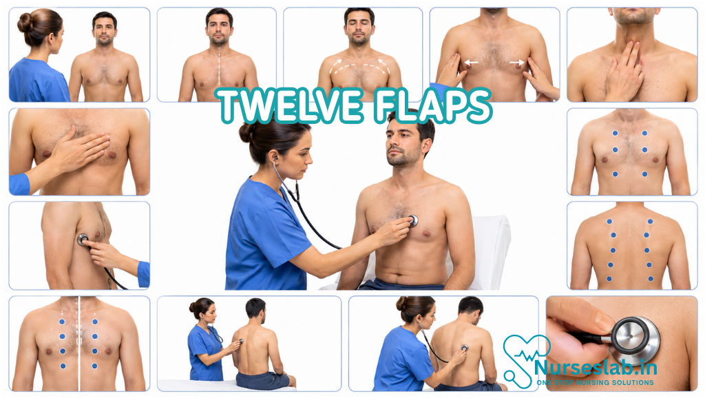
The commonly taught expansion is: Tracheal deviation, Wounds and bleeding, surgical Emphysema, Laryngeal injury or crepitus, neck Veins, Expose and examine, then Feel, Look, Auscultate, Percuss, and examine the Sides and back. Variations in wording and order exist, so nurses should use the version taught by their employer or education provider. The mnemonic structures assessment; it does not replace clinical judgement, local policy, or an ABCDE approach.
Where TWELVE FLAPS Fits
TWELVE FLAPS belongs within the Breathing stage of a primary survey. Deal immediately with catastrophic haemorrhage and airway compromise before beginning a detailed chest examination. If breathing is severely compromised, call for senior or emergency help, provide oxygen when indicated and authorised, attach monitoring, prepare equipment, and support time-critical interventions while continuing assessment.
Do not complete the mnemonic mechanically while the patient deteriorates. A life-threatening finding interrupts the sequence. Treat or escalate it, reassess the response, and then continue. Compare both sides of the chest whenever possible, integrate observations and trends, and repeat the assessment after movement, analgesia, intervention, or any change in condition.
T — Tracheal Deviation
Observe and gently assess whether the trachea appears central. Marked deviation can be associated with significant intrathoracic pressure or volume change, but it is often a late, subtle, or unreliable sign. Neck anatomy, obesity, previous surgery, masses, posture, and chronic disease may make interpretation difficult. Never wait for tracheal deviation before escalating a patient with severe respiratory distress.
Look from the front with the head in a neutral position if safe. Avoid forceful palpation in suspected cervical injury or airway trauma. Document whether the trachea appears central or displaced, the direction of any displacement, associated symptoms, and whether the finding is new.
W — Wounds and Bleeding
Inspect the neck and chest for penetrating wounds, blunt-force marks, bruising, abrasions, patterned injuries, open fractures, burns, foreign objects, and external bleeding. A small wound may hide major internal injury, so correlate appearance with respiratory effort, chest movement, pain, vital signs, and mechanism.
Control significant external bleeding with appropriate direct pressure unless doing so would worsen an embedded object or open chest injury. Do not remove an impaled object; stabilise it and seek urgent help. Follow local guidance for open chest wounds and dressings because an inappropriate seal can create or worsen pressure within the chest.
E — Surgical Emphysema
Surgical emphysema, also called subcutaneous emphysema, occurs when air collects beneath the skin. On gentle palpation it may feel crackly, like bubble wrap or crisp snow. It can occur after thoracic trauma, pneumothorax, airway injury, chest procedures, or mechanical ventilation.
Assess the neck, upper chest, and surrounding area gently. Note the location and apparent extent so progression can be recognised. Rapidly expanding emphysema, voice change, breathing difficulty, swelling, or increasing oxygen requirement needs urgent escalation. Do not massage the area.
L — Laryngeal Injury or Crepitus
Look and listen for hoarseness, weak or altered voice, stridor, pain on speaking or swallowing, difficulty handling secretions, coughing blood, neck swelling, bruising, or an abnormal contour. These may indicate laryngeal or upper-airway injury. Crepitus can reflect fracture or air in the tissues, but repeated manipulation may worsen pain or injury.
Airway compromise may evolve after initially reassuring observations. Keep the patient under continuous review, minimise unnecessary neck movement, call for expert airway support early, and prepare emergency equipment according to role and policy. Do not ask the patient to repeatedly speak if doing so increases distress.
V — Neck Veins
Observe the jugular veins if positioning and circumstances allow. Distension may accompany elevated intrathoracic or right-sided cardiac pressure, while flat veins may occur with severe volume loss. These signs are influenced by posture, ventilation, body habitus, chronic heart disease, and lighting, so they must be interpreted with the whole assessment.
Never diagnose tension pneumothorax, tamponade, or shock from the neck veins alone. Combine the finding with respiratory distress, air entry, chest movement, circulation, skin signs, blood pressure, heart rate, mechanism, and response to treatment.
E — Expose and Examine
Expose enough of the chest to examine it properly, while preserving dignity, privacy, warmth, and cultural needs. Cut clothing when speed or movement restrictions make removal unsafe. Search beneath folds, straps, dressings, and medical devices when clinically appropriate. In trauma, remember that important injuries can be hidden in the axillae, lateral chest, and back.
Prevent hypothermia: expose in stages, use blankets and a warm environment, and re-cover promptly. Ask about pain before moving the patient. Maintain spinal precautions where indicated, and coordinate any roll with enough trained staff and a clear leader.
F — Feel
Palpate gently for tenderness, instability, deformity, crepitus, abnormal movement, temperature change, swelling, and subcutaneous air. Assess whether chest expansion appears equal by placing hands symmetrically when safe. Palpation should be purposeful and should stop if it causes significant pain or threatens an unstable injury.
Do not repeatedly compress a suspected rib or sternal fracture to “confirm” it. Do not deliberately provoke paradoxical movement. Findings such as focal tenderness, a palpable step, instability, or reduced expansion should prompt analgesia, monitoring, escalation, and further investigation according to the clinical setting.
L — Look
Observe respiratory rate, depth, rhythm, effort, symmetry, posture, colour, speech, and mental state. Look for accessory-muscle use, nasal flaring, recession, cyanosis, sweating, exhaustion, asymmetrical movement, paradoxical movement, or a segment that appears to move differently from the rest of the chest.
Count the respiratory rate accurately rather than estimating it. Note whether the patient can speak in full sentences and whether effort is improving or worsening. Pulse oximetry is valuable but can lag behind deterioration and may be affected by poor perfusion, movement, nail products, or equipment limitations. A satisfactory saturation does not overrule visible respiratory distress.
A — Auscultate
Listen systematically to comparable areas on both sides, ideally on bare skin. Assess the presence, equality, and character of breath sounds. Reduced or absent air entry may occur with pneumothorax, haemothorax, pleural effusion, collapse, obstruction, or poor respiratory effort. Wheeze, crackles, stridor, transmitted upper-airway sounds, or added noises provide further clues.
Auscultation can be difficult in noisy settings and should not delay action when severe compromise is obvious. Interpret sounds alongside chest movement, respiratory effort, percussion, observations, and mechanism. If uncertain, state what you heard and where rather than assigning a diagnosis.
P — Percuss
Percussion compares the resonance of the two sides of the chest. Increased resonance can be associated with excess air, while dullness can occur with fluid, blood, consolidation, or other dense tissue. Technique and interpretation require practice, and findings may be difficult to appreciate in obesity, noisy environments, or when positioning is limited.
Percuss gently over intercostal spaces and compare like with like. Avoid painful, unstable, wounded, or device-covered areas. Nurses should only perform percussion if trained, competent, and permitted by local practice. It is one component of assessment, not a stand-alone decision rule.
S — Sides and Back
Examine the lateral and posterior chest because serious injuries and abnormal breath sounds may be missed from the front. Inspect for bruising, penetrating injury, bleeding, burns, swelling, deformity, and pressure damage. Palpate and auscultate comparable areas where safe, and include the axillae.
If the patient requires a log roll, plan it carefully, maintain airway and spinal alignment, secure tubes and drains, assign roles, and monitor throughout. Avoid turning a haemodynamically unstable or severely breathless patient simply to complete the mnemonic unless the expected benefit outweighs the risk.
Red Flags Requiring Immediate Escalation
- Severe or rapidly worsening breathlessness, exhaustion, reduced consciousness, or inability to speak.
- Absent or markedly reduced breath sounds on one side with clinical deterioration.
- Open or penetrating chest wound, major haemorrhage, or an impaled object.
- Stridor, voice change, neck swelling, or suspected laryngeal injury.
- Rapidly spreading surgical emphysema.
- Asymmetrical or paradoxical chest movement.
- Cyanosis, falling oxygen saturation, shock features, or worsening vital signs.
- Any sudden deterioration after chest trauma, a procedure, or positive-pressure ventilation.
These findings may indicate immediately life-threatening chest or airway pathology. Activate the appropriate emergency response, continue ABCDE assessment, provide supportive care within scope, and prepare for definitive intervention. Procedures such as needle or finger decompression, advanced airway management, and insertion of a chest drain require specific authority, training, and governance.
Documentation and Handover
Record time, mechanism or clinical context, respiratory rate and effort, oxygen therapy, saturation, chest symmetry, wound location, air entry, added sounds, palpation and percussion findings, pain, interventions, response, and escalation. Describe laterality and anatomical location precisely. “Reduced air entry at the right base” is more useful than “abnormal chest.”
Use a recognised handover framework such as SBAR or ATMIST according to local practice. State trends and changes: what was found initially, what treatment was given, and what happened afterwards. Include uncertainty honestly and distinguish observed findings from interpretation.
Common Pitfalls
- Using the mnemonic before ABCDE priorities: airway obstruction, apnoea, catastrophic bleeding, and shock cannot wait.
- Treating one sign as diagnostic: no single inspection, neck-vein, percussion, or auscultation finding proves a diagnosis.
- Examining only the front: lateral and posterior injuries are easily missed.
- Using unnecessary force: aggressive palpation can increase pain and risk.
- Ignoring trends: a change from baseline may be more important than one isolated value.
- Over-relying on monitors: observe the patient, not only the screen.
- Exposing without re-warming: hypothermia worsens trauma outcomes and discomfort.
- Working beyond competence: escalate early and follow scope, training, and policy.
A Practical Bedside Sequence
- Confirm safety, call for help, and begin the primary survey.
- Assess airway and immediately treat or escalate compromise.
- Observe breathing, obtain vital signs, and provide indicated support.
- Survey the trachea, wounds, surgical emphysema, larynx, and neck veins.
- Expose in stages while maintaining warmth and dignity.
- Feel and look systematically, comparing both sides.
- Auscultate, then percuss if trained and clinically appropriate.
- Examine the sides and back when safe.
- Treat urgent findings, reassess after every intervention, and document trends.
- Give a concise structured handover and continue monitoring.
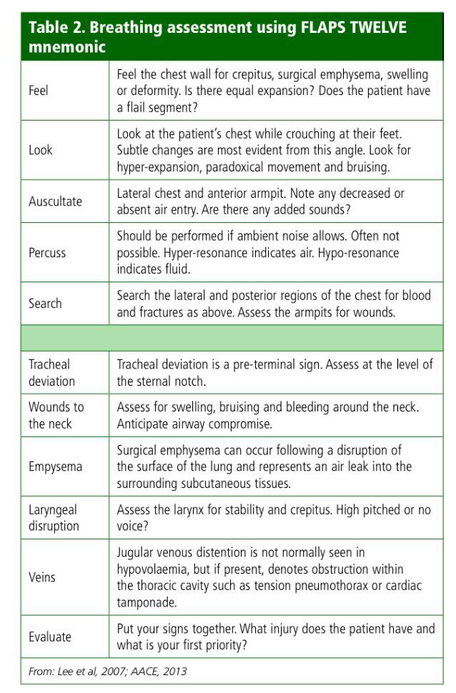
What Each Letter of The TWELVE FLAPS Stands For?
Clinical Example
A patient arrives after a high-speed collision, speaking in short phrases with right-sided chest pain. The nurse begins ABCDE, calls the trauma team, applies monitoring, and supports oxygen delivery according to protocol. The trachea appears central. There is bruising but no obvious open wound; gentle palpation finds tenderness and a small area of surgical emphysema. The right chest moves less, and air entry is markedly reduced on that side. The nurse stops the extended assessment, escalates the findings immediately, prepares indicated equipment, and repeats observations while the senior clinician assesses for time-critical thoracic injury.
This example shows the purpose of TWELVE FLAPS: not to collect every finding before acting, but to reveal dangerous patterns reliably and communicate them clearly.
Final Takeaway
TWELVE FLAPS gives nurses a memorable structure for a focused chest assessment: check the trachea, wounds, subcutaneous air, larynx, and neck veins; expose safely; then feel, look, listen, percuss, and include the sides and back. Its strength is completeness, comparison, and repeatability.
Its safest use is dynamic. Keep ABCDE priorities first, interrupt the sequence for life-threatening findings, work within competence, protect dignity and warmth, reassess after intervention, and communicate trends. Practised alongside simulation, supervised clinical teaching, and local pathways, TWELVE FLAPS can help nurses turn a complex respiratory examination into prompt, organised, patient-centred action.
REFERENCES
- Blair Laird, Paramedic Mnemonics and Memory Aids, Respiratory/chest assessment: TWELVE FLAPS, ·January 26, 2024, https://geekymedics.com/paramedic-mnemonics-and-memory-aids/
- First Aid Acronym, https://explorefirstaid.com/first-aid-acronym/
Stories are the threads that bind us; through them, we understand each other, grow, and heal.
JOHN NOORD
Connect with “Nurses Lab Editorial Team”
I hope you found this information helpful. Do you have any questions or comments? Kindly write in comments section. Subscribe the Blog with your email so you can stay updated on upcoming events and the latest articles.


