


Last Updated on May 27, 2026 by Nurseslab.in Editorial Team
The liver is one of the most vital organs in the human body, playing a central role in metabolism, detoxification, synthesis of important proteins, and storage of nutrients.
Location and Gross Structure of the Liver
Position in the Abdomen
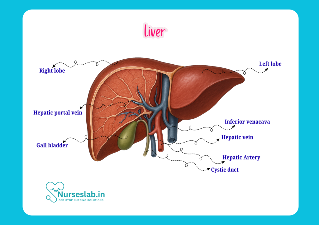
The liver is the largest solid organ in the human body, weighing approximately 1.2 to 1.5 kilograms in adults. It is located in the upper right quadrant of the abdomen, just beneath the diaphragm. The liver extends across the midline towards the left upper quadrant and is partially protected by the lower ribs on both sides. Its upper surface is in contact with the diaphragm, while the lower surface is related to the stomach, duodenum, right kidney, and colon.
Size, Shape, and Surface Anatomy
The liver is wedge-shaped, with a convex superior (diaphragmatic) surface and a relatively flat inferior (visceral) surface. Its anterior border is sharp, while the posterior border is more rounded. The average length of the liver is about 15 to 17 centimetres in adults, though this can vary. The superior surface fits snugly against the diaphragm, while the inferior surface presents several impressions made by neighbouring organs, such as the gallbladder fossa, renal impression, and gastric impression.
Lobes and Segments of the Liver
Right and Left Lobes
Anatomically, the liver is divided into two main lobes: the right lobe and the left lobe. The right lobe is much larger and occupies most of the right hypochondrium, while the left lobe extends into the epigastrium and left hypochondrium. The division between these lobes on the diaphragmatic surface is marked by the falciform ligament.
Caudate and Quadrate Lobes
On the visceral (inferior) surface, two additional smaller lobes are identified: the caudate lobe (situated posteriorly, near the inferior vena cava) and the quadrate lobe (anteriorly, near the gallbladder). Although these are not true anatomical lobes, they are important landmarks for surgeons and clinicians.
Segmental Anatomy (Couinaud Classification)
For clinical and surgical purposes, the liver is further divided into eight segments according to the Couinaud classification. Each segment is supplied by its own branch of the portal vein, hepatic artery, and bile duct. These segments are numbered I to VIII, starting with the caudate lobe as segment I. Understanding segmental anatomy is essential for interpreting imaging studies and planning surgical procedures, such as liver resections or transplantation.
Peritoneal Relations and Ligaments of the Liver
Falciform Ligament
The falciform ligament is a sickle-shaped fold of peritoneum that attaches the liver to the anterior abdominal wall and the diaphragm. It also demarcates the anatomical division between the right and left lobes on the anterior surface.
Coronary Ligament
The coronary ligament consists of peritoneal reflections that attach the posterior aspect of the liver to the diaphragm. It forms the boundaries of the so-called “bare area” of the liver, which is not covered by peritoneum.
Lesser Omentum
The lesser omentum is a double layer of peritoneum that extends from the liver to the lesser curvature of the stomach and the first part of the duodenum. It contains important structures, including the portal triad (hepatic artery, portal vein, and bile duct).
Bare Area of the Liver
The bare area is a region on the posterior surface of the liver that is devoid of peritoneal covering. This area is in direct contact with the diaphragm and is significant because it provides a potential route for the spread of infection or malignancy between the abdomen and the thoracic cavity.
Blood Supply and Drainage of the Liver
Dual Blood Supply
The liver receives a dual blood supply: approximately 75% of its blood comes from the portal vein, which carries nutrient-rich blood from the gastrointestinal tract, while the remaining 25% is supplied by the hepatic artery, which delivers oxygenated blood from the systemic circulation. This unique arrangement allows the liver to process nutrients and toxins absorbed from the gut.
Portal Vein
The portal vein is formed by the union of the superior mesenteric vein and the splenic vein. It enters the liver at the porta hepatis and branches into right and left portal veins, which further subdivide to supply each segment of the liver.
Hepatic Artery
The hepatic artery arises from the common hepatic artery, a branch of the coeliac trunk. It enters the liver alongside the portal vein and bile duct within the lesser omentum, providing oxygenated blood to the liver parenchyma.
Hepatic Veins
Blood from the liver is drained by the hepatic veins—typically right, middle, and left—which empty directly into the inferior vena cava. These veins are valveless and collect deoxygenated blood from the central veins of the liver lobules.
Lymphatic Drainage
Lymph from the liver drains through both superficial and deep lymphatic vessels, eventually reaching the hepatic and coeliac lymph nodes. The liver is a major source of lymph, contributing nearly one-third of the body’s total lymph flow.
Nerve Supply
The liver’s nerve supply is derived from the hepatic plexus, which contains sympathetic and parasympathetic fibres. Sympathetic nerves originate from the coeliac plexus, while parasympathetic fibres are supplied by the vagus nerve. These nerves help regulate blood flow and bile secretion.
Microscopic Anatomy (Histology) of the Liver
Hepatocytes
The main functional cells of the liver are hepatocytes, which make up about 80% of the liver’s mass. Hepatocytes are polygonal cells arranged in plates or cords, radiating from a central vein. They have abundant cytoplasm and numerous organelles to support their metabolic activities.
Liver Lobules
The structural and functional unit of the liver is the hepatic lobule. Each lobule is roughly hexagonal in shape, with a central vein at its core. At the corners of the lobule are portal triads, each containing a branch of the portal vein, hepatic artery, and bile duct.
Sinusoids
Between the plates of hepatocytes are sinusoids—specialised capillaries that allow the mixing of oxygenated and nutrient-rich blood. Sinusoids are lined with endothelial cells and Kupffer cells (liver macrophages) that help filter blood and remove debris or pathogens.
Bile Canaliculi
Hepatocytes secrete bile into tiny channels called bile canaliculi, which run between adjacent hepatocytes. These canaliculi merge to form bile ducts, ultimately draining bile into the gallbladder and duodenum.
Functions of the Liver
Metabolic Functions
The liver is a metabolic powerhouse, responsible for the metabolism of carbohydrates, proteins, and fats. It regulates blood glucose levels by storing glycogen and releasing glucose as needed (glycogenesis and glycogenolysis). It also plays a key role in the synthesis and breakdown of amino acids and lipids.
Synthetic Functions
The liver synthesises various important proteins, including albumin (which maintains blood oncotic pressure), clotting factors (such as fibrinogen, prothrombin), and transport proteins. It also produces cholesterol and certain hormones.
Storage Functions
The liver acts as a storage site for several substances, including glycogen, vitamins (A, D, E, K, and B12), iron, and copper. These reserves are mobilised as needed to maintain homeostasis.
Detoxification
A major function of the liver is detoxification—removal of toxins, drugs, and metabolic waste products from the blood. The liver converts ammonia (a toxic by-product of protein metabolism) into urea, which is then excreted by the kidneys.
Immunological Roles
The liver contains specialised immune cells, such as Kupffer cells, which help defend the body against pathogens and remove old blood cells or debris. The liver also produces acute-phase proteins that are involved in the body’s response to infection or inflammation.
Clinical Relevance for Nurses
Palpation of the Liver
During physical examination, the liver can sometimes be palpated below the right costal margin, especially in children or individuals with hepatomegaly (enlarged liver). Nurses should be familiar with the technique of liver palpation and able to recognise abnormal findings, such as tenderness, nodularity, or enlargement.
Liver Function Tests (LFTs)
Liver function tests are a group of blood tests that assess different aspects of liver health, including levels of enzymes (ALT, AST, ALP), bilirubin, albumin, and clotting factors. Abnormal LFTs can indicate liver injury, inflammation, or dysfunction, and nurses play a key role in monitoring and interpreting these results.
Imaging Studies
Ultrasound, computed tomography (CT), and magnetic resonance imaging (MRI) are commonly used to evaluate liver size, structure, and pathology. Nurses should be familiar with these modalities and support patients through preparation and post-procedure care.
Liver Biopsy
A liver biopsy involves obtaining a small tissue sample for microscopic examination. It is used to diagnose liver diseases, assess the severity of damage, or monitor treatment response. Nurses are essential in patient preparation, monitoring for complications, and providing post-biopsy care.
Common Liver Diseases and Pathologies
Hepatitis
Hepatitis refers to inflammation of the liver, most often caused by viral infections (hepatitis A, B, C, D, E), but also by toxins, drugs, or autoimmune conditions. Symptoms include jaundice, fatigue, abdominal pain, and elevated liver enzymes. Chronic hepatitis can lead to cirrhosis or liver cancer.
Cirrhosis
Cirrhosis is the end-stage of chronic liver disease, characterised by fibrosis (scarring) and the formation of regenerative nodules. The liver becomes firm and shrunken, leading to complications such as portal hypertension, ascites, varices, and hepatic encephalopathy. Nurses must monitor for signs of decompensation and manage complications.
Fatty Liver Disease
Fatty liver disease is the accumulation of fat within liver cells. It can be classified as non-alcoholic fatty liver disease (NAFLD) or alcoholic fatty liver disease. Most cases are asymptomatic, but the condition can progress to inflammation (steatohepatitis), fibrosis, or cirrhosis.
Liver Tumours
Primary liver tumours include hepatocellular carcinoma (HCC) and cholangiocarcinoma, while secondary tumours are metastases from other organs. Liver tumours may present as a mass, weight loss, jaundice, or abnormal LFTs. Early detection and prompt referral are crucial for patient outcomes.
Summary and Key Points for Nurses
- The liver is the largest solid organ, situated in the right upper quadrant of the abdomen, beneath the diaphragm.
- It has two main lobes (right and left), with additional caudate and quadrate lobes visible on the visceral surface.
- The liver is divided into eight functional segments, each with its own blood supply and bile drainage.
- It receives dual blood supply from the portal vein and hepatic artery, and drains blood via the hepatic veins into the inferior vena cava.
- Microscopically, the liver is composed of hepatocytes arranged in lobules with central veins, portal triads, and sinusoids.
- Key functions include metabolism, synthesis of proteins, storage of nutrients, detoxification, and immunological defence.
- Common liver diseases include hepatitis, cirrhosis, fatty liver, and tumours—each with important implications for nursing care.
- Nurses play a vital role in assessment, monitoring, patient education, and support for individuals with liver disorders.
REFERENCES
- Ross and Wilson, Anatomy and Physiology in Health and Illness, Fourteenth Edition, 1 July 2022, ISBN-13: 978-0323834612.
- Roger Watson, Anatomy and Physiology for Nurses, 14th Edition, 12-06-2018, ISBN: 9780702077418
- P.R Asha Latha, Text Book of Applied Anatomy & Physiology for Nurses, 7th Edition,3 January 2024, ISBN-13: 978-9356968622.
- Bryan H. Derikson, Tortora’s Principles of Anatomy and Physiology, 16th Edition, August 2023, ISBN: 978- 1119400066.
- Anatomy.co.uk, Reproductive System, Last updated on April 24, 2025, https://anatomy.co.uk/reproductive-system
Stories are the threads that bind us; through them, we understand each other, grow, and heal.
JOHN NOORD
Connect with “Nurses Lab Editorial Team”
I hope you found this information helpful. Do you have any questions or comments? Kindly write in comments section. Subscribe the Blog with your email so you can stay updated on upcoming events and the latest articles.


