


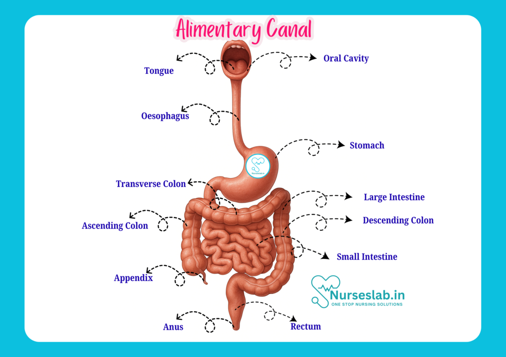
The alimentary canal, also known as the gastrointestinal (GI) tract, is a continuous muscular tube that extends from the mouth to the anus, playing a vital role in digestion, nutrient absorption, and waste elimination.
Introduction
The study of human anatomy is a cornerstone of nursing education, equipping nurses with the foundational knowledge required for effective patient care. Among the various systems in the human body, the alimentary canal—also known as the digestive tract—plays a critical role in nutrition, metabolism, and overall health. For nurses, a thorough understanding of the anatomy and physiology of the alimentary canal is essential for patient assessment, identification of disorders, and implementation of appropriate care strategies.
Overview of the Alimentary Canal
The alimentary canal is a continuous, muscular tube that extends from the mouth to the anus, measuring approximately 8 to 10 metres in length in adults. Its primary function is to ingest, digest, absorb, and expel food and waste materials. The canal comprises several specialised organs and structures, each adapted to perform specific tasks in the process of digestion and absorption. The major components include the mouth, pharynx, oesophagus, stomach, small intestine, and large intestine. In addition, accessory organs such as the liver, pancreas, and gallbladder contribute essential secretions to aid digestion.
General Structure of the Alimentary Canal
Definition
The alimentary canal, also referred to as the gastrointestinal (GI) tract, is defined as the hollow passage through which food travels within the body for digestion and absorption. It begins at the oral cavity and terminates at the anus.
Layers of the Alimentary Canal
The wall of the alimentary canal exhibits a consistent structural organisation throughout its length, comprising four primary layers:
- Mucosa: The innermost layer, consisting of an epithelial lining, lamina propria (connective tissue), and a thin muscularis mucosae. It is involved in secretion, absorption, and protection.
- Submucosa: A layer of connective tissue containing blood vessels, lymphatics, nerves, and glands. It provides support and nourishment to the mucosa.
- Muscularis externa: Typically composed of two layers of smooth muscle—inner circular and outer longitudinal. Responsible for peristalsis and segmentation movements that propel and mix the contents.
- Serosa (or Adventitia): The outermost layer. In areas within the abdominal cavity, it is a serous membrane (serosa), while outside the cavity (e.g., oesophagus), it is a connective tissue layer (adventitia).
Histology Basics
Understanding the histological structure of the alimentary canal is vital for recognising pathological changes that occur in various GI diseases. The mucosa varies in appearance and function depending on the organ—ranging from stratified squamous epithelium in the oesophagus for protection, to simple columnar epithelium in the stomach and intestines for secretion and absorption.
Mouth and Oral Cavity
Anatomy
The mouth, or oral cavity, forms the entry point of the alimentary canal. It is bounded by the lips, cheeks, palate (hard and soft), and tongue. Major structures include:
- Lips and cheeks: Contain muscles for facial expression and assist in food manipulation.
- Teeth: 32 permanent teeth in adults, adapted for cutting, tearing, and grinding food.
- Tongue: A muscular organ involved in taste, speech, and food manipulation.
- Salivary glands: Parotid, submandibular, and sublingual glands secrete saliva, which contains enzymes like amylase for carbohydrate digestion.
Functions
The oral cavity initiates mechanical digestion through mastication (chewing) and chemical digestion via salivary enzymes. It also plays a role in speech, taste, and immune defence (through tonsils and secretory immunoglobulins in saliva).
Clinical Notes
Common oral cavity issues include dental caries, gingivitis, oral thrush, and trauma. Nurses must assess oral hygiene, especially in dependent or unconscious patients, as poor oral health can lead to systemic infections.
Pharynx and Oesophagus
Structure
The pharynx is a muscular tube connecting the oral cavity to the oesophagus and larynx. It is divided into three parts:
- Nasopharynx: Behind the nasal cavity, primarily respiratory in function.
- Oropharynx: Behind the oral cavity, common to both digestive and respiratory tracts.
- Laryngopharynx: Leads to the oesophagus and larynx.
The oesophagus is a 25-cm long muscular tube extending from the pharynx to the stomach, passing through the diaphragm at the oesophageal hiatus. It has upper and lower sphincters to regulate the passage of food and prevent reflux.
Swallowing Mechanism
Swallowing, or deglutition, is a coordinated process involving voluntary and involuntary phases:
- Oral phase: Voluntary movement of the bolus to the oropharynx.
- Pharyngeal phase: Involuntary passage through the pharynx, closure of the larynx to prevent aspiration.
- Oesophageal phase: Involuntary peristaltic waves move the bolus towards the stomach.
Nursing Relevance
Difficulty swallowing (dysphagia) can result from neurological disorders, strictures, or tumours. Nurses must monitor for aspiration risk, especially in stroke or elderly patients, and ensure safe feeding practices.
Stomach
Regions
The stomach is a J-shaped organ located in the upper left abdomen. It consists of four main regions:
- Cardia: Area where the oesophagus enters the stomach.
- Fundus: Dome-shaped upper portion.
- Body (corpus): Central and largest region.
- Pylorus: Lower section leading to the duodenum, containing the pyloric sphincter.
Layers
The stomach wall has the standard four layers, with the muscularis externa featuring an additional inner oblique muscle layer to assist in churning food.
Digestive Functions
The stomach stores food, mixes it with gastric juices (containing hydrochloric acid and digestive enzymes), and initiates protein digestion. It produces chyme, a semi-liquid mixture, which passes into the duodenum in controlled amounts.
Clinical Considerations
Common stomach disorders include gastritis, peptic ulcers, and gastric cancer. Nurses play a vital role in monitoring for symptoms like pain, vomiting, and bleeding, and in managing enteral feeding when required.
Small Intestine
Duodenum, Jejunum, and Ileum
The small intestine is the longest part of the alimentary canal, measuring about 6 metres. It is divided into three regions:
- Duodenum: First 25 cm, C-shaped, receives chyme from the stomach and secretions from the liver and pancreas.
- Jejunum: Middle section, approximately 2.5 metres, specialised for nutrient absorption.
- Ileum: Final 3.5 metres, continues absorption and ends at the ileocaecal valve, which controls entry into the large intestine.
Absorption
The mucosa of the small intestine is highly specialised for absorption, featuring villi and microvilli that increase the surface area. Most nutrients, including carbohydrates, proteins, fats, vitamins, and minerals, are absorbed here.
Nursing Implications
Disorders of the small intestine, such as coeliac disease, Crohn’s disease, and malabsorption syndromes, can lead to significant nutritional deficiencies. Nurses must assess for signs of malnutrition, monitor weight, and educate patients on dietary modifications.
Large Intestine
Sections
The large intestine is about 1.5 metres long and consists of the following parts:
- Caecum: Pouch-like initial segment, with the appendix attached.
- Colon: Divided into ascending, transverse, descending, and sigmoid segments.
- Rectum: Terminal section leading to the anal canal.
- Anal canal: Final passage, with internal and external sphincters controlling defaecation.
Water Absorption and Waste Formation
The large intestine absorbs water and electrolytes from indigestible food residues, converting them into semi-solid faeces. It also houses beneficial bacteria that synthesise certain vitamins and aid in fermentation.
Clinical Notes
Nurses frequently encounter conditions like constipation, diarrhoea, irritable bowel syndrome, and colorectal cancer. Regular bowel assessment, patient education on fibre intake, and early identification of abnormal findings are essential aspects of nursing care.
Accessory Organs of Digestion
Liver
The liver, the largest gland in the body, is located in the right upper quadrant of the abdomen. It performs multiple functions, including:
- Production and secretion of bile for fat digestion
- Metabolism of carbohydrates, proteins, and lipids
- Detoxification of drugs and toxins
- Storage of glycogen, vitamins, and minerals
Bile produced in the liver is stored and concentrated in the gallbladder before being released into the duodenum.
Pancreas
The pancreas is both an exocrine and endocrine gland. The exocrine portion produces digestive enzymes (amylase, lipase, proteases) and bicarbonate, delivered to the duodenum via the pancreatic duct. The endocrine portion secretes hormones like insulin and glucagon, which regulate blood glucose.
Gallbladder
The gallbladder is a small, pear-shaped organ beneath the liver. It stores and concentrates bile, releasing it into the duodenum in response to fatty meals.
Roles in Digestion
These accessory organs are crucial for effective digestion and absorption. Disorders such as hepatitis, pancreatitis, and gallstones are common and require prompt nursing recognition and intervention.
Nervous and Vascular Supply
Blood Supply
The alimentary canal receives a rich blood supply from branches of the aorta—primarily the coeliac trunk, superior mesenteric artery, and inferior mesenteric artery. Venous blood drains into the portal vein, which carries nutrients to the liver for processing.
Innervation
The enteric nervous system, often called the “second brain,” regulates most digestive functions independently. Autonomic nerves (sympathetic and parasympathetic) modulate motility, secretion, and blood flow. This neural control is vital for coordinated digestion and adaptation to stress.
Relevance to Nursing
Knowledge of vascular and nervous supply aids nurses in recognising signs of ischaemia, bleeding, or neuropathic complications, and in monitoring patients after surgical interventions.
Common Disorders of the Alimentary Canal
Peptic Ulcers
Peptic ulcers are erosions of the stomach or duodenal lining, often due to Helicobacter pylori infection or non-steroidal anti-inflammatory drug (NSAID) use. Symptoms include epigastric pain, nausea, and sometimes bleeding. Nursing management includes pain assessment, monitoring for complications (like perforation), and patient education on medication adherence and lifestyle modifications.
Gastro-Oesophageal Reflux Disease (GERD)
GERD is a chronic condition where stomach acid flows back into the oesophagus, causing heartburn, regurgitation, and sometimes respiratory symptoms. Nurses should advise patients on dietary modifications, positioning, and medication adherence, and monitor for complications like oesophagitis.
Malabsorption Syndromes
Conditions such as coeliac disease and lactose intolerance impair the absorption of nutrients, leading to symptoms like diarrhoea, weight loss, and anaemia. Nurses play a critical role in nutritional assessment, patient education, and monitoring for complications.
Clinical Relevance for Nurses
Assessment
Comprehensive assessment of the digestive system includes history-taking (symptoms, dietary patterns), physical examination (inspection, palpation, auscultation), and monitoring of laboratory and diagnostic results. Early identification of GI symptoms can significantly impact patient outcomes.
Patient Education
Nurses are responsible for educating patients about healthy dietary practices, the importance of hydration, and recognition of warning signs (such as persistent pain, bleeding, or sudden weight loss). Education improves compliance and empowers patients in self-care.
Care Strategies
Nursing care strategies include maintaining adequate nutrition and hydration, preventing aspiration, managing enteral or parenteral feeding, monitoring for signs of infection, and providing emotional support to patients with chronic GI disorders.
REFERENCES
- Ross and Wilson, Anatomy and Physiology in Health and Illness, Fourteenth Edition, 1 July 2022, ISBN-13: 978-0323834612.
- Roger Watson, Anatomy and Physiology for Nurses, 14th Edition, 12-06-2018, ISBN: 9780702077418
- P.R Asha Latha, Text Book of Applied Anatomy & Physiology for Nurses, 7th Edition,3 January 2024, ISBN-13: 978-9356968622.
- Bryan H. Derikson, Tortora’s Principles of Anatomy and Physiology, 16th Edition, August 2023, ISBN: 978- 1119400066.
- Anatomy.co.uk, Reproductive System, Last updated on April 24, 2025, https://anatomy.co.uk/reproductive-system
Stories are the threads that bind us; through them, we understand each other, grow, and heal.
JOHN NOORD
Connect with “Nurses Lab Editorial Team”
I hope you found this information helpful. Do you have any questions or comments? Kindly write in comments section. Subscribe the Blog with your email so you can stay updated on upcoming events and the latest articles.


