


Last Updated on May 28, 2026 by Nurseslab.in Editorial Team
Introduction:
The central nervous system (CNS) is the control centre of the human body, responsible for regulating all voluntary and involuntary actions. For nurses, understanding the protective structures that envelop and safeguard the CNS is crucial in clinical practice. Among these protective layers, the meninges play a pivotal role.
Definition and Overview: What Are Meninges?
The term meninges refers to the three membranous layers that envelop the brain and spinal cord, forming a critical protective barrier for the CNS. These layers—dura mater, arachnoid mater, and pia mater—act as both physical and biochemical shields. They help maintain the homeostasis of the CNS, provide a supportive framework for blood vessels, and contain cerebrospinal fluid (CSF), which cushions neural tissues. The meninges are essential for protecting the delicate structures of the brain and spinal cord from injury, infection, and other pathological processes.
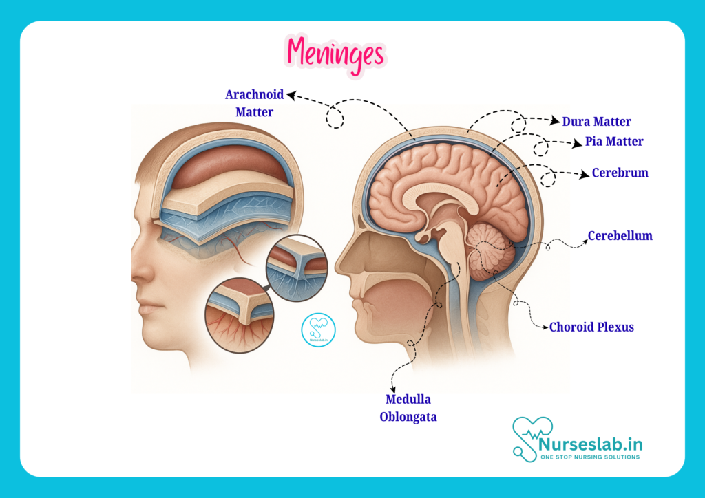
Layers of the Meninges
The meninges are composed of three distinct layers, each with unique structural and functional characteristics. These layers, listed from outermost to innermost, are:
- Dura Mater
- Arachnoid Mater
- Pia Mater
Dura Mater
The dura mater is the outermost and toughest layer of the meninges. Its name is derived from Latin, meaning “tough mother,” reflecting its dense and fibrous nature. The dura mater forms a protective sheath around the brain and spinal cord, providing a strong barrier against mechanical injury. In the cranial cavity, it consists of two layers: the periosteal layer (attached to the inner surface of the skull) and the meningeal layer (closer to the brain). These layers are fused in most areas but separate to form dural venous sinuses, which drain venous blood from the brain.
Arachnoid Mater
Situated beneath the dura mater is the arachnoid mater, a delicate, web-like membrane. The term “arachnoid” is derived from the Greek word for spider, referencing its intricate, trabecular structure. The arachnoid mater is avascular and lies loosely over the brain and spinal cord, separated from the pia mater by the subarachnoid space. This space is filled with CSF and traversed by fine strands of connective tissue known as arachnoid trabeculae.
Pia Mater
The innermost layer, the pia mater, is a thin, transparent membrane that closely invests the surface of the brain and spinal cord. It follows the contours of the CNS, dipping into sulci and fissures, and is richly supplied with blood vessels. The pia mater serves as the final barrier between the CNS tissue and the surrounding environment, playing a role in nourishing the neural tissue and supporting metabolic exchange.
Structure and Composition
Each meningeal layer exhibits distinct structural and compositional features, both at the macroscopic and microscopic levels. Understanding these features is essential for recognising their specific roles in CNS protection and function.
Dura Mater: Structure and Composition
The dura mater is composed of dense, irregular connective tissue rich in collagen fibres, which confer its strength and durability. In the cranial region, the dura mater’s two layers—the periosteal and meningeal—are tightly bound, except at certain points where they separate to create the dural venous sinuses. These sinuses play a crucial role in draining blood from the brain.
The spinal dura mater, in contrast, consists of only the meningeal layer and is separated from the vertebral column by the epidural space, which contains fat and blood vessels. This distinction is clinically important, particularly in procedures such as epidural anaesthesia.
Arachnoid Mater: Structure and Composition
The arachnoid mater is a thin, avascular membrane composed of flattened cells and delicate connective tissue. Its inner surface gives rise to numerous fine, web-like trabeculae that extend across the subarachnoid space to the pia mater. The arachnoid barrier cells form tight junctions, contributing to the regulation of substance exchange between the CSF and the blood.
The subarachnoid space is clinically significant, as it contains CSF, major blood vessels, and cranial nerves. Enlargements in this space, known as cisterns, serve as reservoirs for CSF and can be sites for pathological accumulation of blood or infection.
Pia Mater: Structure and Composition
The pia mater is composed of a single layer of flattened cells interspersed with fine collagen and elastic fibres. It is highly vascular, with capillaries that supply the underlying neural tissue. The pia mater is intimately associated with the surface of the brain and spinal cord, following all their contours and providing a conduit for blood vessels entering the CNS.
Microscopically, the pia mater is separated from the neural tissue by a thin layer of glial cells called the glia limitans, which forms part of the blood-brain barrier and helps regulate the movement of substances into the CNS.
Functions of the Meninges
The meninges serve several vital functions that are essential for the integrity and optimal functioning of the CNS. These include:
- Protection: The meninges act as a physical barrier, safeguarding the brain and spinal cord from mechanical injury, infection, and the effects of toxic substances.
- Support: The dura mater provides structural support, anchoring the brain within the skull and the spinal cord within the vertebral column. The subarachnoid trabeculae help maintain the position of the CNS within the meningeal sac.
- Cerebrospinal Fluid Containment: The arachnoid and pia mater enclose the subarachnoid space, which contains CSF. This fluid cushions the CNS, absorbs shock, and provides a medium for the exchange of nutrients and waste products.
- Blood Vessel Support: The meninges provide a framework for the passage of blood vessels that supply the CNS, ensuring continuous oxygen and nutrient delivery.
- Participation in the Blood-Brain Barrier: The pia mater and associated glial cells contribute to the selective permeability of the blood-brain barrier, protecting the CNS from harmful agents.
Blood Supply and Innervation
A thorough understanding of the blood supply and nerve innervation of the meninges is essential for nurses, particularly when caring for patients with neurological or vascular conditions.
Arterial Supply
The arterial supply to the meninges is primarily provided by branches of the external and internal carotid arteries. The most significant of these is the middle meningeal artery, a branch of the maxillary artery, which supplies the dura mater of the cranial vault. Other arteries include the anterior and posterior meningeal arteries, which supply the anterior and posterior cranial fossae, respectively.
The spinal meninges receive their blood supply from segmental spinal arteries, which branch into anterior and posterior radicular arteries, supplying the dura and pia mater along the length of the spinal cord.
Venous Drainage
Venous drainage from the cranial meninges occurs through the dural venous sinuses, which are endothelial-lined channels located between the layers of the cranial dura mater. These sinuses collect blood from the brain and meninges and drain into the internal jugular veins. The major sinuses include the superior sagittal sinus, transverse sinuses, straight sinus, and cavernous sinus.
The spinal meningeal veins drain into the internal vertebral venous plexus, which ultimately communicates with systemic venous circulation.
Nerve Innervation
The dura mater is richly innervated by sensory nerves, making it sensitive to pain. The major sources of innervation include the trigeminal nerve (cranial nerve V) for the supratentorial dura and the upper cervical nerves (C1–C3) for the infratentorial dura. The spinal dura is innervated by recurrent meningeal branches of spinal nerves. The arachnoid and pia mater have minimal sensory innervation.
Clinical Significance
The meninges and their associated spaces are of great clinical importance, especially in the context of neurological assessment, diagnosis, and intervention.
Meningeal Spaces
There are three potential spaces associated with the meninges:
- Epidural Space: Located between the dura mater and the skull or vertebral canal. In the spine, this space is real and contains fat and blood vessels; in the cranium, it is a potential space that may become apparent in cases of trauma or bleeding.
- Subdural Space: A potential space between the dura mater and arachnoid mater. Pathological processes, such as subdural haematomas, can create or expand this space.
- Subarachnoid Space: Located between the arachnoid mater and pia mater, this is a real space filled with CSF and traversed by blood vessels. It is the site of subarachnoid haemorrhage and is accessed during lumbar puncture.
Blood-Brain Barrier
The meninges, particularly the pia mater and associated glial cells, contribute to the formation and maintenance of the blood-brain barrier (BBB). The BBB is a selective barrier that regulates the passage of substances from the blood into the CNS, protecting neural tissue from toxins, pathogens, and fluctuations in blood composition. Disruption of the BBB can lead to serious neurological complications.
Lumbar Puncture Relevance
A lumbar puncture (spinal tap) is a common clinical procedure in which a needle is inserted into the subarachnoid space of the lumbar spine to obtain CSF for diagnostic or therapeutic purposes. Knowledge of meningeal anatomy is essential for safe and effective lumbar puncture, as the needle must pass through the dura mater and arachnoid mater without damaging the spinal cord or nerve roots.
Common Pathologies of the Meninges
Several pathological conditions can affect the meninges, each with significant clinical implications for nursing care.
Meningitis
Meningitis is an inflammation of the meninges, most commonly caused by bacterial, viral, or fungal infections. The classic symptoms include headache, fever, neck stiffness, photophobia, and altered mental status. Meningitis is a medical emergency, and prompt recognition, initiation of antibiotics, and supportive care are critical. Nurses play a vital role in monitoring neurological status, administering medications, and providing supportive care to prevent complications.
Subdural Haematoma
A subdural haematoma occurs when blood accumulates in the subdural space, usually as a result of head trauma. This can lead to increased intracranial pressure, neurological deficits, and, if untreated, death.
Meningioma
Meningiomas are tumours that arise from the meninges, most often the dura mater. They are typically benign but can cause symptoms by compressing adjacent brain tissue. Clinical manifestations depend on the location and size of the tumour. Nursing care includes preoperative and postoperative management, monitoring for neurological changes, and providing patient education regarding prognosis and treatment options.
Nursing Considerations
Nurses are at the forefront of patient assessment, monitoring, and care for individuals with meningeal disorders. Key nursing considerations include:
- Assessment: Conduct thorough neurological assessments, including evaluation of consciousness, motor and sensory function, cranial nerve function, and signs of meningeal irritation (e.g., neck stiffness, photophobia, Kernig’s and Brudzinski’s signs).
- Monitoring: Observe for signs of increased intracranial pressure (headache, vomiting, altered mental status), infection (fever, neck rigidity), and neurological deterioration.
- Medication Administration: Administer prescribed antibiotics, antivirals, corticosteroids, or other medications promptly and monitor for adverse effects.
- Infection Control: Implement infection prevention measures, especially in cases of suspected or confirmed meningitis.
- Patient and Family Education: Provide information regarding the nature of meningeal disorders, treatment options, and signs that require immediate medical attention.
- Supportive Care: Ensure patient comfort, manage pain, and assist with activities of daily living as needed.
- Collaboration: Work closely with physicians, neurologists, and other healthcare professionals to ensure comprehensive care.
Summary and Key Takeaways
The meninges are three vital layers—dura mater, arachnoid mater, and pia mater—that protect and support the brain and spinal cord. For nurses, a solid understanding of meningeal anatomy is essential for effective assessment, monitoring, and care of patients with neurological conditions. Key points include:
- Meninges provide protection, support, and containment of CSF for the CNS.
- Each layer—dura mater, arachnoid mater, pia mater—has unique structural and functional roles.
- Blood supply and innervation of the meninges are clinically significant, particularly in pain perception and vascular disorders.
- Common pathologies such as meningitis, subdural haematoma, and meningioma require prompt recognition and specialised nursing care.
- Nursing considerations focus on assessment, monitoring, medication administration, infection control, education, and supportive care.
By mastering the anatomy and clinical relevance of the meninges, nursing professionals can contribute significantly to the prevention, diagnosis, and management of CNS disorders, ultimately improving patient outcomes.
REFERENCES
- Ross and Wilson, Anatomy and Physiology in Health and Illness, Fourteenth Edition, 1 July 2022, ISBN-13: 978-0323834612.
- Roger Watson, Anatomy and Physiology for Nurses, 14th Edition, 12-06-2018, ISBN: 9780702077418
- P.R Asha Latha, Text Book of Applied Anatomy & Physiology for Nurses, 7th Edition,3 January 2024, ISBN-13: 978-9356968622.
- Bryan H. Derikson, Tortora’s Principles of Anatomy and Physiology, 16th Edition, August 2023, ISBN: 978- 1119400066.
- Anatomy.co.uk, Reproductive System, Last updated on April 24, 2025, https://anatomy.co.uk/reproductive-system
Stories are the threads that bind us; through them, we understand each other, grow, and heal.
JOHN NOORD
Connect with “Nurses Lab Editorial Team”
I hope you found this information helpful. Do you have any questions or comments? Kindly write in comments section. Subscribe the Blog with your email so you can stay updated on upcoming events and the latest articles.


