


Last Updated on May 28, 2026 by Nurseslab.in Editorial Team
The oral cavity, commonly referred to as the mouth, is not only the gateway to the digestive system but also plays a crucial role in communication, respiration, and protection against pathogens.
Introduction
Nurses frequently encounter patients with oral health issues, ranging from minor discomforts to serious systemic diseases. The oral cavity can reflect underlying health conditions, serve as a route for medication administration, and is integral to patient nutrition and communication. Knowledge of oral anatomy enables nurses to:
- Conduct thorough oral assessments.
- Recognise signs of systemic and local diseases.
- Educate patients about oral hygiene.
- Collaborate effectively with dental and medical teams.
Overview and Boundaries of the Oral Cavity
Definition
The oral cavity is the initial part of the alimentary canal, serving as the entrance to both the digestive and respiratory tracts. It is a complex anatomical space housing numerous structures essential for vital functions.
Anatomical Boundaries
Understanding the boundaries of the oral cavity is foundational for nursing assessment and clinical practice. The oral cavity is divided into two main regions:
- Vestibule: The space between the lips/cheeks and the teeth/gums.
- Oral Cavity Proper: The area internal to the teeth and gums, extending posteriorly to the oropharynx.
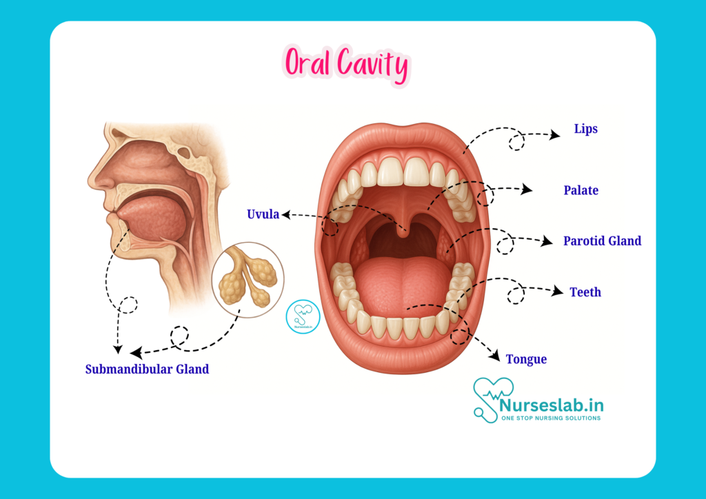
The boundaries of the oral cavity are as follows:
- Anterior: Lips
- Lateral: Cheeks
- Superior: Palate (hard and soft)
- Inferior: Floor of the mouth (formed by the tongue and underlying muscles)
- Posterior: Oropharyngeal isthmus (opening to the oropharynx)
Structures of the Oral Cavity
Lips
The lips (labia oris) are muscular folds that mark the entrance to the oral cavity. They play a vital role in speech, facial expression, mastication, and maintaining the oral seal. The lips consist of:
- External surface: Covered by skin.
- Vermilion border: The reddish transition zone.
- Internal surface: Lined by mucous membrane.
The orbicularis oris muscle forms the bulk of the lips, enabling their mobility.
Cheeks
The cheeks (buccae) form the lateral walls of the oral cavity. They are composed of skin externally, buccinator muscle centrally, and mucous membrane internally. The cheeks assist in mastication by keeping food between the teeth and contribute to facial expressions.
Palate
The palate separates the oral cavity from the nasal cavity and is divided into two parts:
- Hard Palate: The bony anterior portion, formed by the maxilla and palatine bones. It provides a rigid surface for food manipulation.
- Soft Palate: The muscular posterior portion, ending in the uvula. It moves during swallowing and speech, closing off the nasopharynx.
Floor of the Mouth
The floor is formed by the mylohyoid, geniohyoid, and digastric muscles, with the tongue lying above. The mucosa of the floor contains the openings of the submandibular and sublingual salivary glands. This area is clinically significant for the absorption of medications and rapid spread of infections.
Tongue
The tongue is a muscular organ occupying most of the oral cavity floor. It is essential for taste, mastication, swallowing, and speech.
Parts of the Tongue
- Anterior two-thirds (oral part): Lies within the oral cavity.
- Posterior one-third (pharyngeal part): Lies within the oropharynx.
Muscles of the Tongue
The tongue has two groups of muscles:
- Intrinsic muscles: Alter the shape of the tongue (e.g., superior longitudinal, inferior longitudinal, transverse, vertical).
- Extrinsic muscles: Move the tongue (e.g., genioglossus, hyoglossus, styloglossus, palatoglossus).
Papillae of the Tongue
Papillae are small projections on the tongue’s surface, and include:
- Filiform: Numerous, thread-like, for mechanical purposes.
- Fungiform: Mushroom-shaped, contain taste buds.
- Circumvallate: Large, arranged in a V-shape posteriorly, contain taste buds.
- Foliate: Located on the lateral borders, contain taste buds.
Teeth
Teeth are specialised hard structures embedded in the alveolar processes of the maxilla and mandible. They are vital for mastication, speech, and aesthetics.
Types of Teeth
- Incisors: Cutting teeth (four in each jaw).
- Canines: Tearing teeth (two in each jaw).
- Premolars: Grinding teeth (four in each jaw).
- Molars: Grinding teeth (six in each jaw for adults).
Structure of a Tooth
- Crown: Visible part above the gum.
- Root: Embedded in the jawbone.
- Neck: Junction between crown and root.
- Enamel: Hard, protective outer layer.
- Dentin: Main bulk of the tooth.
- Pulp: Soft tissue containing nerves and blood vessels.
Dental Formula
The adult human dental formula is:
2 incisors, 1 canine, 2 premolars, 3 molars (per quadrant)
Total: 32 teeth.
Gingiva
The gingiva, or gums, are the soft tissues surrounding the teeth and covering the alveolar bone. Healthy gums are essential for tooth stability and protection against infection.
Salivary Glands
Salivary glands produce saliva, which lubricates the oral cavity, initiates digestion, and protects against pathogens. There are major and minor salivary glands:
Major Salivary Glands
- Parotid Glands: Largest, located in front of the ears, open into the mouth via Stensen’s duct opposite the upper second molar.
- Submandibular Glands: Located beneath the mandible, open via Wharton’s duct at the floor of the mouth near the lingual frenulum.
- Sublingual Glands: Smallest, located under the tongue, open via numerous small ducts.
Minor Salivary Glands
Numerous minor glands are scattered throughout the oral mucosa (e.g., labial, buccal, palatal glands). They contribute to mucous secretion and oral moisture.
Functions of Saliva
- Lubrication of food and oral tissues.
- Initiation of starch digestion (via amylase).
- Antimicrobial activity.
- Facilitation of taste.
Clinical Relevance
Salivary gland disorders include infections (mumps), stones (sialolithiasis), tumours, and Sjögren’s syndrome (autoimmune dry mouth). Nurses should monitor for swelling, pain, dry mouth, and changes in saliva consistency.
Blood Supply and Innervation
Arterial Supply
The oral cavity receives its arterial supply mainly from branches of the external carotid artery:
- Lingual artery: Supplies the tongue.
- Facial artery: Supplies the lips and cheeks.
- Maxillary artery: Supplies the teeth, palate, and upper jaw.
Venous Drainage
Venous blood from the oral cavity drains into the internal jugular and facial veins, facilitating the removal of waste products and maintaining tissue health.
Nerve Supply
Sensory and motor innervation of the oral cavity is vital for function and sensation.
- Sensory:
- Trigeminal nerve (CN V): Main sensory supply to the teeth, gums, palate, and tongue (anterior two-thirds).
- Glossopharyngeal nerve (CN IX): Sensory supply to the posterior one-third of the tongue and oropharynx.
Motor:
Hypoglossal nerve (CN XII): Motor supply to tongue muscles.
Facial nerve (CN VII): Motor supply to muscles of facial expression, including lips and cheeks.
Taste:
Chorda tympani branch of facial nerve (CN VII): Taste sensation from anterior two-thirds of the tongue.
Glossopharyngeal nerve (CN IX): Taste sensation from posterior one-third.
Lymphatic Drainage
Lymph from the oral cavity drains into submental, submandibular, and deep cervical lymph nodes. This pathway is crucial for the spread of infections and malignancies.
Functions of the Oral Cavity
The oral cavity serves multiple functions vital to human health:
- Mastication: Chewing food with the help of teeth, tongue, cheeks, and saliva.
- Speech: Articulation of sounds and words through coordinated movements of lips, tongue, palate, and teeth.
- Taste: Detection of flavours—sweet, sour, salty, bitter, umami—by taste buds primarily on the tongue.
- Swallowing (Deglutition): Coordinated movement of food from the oral cavity to the pharynx, aided by the tongue and soft palate.
- Protection: Saliva contains antimicrobial agents; oral mucosa acts as a physical barrier; tonsils provide immune defence.
Clinical Relevance for Nurses
Common Oral Pathologies
Nurses must be vigilant for signs and symptoms of oral diseases, some of which include:
- Dental caries: Decay of tooth structure due to bacterial action.
- Periodontal disease: Inflammation and infection of gums and supporting structures.
- Oral candidiasis: Fungal infection, often seen in immunocompromised patients.
- Herpetic lesions: Viral infections causing painful blisters.
- Oral cancers: Malignancies affecting lips, tongue, cheeks, and palate.
- Xerostomia: Dry mouth due to reduced saliva production.
- Trauma: Injuries from accidents, falls, or ill-fitting dental appliances.
Assessment Techniques
Effective assessment of the oral cavity includes:
- Inspection of lips, mucosa, gums, teeth, tongue, palate, and floor of mouth for colour, lesions, swelling, and ulceration.
- Palpation for tenderness, masses, or induration.
- Assessment of saliva quantity and quality.
- Evaluation of speech, swallowing, and taste disturbances.
- Use of tongue depressor, torch, and gloves for thorough examination.
Implications for Nursing Care
- Early identification and referral of oral pathologies.
- Education on oral hygiene practices, including brushing, flossing, and use of mouthwashes.
- Management of oral side effects due to medications (e.g., dry mouth, ulcers).
- Assistance with oral care for dependent or critically ill patients.
- Prevention of aspiration in patients with swallowing difficulties.
- Support for nutrition and hydration through oral health maintenance.
REFERENCES
- Ross and Wilson, Anatomy and Physiology in Health and Illness, Fourteenth Edition, 1 July 2022, ISBN-13: 978-0323834612.
- Roger Watson, Anatomy and Physiology for Nurses, 14th Edition, 12-06-2018, ISBN: 9780702077418
- P.R Asha Latha, Text Book of Applied Anatomy & Physiology for Nurses, 7th Edition,3 January 2024, ISBN-13: 978-9356968622.
- Bryan H. Derikson, Tortora’s Principles of Anatomy and Physiology, 16th Edition, August 2023, ISBN: 978- 1119400066.
- Anatomy.co.uk, Reproductive System, Last updated on April 24, 2025, https://anatomy.co.uk/reproductive-system
Stories are the threads that bind us; through them, we understand each other, grow, and heal.
JOHN NOORD
Connect with “Nurses Lab Editorial Team”
I hope you found this information helpful. Do you have any questions or comments? Kindly write in comments section. Subscribe the Blog with your email so you can stay updated on upcoming events and the latest articles.


