


Last Updated on May 28, 2026 by Nurseslab.in Editorial Team
The human nervous system is a marvel of biological engineering, and understanding its structure is crucial for anyone entering the medical field. For nursing students, a solid grasp of neuroanatomy not only enhances clinical competence but also aids in the effective assessment and care of patients with neurological conditions. Among the key components of the central nervous system (CNS) are grey matter and white matter—two distinct tissue types that play vital roles in brain and spinal cord function.
Introduction
Neuroanatomy forms the foundation for understanding how the nervous system operates and interacts with the rest of the body. For nurses, knowledge of the CNS—particularly grey and white matter—is indispensable for patient assessment, monitoring neurological status, and recognising early signs of neurological disorders.
Basic Structure of the Central Nervous System (CNS)
The central nervous system comprises the brain and spinal cord, serving as the primary command centre for the body. The brain, encased within the skull, is divided into several regions: the cerebrum, cerebellum, and brainstem. The spinal cord, housed within the vertebral column, extends from the base of the brain to the lower back. Both brain and spinal cord are composed of two main types of tissue: grey matter and white matter. Understanding their organisation is fundamental to grasping CNS function.
- Brain: Includes cerebral hemispheres, diencephalon, cerebellum, and brainstem.
- Spinal Cord: Extends from the medulla oblongata to the lumbar region; acts as a conduit for signals between brain and body.
- Grey Matter and White Matter: Integral components found throughout the CNS, differing in structure and function.
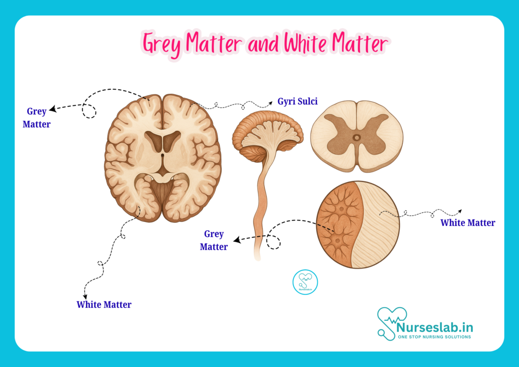
Grey Matter
Definition
Grey matter refers to regions of the CNS that are primarily composed of neuronal cell bodies, dendrites, unmyelinated axons, and various glial cells. It appears greyish in fresh tissue due to the absence of myelin, which gives white matter its characteristic colour.
Cellular Composition
- Neurons: The principal cells responsible for transmitting electrical and chemical signals. Neuronal cell bodies are abundant in grey matter.
- Dendrites: Extensions from neurons that receive synaptic inputs from other cells.
- Glial Cells: Supportive cells including astrocytes, oligodendrocytes (in the CNS), and microglia, which maintain homeostasis and protect neurons.
- Unmyelinated Axons: Axons lacking a myelin sheath, enabling local processing of information.
Location in Brain and Spinal Cord
In the brain, grey matter is predominantly found in the outer layer known as the cerebral cortex, as well as in deep nuclei such as the basal ganglia, thalamus, and hypothalamus. In the spinal cord, grey matter forms an H-shaped or butterfly-shaped region in the centre, surrounded by white matter.
Structural Features
- Cerebral Cortex: The outermost layer of the cerebrum, involved in higher cognitive functions.
- Basal Ganglia: Deep grey matter clusters involved in movement regulation.
- Spinal Cord Grey Matter: Organised into dorsal (sensory), ventral (motor), and lateral horns.
White Matter
Definition
White matter comprises bundles of myelinated axons, which connect various grey matter areas and facilitate rapid transmission of nerve impulses. The white appearance is due to the high lipid content of myelin.
Cellular Composition
- Myelinated Axons: Axons covered with myelin sheaths, speeding up electrical signal conduction.
- Glial Cells: Oligodendrocytes produce myelin in the CNS; astrocytes and microglia provide support and immune protection.
Location in Brain and Spinal Cord
In the brain, white matter lies beneath the cerebral cortex, forming tracts that connect cortical and subcortical regions. In the spinal cord, white matter surrounds the central grey matter, divided into anterior, lateral, and posterior columns.
Structural Features
- Tracts: Bundles of axons carrying signals between different parts of the CNS.
- Corpus Callosum: The largest white matter structure, connecting the two cerebral hemispheres.
- Spinal Cord Columns: Organised white matter pathways facilitating sensory and motor transmission.
Differences Between Grey Matter and White Matter
| Feature | Grey Matter | White Matter |
| Colour | Greyish (due to lack of myelin) | Whitish (due to myelin) |
| Main Components | Neuronal cell bodies, dendrites, glial cells | Myelinated axons, glial cells |
| Location | Cerebral cortex, deep nuclei, central spinal cord | Subcortical brain regions, outer spinal cord |
| Function | Processing and integration of information | Transmission of signals |
| Clinical Relevance | Affected in neurodegenerative diseases, stroke | Affected in demyelinating disorders (e.g., MS) |
Functions of Grey Matter and White Matter
Grey Matter Functions
- Sensory Processing: Receives and integrates sensory information from the body and environment.
- Motor Control: Initiates and regulates voluntary movements via motor neurons.
- Cognition: Supports functions such as memory, attention, language, and decision-making.
- Local Communication: Facilitates synaptic interactions among neurons.
White Matter Functions
- Signal Transmission: Rapidly conducts electrical impulses between grey matter regions.
- Inter-regional Communication: Connects different brain areas, enabling coordinated function.
- Supports Learning: Efficient transmission aids in learning and adaptation.
- Motor and Sensory Pathways: Carries signals for movement and sensation to and from the brain.
Clinical Significance
Common Disorders Affecting Grey Matter
- Stroke: Loss of blood supply can cause grey matter damage, manifesting as sensory, motor, or cognitive deficits.
- Neurodegenerative Diseases: Conditions such as Alzheimer’s disease, Parkinson’s disease, and Huntington’s disease primarily affect grey matter regions, leading to progressive loss of function.
- Epilepsy: Abnormal neuronal activity in grey matter can result in seizures.
Common Disorders Affecting White Matter
- Multiple Sclerosis (MS): An autoimmune disorder leading to demyelination of white matter, causing neurological impairment.
- Leukodystrophies: Genetic disorders affecting myelin formation, leading to progressive white matter degeneration.
- Traumatic Brain Injury: Diffuse axonal injury often affects white matter tracts, impacting signal transmission.
Diagnostic Imaging Relevance
Modern imaging techniques, such as Magnetic Resonance Imaging (MRI) and Computed Tomography (CT), enable detailed visualisation of grey and white matter. MRI is particularly effective in differentiating between these tissues and identifying pathological changes, such as demyelination in MS or infarcts in stroke. For nurses, understanding imaging findings is crucial for interpreting patient status and collaborating effectively with the healthcare team.
Nursing Implications
A thorough understanding of grey and white matter anatomy is essential for nurses involved in neurological care. This knowledge aids in:
- Assessment: Recognising signs of neurological impairment, such as altered consciousness, weakness, or sensory deficits.
- Monitoring: Tracking changes in neurological status and reporting significant findings promptly.
- Patient Education: Explaining neurological conditions and their implications to patients and families in an accessible manner.
- Collaboration: Working with physicians, physiotherapists, and other healthcare professionals to optimise patient outcomes.
- Rehabilitation: Supporting recovery and adaptation following CNS injury or disease.
Nurses play a pivotal role in early detection, ongoing assessment, and rehabilitation of patients with CNS disorders. Familiarity with the structure and function of grey and white matter enhances clinical judgement and patient advocacy.
REFERENCES
- Ross and Wilson, Anatomy and Physiology in Health and Illness, Fourteenth Edition, 1 July 2022, ISBN-13: 978-0323834612.
- Roger Watson, Anatomy and Physiology for Nurses, 14th Edition, 12-06-2018, ISBN: 9780702077418
- P.R Asha Latha, Text Book of Applied Anatomy & Physiology for Nurses, 7th Edition,3 January 2024, ISBN-13: 978-9356968622.
- Bryan H. Derikson, Tortora’s Principles of Anatomy and Physiology, 16th Edition, August 2023, ISBN: 978- 1119400066.
- Anatomy.co.uk, Reproductive System, Last updated on April 24, 2025, https://anatomy.co.uk/reproductive-system
Stories are the threads that bind us; through them, we understand each other, grow, and heal.
JOHN NOORD
Connect with “Nurses Lab Editorial Team”
I hope you found this information helpful. Do you have any questions or comments? Kindly write in comments section. Subscribe the Blog with your email so you can stay updated on upcoming events and the latest articles.


