


Last Updated on May 28, 2026 by Nurseslab.in Editorial Team
Introduction
The spinal cord is a central structure in the human nervous system, acting as a highway for messages between the brain and the body. For nurses and nursing students, a robust understanding of spinal cord anatomy is essential for assessing neurological health, interpreting patient symptoms, and delivering effective care.
Gross Anatomy of the Spinal Cord
Location and Length
The spinal cord is a cylindrical bundle of nervous tissue, positioned within the vertebral canal. It extends from the lower end of the medulla oblongata (at the base of the brain) down to the level of the first or second lumbar vertebra (L1/L2) in adults. Its average length is about 45 cm in men and 42 cm in women, and it is approximately 1 cm in diameter. The cord is protected by the vertebral column, which acts as a bony shield against external trauma.
External Features
Externally, the spinal cord appears as a white, glistening structure. It is slightly flattened anterior-posteriorly and exhibits two notable enlargements:
- Cervical enlargement: Found between C4 and T1, this region gives rise to nerves supplying the upper limbs.
- Lumbar enlargement: Located between L2 and S3, this area provides nerves to the lower limbs.
At its inferior end, the spinal cord tapers to form the conus medullaris, from which the filum terminale (a fibrous extension) continues downward, anchoring the cord to the coccyx.
Meninges
The spinal cord is enveloped by three protective layers known as meninges:
- Dura mater: The outermost, tough, and fibrous layer.
- Arachnoid mater: The middle, web-like layer.
- Pia mater: The innermost, delicate layer that closely adheres to the cord.
Between the arachnoid and pia mater lies the subarachnoid space, which contains cerebrospinal fluid (CSF) that cushions and nourishes the spinal cord.
Regions and Segments of the Spinal Cord
Regional Organisation
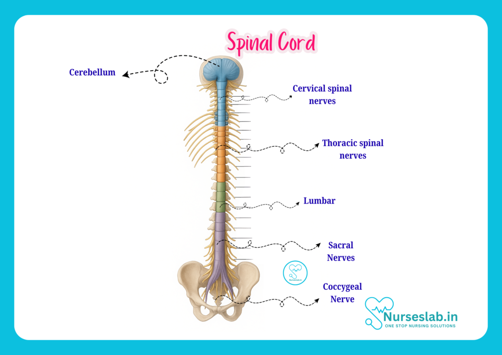
The spinal cord is divided into five major regions based on the vertebral column:
- Cervical (C1–C8): Supplies the head, neck, shoulders, arms, and hands.
- Thoracic (T1–T12): Innervates the chest and abdominal muscles.
- Lumbar (L1–L5): Controls the lower back and parts of the legs.
- Sacral (S1–S5): Regulates the pelvic organs and lower limbs.
- Coccygeal (Co1): The terminal region, with minimal functional role.
Segmental Organisation
Each region consists of segments that correspond to pairs of spinal nerves. There are 31 pairs in total:
- 8 cervical
- 12 thoracic
- 5 lumbar
- 5 sacral
- 1 coccygeal
Each segment is defined by the origin of a pair of spinal nerves, which emerge from the cord via dorsal (sensory) and ventral (motor) roots. These nerves serve specific areas of the body known as dermatomes (sensory distribution) and myotomes (motor distribution), information that is crucial for nurses when assessing neurological function.
Internal Structure of the Spinal Cord
Gray Matter
At the centre of the spinal cord lies the gray matter, shaped like a butterfly or the letter ‘H’ in cross-section. The gray matter contains cell bodies of neurons, interneurons, and glial cells. It is organised into:
- Anterior (ventral) horns: Contain motor neurons that send impulses to skeletal muscles.
- Posterior (dorsal) horns: House sensory neurons that receive input from peripheral nerves.
- Lateral horns: Present in thoracic and upper lumbar segments, containing neurons of the autonomic nervous system.
White Matter
Surrounding the gray matter is white matter, composed of myelinated nerve fibres (axons) that form ascending and descending tracts. These tracts are responsible for transmitting sensory and motor information between the body and the brain. The white matter is organised into three columns (funiculi):
- Anterior (ventral) funiculus
- Posterior (dorsal) funiculus
- Lateral funiculus
Each funiculus contains specific tracts for different functions, such as pain, temperature, touch, and voluntary movement.
Central Canal
Running through the centre of the gray matter is the central canal, a small channel filled with cerebrospinal fluid. This canal helps maintain the internal environment of the spinal cord and is continuous with the ventricular system of the brain.
Major Tracts
The spinal cord contains several important tracts, including:
- Corticospinal tract: Carries voluntary motor signals from the brain to the body.
- Spinothalamic tract: Transmits pain and temperature sensations.
- Dorsal column-medial lemniscal tract: Conveys fine touch, vibration, and proprioception.
- Spinocerebellar tracts: Carry information about muscle and joint position to the cerebellum.
A clear understanding of these pathways is essential for nurses when evaluating motor and sensory deficits.
Functions of the Spinal Cord
Sensory Pathways
Sensory information from the body is transmitted to the spinal cord via the dorsal roots and then relayed to the brain through ascending tracts. These pathways enable the perception of touch, pressure, pain, temperature, and proprioception (body position awareness).
Motor Pathways
Motor impulses originate in the brain and descend through the spinal cord’s tracts, exiting via the ventral roots to stimulate muscles and glands. This allows for voluntary movement and reflex actions.
Reflexes
The spinal cord is the centre for many reflexes—automatic, rapid responses to stimuli. Examples include the knee-jerk (patellar) reflex and withdrawal reflexes. Reflex arcs consist of sensory input, integration within the spinal cord, and motor output, often bypassing the brain for speed.
Autonomic Functions
The spinal cord also hosts autonomic pathways that regulate involuntary functions such as heart rate, blood pressure, digestion, and bladder control. These functions are especially prominent in the thoracic and lumbar regions, which contain sympathetic and parasympathetic fibres.
Blood Supply and Protective Structures
Arterial Supply
The spinal cord receives blood from a network of arteries:
- Anterior spinal artery: Supplies the anterior two-thirds of the cord.
- Posterior spinal arteries: Serve the posterior one-third.
- Radicular arteries: Arise from segmental arteries along the length of the cord, reinforcing the main spinal arteries.
Adequate blood supply is vital for the cord’s function and viability; compromise can lead to serious neurological deficits.
Venous Drainage
Venous blood from the spinal cord is drained by a series of veins that mirror the arterial supply, eventually emptying into the systemic circulation. Disruption of venous drainage can contribute to cord oedema and dysfunction.
Meninges and Cerebrospinal Fluid
As discussed earlier, the meninges surround and protect the cord, while cerebrospinal fluid provides cushioning and removes metabolic waste. The integrity of these structures is crucial for spinal cord health and resilience to injury.
Clinical Relevance
Common Spinal Cord Injuries
Spinal cord injuries (SCIs) occur due to trauma, compression, infection, or vascular events. Common causes include road traffic accidents, falls, sports injuries, and violence. SCIs are classified as complete (total loss of function below the injury) or incomplete (partial preservation of function).
Symptoms depend on the level and extent of injury:
- Cervical injuries: Can cause quadriplegia (paralysis of all limbs), respiratory difficulties.
- Thoracic injuries: May result in paraplegia (paralysis of lower limbs), loss of abdominal muscle function.
- Lumbar/sacral injuries: Affect leg movement, bladder, and bowel control.
Early recognition and intervention are critical to prevent permanent disability and complications.
Diseases Affecting the Spinal Cord
Various diseases can impact the spinal cord, including:
- Multiple sclerosis: An autoimmune condition causing demyelination and neurological deficits.
- Transverse myelitis: Inflammation of the cord, leading to sensory and motor impairment.
- Tumours: Can compress the cord and disrupt function.
- Infections: Such as spinal epidural abscesses and tuberculosis of the spine.
Symptoms and Diagnostic Approaches
Symptoms of spinal cord pathology include:
- Weakness or paralysis
- Loss of sensation
- Bladder and bowel dysfunction
- Neuropathic pain
Diagnosis may involve neurological examination, imaging (MRI, CT scan), and laboratory tests. Nurses play a vital role in recognising early signs and facilitating prompt medical attention.
Nursing Implications
Assessment
Nurses must conduct thorough neurological assessments, including evaluation of motor strength, sensory function, reflexes, and autonomic status. Use of dermatomal and myotomal charts aids in pinpointing the level of cord involvement.
Monitoring
Continuous monitoring is crucial in patients with spinal cord injuries or diseases. Nurses should watch for changes in motor ability, sensation, vital signs, and signs of autonomic dysreflexia (a potentially life-threatening complication characterised by sudden hypertension and bradycardia).
Care Strategies
Nursing care focuses on:
- Maintaining airway, breathing, and circulation
- Preventing pressure ulcers through regular repositioning
- Managing bladder and bowel function
- Promoting mobility and preventing contractures
- Providing pain management and psychological support
- Educating patients and families about spinal cord health
Individualised care plans are essential, considering each patient’s unique needs and level of impairment.
Patient Education
Empowering patients and families with knowledge about spinal cord anatomy, function, and prevention of complications is a key nursing responsibility. This includes instruction on skin care, self-catheterisation, transfer techniques, and recognising warning signs of infection or autonomic dysfunction.
REFERENCES
- Ross and Wilson, Anatomy and Physiology in Health and Illness, Fourteenth Edition, 1 July 2022, ISBN-13: 978-0323834612.
- Roger Watson, Anatomy and Physiology for Nurses, 14th Edition, 12-06-2018, ISBN: 9780702077418
- P.R Asha Latha, Text Book of Applied Anatomy & Physiology for Nurses, 7th Edition,3 January 2024, ISBN-13: 978-9356968622.
- Bryan H. Derikson, Tortora’s Principles of Anatomy and Physiology, 16th Edition, August 2023, ISBN: 978- 1119400066.
- Anatomy.co.uk, Reproductive System, Last updated on April 24, 2025, https://anatomy.co.uk/reproductive-system
Stories are the threads that bind us; through them, we understand each other, grow, and heal.
JOHN NOORD
Connect with “Nurses Lab Editorial Team”
I hope you found this information helpful. Do you have any questions or comments? Kindly write in comments section. Subscribe the Blog with your email so you can stay updated on upcoming events and the latest articles.


