


Calcium is an electrolyte necessary for numerous cellular and enzymatic processes. 99% of the total amount of calcium in the body is found in the skeleton and it is a crucial part of bone ossification. Soft tissues and extracellular fluids contain the other 1%. Calcium is vital for bone structure and integrity, muscle contraction, blood coagulation, and other processes. The parathyroid hormone regulates calcium levels.
Normal serum calcium concentrations are 9 to 10.5 mg/dL (2.25 – 2.75 mmol/L)
- Hypocalcemia- serum calcium level < 9 mg/dL (2.25 mmol/L)
- Hypercalcemia- serum calcium level > 10.5 mg/dL (2.75 mmol/L)
(Specific normal or abnormal values will depend on each lab’s guidelines)
Causes of Hypocalcaemia
Possible causes of hypocalcemia include the following:
- Inhibition of calcium absorption from the gastrointestinal tract
- Inadequate oral calcium intake
- Lactose intolerance
- Syndromes of malabsorption like celiac disease or Crohn’s disease
- Inadequate vitamin D consumption
- Advanced renal disease
- Increased excretion of calcium
- Polyuric (increased urine) phase of kidney disease
- Diarrhea
- Steatorrhea
- Discharge from wounds, particularly gastrointestinal
- Conditions that lower the calcium ionized fraction
- Hyperproteinemia
- Alkalosis
- Medications like calcium chelators or binders
- Acute pancreatitis
- Hyperphosphatemia
- Removal or damage of the parathyroid glands
Causes of Hypercalcemia
Possible causes of hypercalcemia include the following:
- Increased absorption of calcium
- Excessive oral calcium consumption
- Excessive oral vitamin D consumption
- Decreased excretion of calcium
- Kidney illness
- Use of thiazide diuretics
- Increased calcium loss from the bones
- Hyperparathyroidism
- Hyperthyroidism
- Malignancy (bone destruction by metastatic tumors)
- Immobility
- Hemoconcentration
- Dehydration
- Use of lithium
- Lack of adrenal function
Signs and Symptoms
Signs and symptoms of calcium imbalance include:
- Hypocalcemia
- Decreased heart rate (bradycardia), hypotension, diminished peripheral pulses.
- Muscle tetany or seizures can reduce respiratory movement leading to respiratory failure or arrest
- Anxiety, irritability
- Muscle twitches, cramps, tetany, and seizures
- Hyperactive deep tendon reflexes, paresthesia
- Positive Trousseau’s and Chvostek’s signs
- Increased gastric motility, hyperactive bowel sounds, abdominal cramps, and diarrhea
- ECG changes: Prolonged ST interval, prolonged QT interval
- Hypercalcemia
- Increased heart rate (tachycardia) in the early phase, bradycardia in the late phase
- Hypertension, bounding peripheral pulses
- Severe skeletal and muscular weakening leading to ineffective respiratory movement
- Disorientation, lethargy, coma
- Profound muscle weakness and diminished or absent deep tendon reflexes
- Decreased gastric motility, hypoactive bowel sounds, anorexia, nausea, constipation
- ECG changes: shortened ST segment, widened T wave, heart block
Nursing Process
The body must maintain calcium homeostasis to ensure appropriate functional processes, especially of the muscles and bones. A variety of medical treatments, conditions, and interventions can impact calcium levels. Therefore, interdisciplinary teams must collaborate and communicate to discuss treatment and ensure patient safety.
Interprofessional patient care comprising doctors, nurses, pharmacists, and dieticians can coordinate efforts to deliver corrective actions addressing calcium levels to produce the best patient outcomes. Nurses must regularly check patients’ calcium blood levels and manage their imbalances to avoid complications.
Nursing Assessment
Effective nursing assessment and intervention are crucial in managing these conditions to prevent complications and promote patient well-being.

Hypocalcemia
The initial evaluation of a patient with suspected hypocalcemia involves a thorough medical history and physical examination. Key aspects to assess include:
- Medical History: Review the patient’s history for risk factors such as recent surgeries, particularly thyroid or parathyroid surgery, chronic kidney disease, or vitamin D deficiency. Also, inquire about symptoms such as muscle cramps, tingling sensations, fatigue, and confusion.
- Physical Examination: Perform a comprehensive physical examination focusing on signs of neuromuscular irritability, such as Chvostek’s sign (facial muscle twitching in response to tapping) and Trousseau’s sign (carpal spasm induced by inflating a blood pressure cuff).
- Laboratory Tests: Obtain baseline laboratory tests, including serum calcium, ionized calcium, magnesium, phosphate, parathyroid hormone (PTH), and vitamin D levels. These tests help confirm the diagnosis and identify the underlying cause of hypocalcemia.
Hypercalcemia
The initial evaluation of a patient with suspected hypercalcemia involves a thorough medical history and physical examination. Key aspects to assess include:
- Medical History: Review the patient’s history for risk factors such as malignancies, hyperparathyroidism, prolonged immobilization, or excessive intake of calcium or vitamin D supplements. Also, inquire about symptoms such as fatigue, weakness, nausea, vomiting, constipation, and confusion.
- Physical Examination: Perform a comprehensive physical examination focusing on signs of dehydration, altered mental status, and cardiovascular abnormalities, such as hypertension and arrhythmias.
- Laboratory Tests: Obtain baseline laboratory tests, including serum calcium, ionized calcium, phosphate, PTH, and renal function tests. These tests help confirm the diagnosis and identify the underlying cause of hypercalcemia.
Nursing Intervention
Nurses play a vital role in improving patient outcomes and preventing complications. Ongoing vigilance and adherence to evidence-based practices are essential in delivering high-quality, safe, and effective care for patients with these electrolyte imbalances.
Hypocalcaemia
Acute Management
Acute management of hypocalcemia aims to correct the calcium deficit and address the underlying cause. Nursing interventions include:
- Administering Calcium Supplements: Provide intravenous calcium gluconate or calcium chloride for severe cases, and oral calcium supplements for mild to moderate cases. Monitor for signs of improvement and potential side effects, such as arrhythmias.
- Correcting Magnesium Deficiency: If hypomagnesemia is present, administer magnesium supplements to enhance the effectiveness of calcium treatment.
- Monitoring Electrolytes: Regularly monitor serum calcium, magnesium, and phosphate levels to ensure effective correction and prevent complications.
Chronic Management
Chronic management focuses on maintaining normal calcium levels and preventing recurrence. Nursing interventions include:
- Dietary Modifications: Encourage a diet rich in calcium and vitamin D, including dairy products, leafy greens, and fortified foods.
- Vitamin D Supplementation: Provide vitamin D supplements to enhance calcium absorption and maintain adequate levels.
- Patient Education: Educate patients on the importance of adhering to prescribed treatments, recognizing symptoms of hypocalcaemia, and seeking prompt medical attention if symptoms recur.
Hypercalcaemia
Acute Management
Acute management of hypercalcemia aims to rapidly lower serum calcium levels and address the underlying cause. Nursing interventions include:
- Hydration: Provide intravenous fluids to promote renal excretion of calcium and prevent dehydration. Monitor fluid status and urine output.
- Medications: Administer medications such as bisphosphonates, calcitonin, or corticosteroids to reduce calcium levels. Monitor for potential side effects and signs of improvement.
- Monitoring Electrolytes: Regularly monitor serum calcium, phosphate, and renal function tests to ensure effective correction and prevent complications.
Chronic Management
Chronic management focuses on maintaining normal calcium levels and preventing recurrence. Nursing interventions include:
- Dietary Modifications: Encourage a balanced diet with appropriate calcium and vitamin D intake. Avoid excessive calcium or vitamin D supplementation without medical supervision.
- Monitoring and Follow-up: Schedule regular follow-up appointments to monitor calcium levels, renal function, and overall health status.
- Patient Education: Educate patients on the importance of adhering to prescribed treatments, recognizing symptoms of hypercalcemia, and seeking prompt medical attention if symptoms recur.
Nursing Care Plans
Once the nurse identifies nursing diagnoses for hypocalcemia or hypercalcemia, nursing care plans help prioritize assessments and interventions for both short and long-term goals of care. In the following section, you will find nursing care plan examples for hypocalcemia and hypercalcemia.
Electrolyte Imbalance
Electrolyte imbalance associated with calcium imbalance (hypocalcemia/hypercalcemia) can be caused by conditions affecting the regulation, intake and excretion, and movement of calcium in the cellular space.
Nursing Diagnosis: Electrolyte Imbalance
Related to:
- Changes in the regulation of calcium
- Changes in the intake of calcium
- Difficulty excreting calcium
- Conditions that affect the movement of calcium in the cellular space
- Conditions that affect calcium metabolism
As evidenced by:
- Alterations in the electrical conductivity of the heart
- Ineffective respirations
- Muscle irregularities (such as muscle tetany and seizures for hypocalcemia, muscle weakness for hypercalcemia)
- Neuromuscular alterations
- Changes in bowel habits
Expected outcomes:
- Patient will demonstrate serum calcium levels within normal limits.
- Patient will manifest an absence of muscle symptoms (such as muscle twitching, cramps, or paresthesias).
- Patient will display no ECG irregularities.
Assessment:
1. Closely monitor blood calcium levels.
For optimum extracellular and intracellular function, serum calcium values must be kept within a narrow range. Serum calcium levels should be continuously monitored and drawn as ordered to determine calcium status.
2. Check albumin levels.
Serum calcium is associated with blood proteins, primarily albumin. Low albumin levels can result in an inaccurate low total serum calcium level.
3. Determine the patient’s current medications.
Several medications, such as diuretics, aluminum hydroxide, and vitamin D intake, can result in imbalanced calcium levels.
4. Assess the patient’s calcium intake.
The body cannot make calcium and it must be ingested through food and dietary supplements. The amount of intake can significantly affect calcium levels.
5. Review the patient’s health history.
Assess for conditions affecting calcium metabolism, such as pregnancy, menopause, and skeletal disorders (like osteoporosis and fracture).
6. Consider the patient’s life stage.
The amount of calcium the body needs for bone development and remodeling depends on the stage of life. The two main physiological processes are bone formation during skeletal development and bone mass conservation once development is complete. When bone growth can no longer keep up with bone resorption later in life, net calcium is lost from the body.
7. Assess Chvostek’s sign and Trousseau’s sign for hypocalcemia.
Trousseau’s sign and Chvostek’s sign are tests for hypocalcemia. A gentle tap across the facial nerve in front of the ear causes the facial muscles to twitch, producing Chvostek’s sign as compared to Trousseau’s sign, which is a carpal spasm brought on by briefly inflating a blood pressure cuff past the systolic pressure.
8. Determine the causative factors of the calcium imbalance.
The main determinants of serum Ca concentration are parathyroid hormone (PTH), calcitriol, ionized calcium, vitamin D, and blood phosphate level. The most common cause of serum calcium imbalance is a disturbed regulation and metabolism of the PTH and vitamin D interaction.
Interventions:
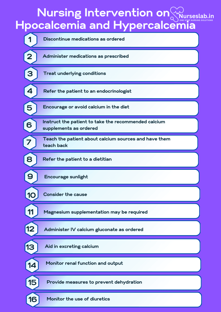
1. Discontinue medications as ordered.
Aluminum hydroxide raises calcium levels as a result of lowering phosphorus levels. Discontinue thiazide diuretics (calcium-sparing) and replace them with diuretics that enhance calcium excretion.
2. Administer medications as prescribed.
Calcium from the digestive system can be absorbed more easily with vitamin D. Give calcium supplements with vitamin D for hypocalcemia.
3. Treat underlying conditions.
Malignancy, hypoparathyroidism, renal, and skeletal diseases can all contribute to calcium imbalances. Monitoring and preventing hypo/hypercalcemia are essential parts of treating these disorders.
4. Refer the patient to an endocrinologist.
The most frequent reason for changes in calcium levels is an endocrine disorder. Endocrinologists can identify and classify the numerous causes of calcium imbalances and assist with treating and managing these conditions.
Imbalanced Nutrition: Less Than Body Requirements
Imbalanced nutrition associated with calcium imbalance is caused by an inadequate or excessive amount of calcium in the diet and improper use of calcium and vitamin D supplements.
Nursing Diagnosis: Imbalanced Nutrition
Related to:
- Inadequate calcium intake
- Excessive calcium intake
- Improper use of calcium supplements
- Lacking vitamin D
As evidenced by:
- Alterations in the electrical conductivity of the heart
- Ineffective respirations
- Muscle irregularities (such as tetany, paresthesias, weakness)
- Neuromuscular alterations
- Changes in bowel habits
Expected outcomes:
- Patient will participate in meal planning.
- Patient will be able to achieve an increase/decrease in calcium intake.
- Patient will be able to list 5 sources of calcium.
Assessment:
1. Assess the patient’s calcium intake.
Dietary calcium is the main factor influencing bone development and calcium levels in the body. Calcium will be transported from the bones into the bloodstream if dietary calcium intake is insufficient, making bones more brittle.
2. Consider the patient’s age and life stage.
The amount of calcium needed varies by age group and is higher during rapid growth, like adolescence, when the need is roughly 1,300 mg/day. Pregnant patients need 1,000 mg/day.
3. Determine the patient’s knowledge about calcium sources.
Assessing the patient’s knowledge about calcium sources can drive health teaching for the patient’s individual needs.
Interventions:
1. Encourage or avoid calcium in the diet.
Calcium is associated with dairy products; milk, yogurt, and cheese are excellent sources of calcium. Encourage or avoid calcium depending on the condition.
2. Instruct the patient to take the recommended calcium supplements as ordered.
Calcium carbonate and calcium citrate are the two most common calcium supplements.
3. Teach the patient about calcium sources and have them teach back.
Calcium sources include:
- Cheese, milk, yogurt, and other dairy products
- Green leafy vegetables
- Soy beverages with calcium
- Fish with bones, such as sardines
4. Refer the patient to a dietitian.
Dietitians can provide information about the recommended calcium intake for the patient and help them plan meals.
5. Encourage sunlight.
Sunlight is a natural source of vitamin D, which is necessary to absorb calcium. Encourage safe sun exposure.
Ineffective Tissue Perfusion
Alterations in calcium levels affect the cardiac, skeletal, and respiratory muscles and the brain.
Nursing Diagnosis: Ineffective Tissue Perfusion
Related to:
- Impaired oxygen transport
- Interruption in blood flow
- Alteration in serum calcium level
- Insufficient knowledge of hypocalcemia/hypercalcemia and its management
As evidenced by:
- Muscle cramping
- Muscle weakness
- Ineffective respirations
- Paresthesia
- Scaly skin
- Brittle nails
- Coarse hair
- Hyperactive reflexes
- Diminished or absent reflexes
- Tachycardia
- Bradycardia
- Weak peripheral pulses
- ECG changes
- Disorientation
- Lethargy
- Seizures
Expected outcomes:
- Patient will maintain optimal tissue perfusion as evidenced by the following:
- Strong, palpable pulses
- Absence of paresthesias
- 2+ reflexes
- Patient will not experience seizure activity.
Assessment:
1. Assess causative and contributing factors.
Hypocalcaemia/hypercalcemia has a variety of causes. Identifying the underlying condition that leads to the compromise in tissue perfusion directs the treatment plan. Causes can vary from cancer to renal disease to pancreatitis.
2. Assess pulses.
Hypocalcaemia causes bradycardia, hypotension, and diminished peripheral pulses, while hypercalcemia causes tachycardia (initially), hypertension, and bounding peripheral pulses.
3. Assess for paresthesias, muscle cramps, and changes in reflexes.
Hypocalcaemia causes muscle tetany (spasms), cramps, and hyperactive deep tendon reflexes (4+ on the grading scale). Hypercalcemia causes muscle weakness and diminishes or absent deep tendon reflexes.
Interventions:
1. Consider the cause.
Hypoparathyroidism responds well to IV calcium gluconate. Patients with chronic hypocalcemia may need to take a combination of oral calcium with vitamin D. Patients with renal failure benefit from calcitriol.
2. Magnesium supplementation may be required.
Hypomagnesemia causes hypocalcaemia and IV or oral magnesium may be necessary.
3. Administer IV calcium gluconate as ordered.
IV calcium gluconate is the therapy of choice for acute symptomatic hypocalcemia.
4. Aid in excreting calcium.
Hypercalcemia may require methods to eliminate excess calcium through IV saline infusions, diuretics, haemodialysis, and steroids.
Risk for Imbalanced Fluid Volume
Alterations in fluid balance can affect calcium levels.
Nursing Diagnosis: Risk for Imbalanced Fluid Volume
Related to:
- Compromised calcium transport
- Renal failure
- Malignancy
- Active fluid volume loss
- Sepsis
As evidenced by:
A risk diagnosis is not evidenced by signs and symptoms as the problem has not yet occurred, and the goal of nursing interventions is aimed at prevention.
Expected outcomes:
- Patient will demonstrate adequate fluid balance as evidenced by the following:
- Stable vital signs
- Strong, palpable pulses
- Even and unlabored respiratory pattern
- Urinary output 0.5 to 1.5 cc/kg/hour
- Alert and oriented mental status
Assessment:
1. Monitor vital signs trends.
Note any increase or decrease in blood pressure, heart rate, and respiratory rate as vital signs correspond with either fluid excess or deficit.
2. Measure ionized calcium.
This lab value demonstrates the active, free form of calcium in the blood and is more accurate in determining imbalances.
Interventions:
1. Monitor renal function and output.
Abnormalities in urine output or BUN/creatinine lead to fluid imbalances and alterations in calcium.
2. Provide measures to prevent dehydration.
Fluid loss through severe vomiting, diarrhea, or fever increases the level of calcium. The nurse may administer antidiarrheal or antiemetics to prevent fluid loss. Antipyretics reduce fever and fluid losses due to diaphoresis.
3. Monitor the use of diuretics.
Loop diuretics increase calcium excretion, while thiazide diuretics reduce calcium excretion. If the patient displays a calcium imbalance, consider if diuretics need to be added or discontinued.
4. Take precautions with major blood transfusions.
If a patient requires major blood transfusions, remember that hypocalcemia may occur from the citrate preservative in blood products. Some facility protocols may include calcium supplementation with major trauma transfusions.
Risk for Injury
Risk for injury associated with calcium imbalance is caused by alterations in muscle contraction, resulting in muscle weakness and CNS changes for hypercalcemia or tetany and seizure for hypocalcemia.
Nursing Diagnosis: Risk For Injury
Related to:
- Alterations in muscle activity (contraction and relaxation)
- Changes in the levels of consciousness
- Fragile bones (demineralized bones)
- Muscle weakness
- Tetany
- Seizures
As evidenced by:
A risk for diagnosis is not evidenced by signs and symptoms as the problem has not yet occurred, and nursing interventions are aimed at prevention.
Expected outcomes:
- Patient will not have any incidence of injury.
- Patient will demonstrate good muscle tone.
- Patient will be able to verbalize an understanding of how hypo/hypercalcemia can lead to injuries.
Assessment:
1. Perform a seizure risk assessment.
A very low serum calcium level increases the risk of seizures. When this is identified, seizure precautions should be implemented.
2. Assess the patient’s neuromuscular status.
Calcium aids in the transmission of electrical signals and affects cell development, metabolism, and memory formation. Assess deep tendon reflexes, cranial nerves, coordination, and more.
3. Determine the patient’s independence in performing activities.
The patient should be allowed to ambulate and perform tasks within their capabilities as this encourages muscle strength and independence in self-care.
4. Check for safety hazards in the patient’s environment.
Sturdy stair railings, bathroom grab bars, and adequate room lighting can prevent falls. Secure tripping hazards such as rugs or cords.
5. Review the patient’s current medications.
Drugs may interact with muscle relaxants and general anaesthetics by depressing muscle contractility or acting as a local anaesthetic. Calcium salts can interact with aminoglycosides. Tetracyclines, polymyxins, aminoglycosides, and lacosamide’s have neuromuscular blocking effects.
6. Determine bone density.
Too much bone loss in the body results in low bone density. As a result, bones become brittle and more likely to break.
7. Check if the patient has any history or current fractures.
A higher risk of injury can result from previous fractures that may have healed improperly.
Interventions:
1. Administer medications as ordered.
Consuming sufficient amounts of calcium and vitamin D can preserve bone strength and lower the risk of injury.
2. Implement seizure precautions.
Seizure precautions are intended to protect the patient from injury. To prevent injury from seizures, the patient’s bed should be in the lowest position with padded side rails, or, if possible, the mattress or rubber mats should be placed on the floor.
3. Ensure safety when administering calcium IV.
Monitor for infiltration, hypercalcemia, and ECG changes during administration.
4. Prepare the patient for possible dialysis.
If treatment for severe hypercalcemia fails to lower serum calcium levels, prepare the patient for dialysis to remove the excess calcium.
Nursing Diagnoses and Rationales for Hypocalcaemia and Hypercalcemia
Hypocalcaemia
1. Risk for Tetany
Rationale: Hypocalcemia can increase neuromuscular excitability, leading to muscle spasms or tetany. Monitoring serum calcium levels and assessing for signs of tetany, such as Chvostek’s and Trousseau’s signs, are crucial. Calcium supplementation and dietary modifications may be necessary.
2. Risk for Seizures
Rationale: Low calcium levels can cause neuronal instability, leading to an increased risk of seizures. Continuous monitoring of neurological status and maintaining optimal calcium levels can help mitigate this risk. Administering anticonvulsants may also be required in severe cases.
3. Impaired Cardiac Output
Rationale: Hypocalcemia can affect cardiac muscle function, leading to arrhythmias and decreased cardiac output. Regular cardiac monitoring, along with electrocardiogram (ECG) assessments, can detect early signs of cardiac impairment. Appropriate calcium replacement therapy should be initiated.
4. Risk for Impaired Physical Mobility
Rationale: Muscle cramps and spasms associated with hypocalcemia can limit physical mobility. Encouraging gentle exercises, providing pain relief, and ensuring a safe environment can help improve mobility and prevent falls.
5. Risk for Impaired Gas Exchange
Rationale: Severe muscle spasms can affect respiratory muscles, leading to impaired breathing. Monitoring respiratory status and providing oxygen therapy if necessary can help maintain adequate gas exchange.
6. Risk for Imbalanced Nutrition: Less than Body Requirements
Rationale: Dietary calcium deficiency can contribute to hypocalcemia. Assessing dietary intake, educating patients on calcium-rich foods, and considering dietary supplements can help ensure adequate calcium levels.
7. Risk for Acute Pain
Rationale: Muscle spasms and cramps can cause significant discomfort. Providing analgesics and muscle relaxants, along with non-pharmacological pain management strategies, can help alleviate pain.
8. Deficient Knowledge
Rationale: Patients may lack understanding about the causes, symptoms, and management of hypocalcemia. Educating patients and their families about the importance of calcium, the need for regular monitoring, and adherence to treatment plans can empower them to manage the condition effectively.
Hypercalcemia
1. Risk for Cardiac Dysrhythmias
Rationale: Elevated calcium levels can interfere with the heart’s electrical activity, leading to dysrhythmias. Continuous cardiac monitoring and regular ECG assessments are essential. Treatment may include hydration, diuretics, and medications like bisphosphonates to reduce calcium levels.
2. Risk for Fluid Volume Deficit
Rationale: Hypercalcemia can cause polyuria, leading to dehydration. Monitoring fluid balance, encouraging adequate hydration, and administering intravenous fluids if necessary can help maintain fluid volume.
3. Risk for Constipation
Rationale: High calcium levels can slow gastrointestinal motility, leading to constipation. Encouraging a high-fiber diet, ensuring adequate fluid intake, and providing stool softeners or laxatives can alleviate constipation.
4. Risk for Impaired Urinary Elimination
Rationale: Hypercalcemia can lead to the formation of kidney stones. Monitoring urinary output, ensuring adequate hydration, and educating patients about the importance of fluid intake can help prevent stone formation.
5. Risk for Activity Intolerance
Rationale: Muscle weakness and fatigue are common in hypercalcemia. Assessing energy levels, encouraging regular but gentle physical activity, and providing rest periods can help manage activity intolerance.
6. Risk for Confusion
Rationale: Elevated calcium levels can affect cognitive function, leading to confusion and altered mental status. Regularly assessing neurological status and providing a safe environment can help manage confusion.
7. Risk for Imbalanced Nutrition: Less than Body Requirements
Rationale: Hypercalcemia can cause anorexia and nausea, leading to reduced food intake. Monitoring nutritional status, providing small, frequent meals, and considering nutritional supplements can help ensure adequate nutrition.
8. Deficient Knowledge
Rationale: Patients may not fully understand the implications of hypercalcemia. Educating patients and their families about the condition, its causes, symptoms, and management strategies can help them better manage their health.
9. Risk for Impaired Skin Integrity
Rationale: Dehydration and immobility associated with hypercalcemia can increase the risk of pressure ulcers. Regular skin assessments, encouraging mobility, and providing appropriate skin care can help maintain skin integrity.
10. Risk for Complications
Rationale: Chronic hypercalcemia can lead to serious complications such as kidney damage, bone pain, and cardiovascular issues. Educating patients about the importance of regular follow-up appointments, adherence to prescribed treatments, and lifestyle modifications can help prevent and manage these complications.
REFERENCES
- Ackley, B.J., Ladwig, G.B.,& Makic, M.B.F. (2017). Nursing diagnosis handbook: An evidence-based guide to planning care (11th ed.). Elsevier.
- Carpenito, L.J. (2013). Nursing diagnosis: Application to clinical practice. (14th ed.). Lippincott Williams & Wilkins.
- Cleveland Clinic. (2022). Hypercalcemia: Causes, symptoms, diagnosis, treatments, prevention. https://my.clevelandclinic.org/health/diseases/14597-hypercalcemia
- Doenges, M. E., Moorhouse, M. F., & Murr, A. C. (2019). Nurse’s pocket guide: Diagnoses, interventions, and rationales (15th ed.). F A Davis Company.
- Drake, T. M. (2022). Calcium. StatPearls. https://www.statpearls.com/ArticleLibrary/viewarticle/80649
- Mayo Clinic. (2022). Hypercalcemia – Symptoms and causes. https://www.mayoclinic.org/diseases-conditions/hypercalcemia/symptoms-causes/syc-20355523
- Ross, A. C., Taylor, C. L., & Yaktine, A. L. (n.d.). Overview of calcium – Dietary reference intakes for calcium and vitamin D – NCBI bookshelf. National Center for Biotechnology Information. https://www.ncbi.nlm.nih.gov/books/NBK56060/
- Sadiq, N.M., Naganathan, S.,& Badireddy, M. (2022). Hypercalcemia. StatPearls https://www.ncbi.nlm.nih.gov/books/NBK430714/
- Silvestri, L. A., Silvestri, A. E., & Grimm, J. (2022). Saunders comprehensive review for the NCLEX-RN examination (9th ed.). Elsevier Inc.
- Steele, T., Kolamunnage-Dona, R., Downey, C., Toh, H., & Welters, I. (2013). Assessment and clinical course of hypocalcemia in critical illness. Critical Care, 17(3), R106. https://doi.org/10.1186/cc12756
- Tinawi, M. (2021). Disorders of calcium metabolism: Hypocalcemia and hypercalcemia. PubMed Central (PMC). https://www.ncbi.nlm.nih.gov/pmc/articles/PMC7849212/
Stories are the threads that bind us; through them, we understand each other, grow, and heal.
JOHN NOORD
Connect with “Nurses Lab Editorial Team”
I hope you found this information helpful. Do you have any questions or comments? Kindly write in comments section. Subscribe the Blog with your email so you can stay updated on upcoming events and the latest articles.


