


Last Updated on May 27, 2026 by Nurseslab.in Editorial Team
Introduction
In the domain of neurological nursing, a sound understanding of the anatomy and physiology of the ventricular system and cerebrospinal fluid (CSF) is essential. The brain’s ventricles and CSF play vital roles in maintaining central nervous system (CNS) health, protecting delicate neural tissues, and facilitating the diagnosis and management of numerous neurological conditions.
Ventricular System Overview
Definition and Location
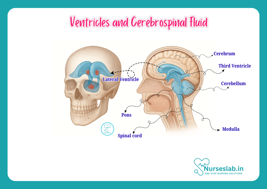
The ventricular system is a series of interconnected cavities located deep within the brain. These chambers are filled with cerebrospinal fluid and are integral to the CNS. The system comprises four main ventricles: two lateral ventricles, the third ventricle, and the fourth ventricle. They are positioned in such a way that they allow the flow and circulation of CSF throughout the brain and spinal cord.
General Function
The primary function of the ventricular system is to produce, circulate, and regulate cerebrospinal fluid. CSF cushions the brain, removes waste, provides nutrients, and maintains intracranial pressure. The ventricles also serve as pathways for CSF movement and help maintain the delicate homeostasis of the CNS environment.
Detailed Anatomy of the Ventricles
Lateral Ventricles
The lateral ventricles are the largest of the ventricular system and are paired—one in each cerebral hemisphere. They are roughly C-shaped and consist of several regions: the anterior (frontal) horn, body, posterior (occipital) horn, and inferior (temporal) horn. The lateral ventricles are separated by a thin membrane called the septum pellucidum and are located beneath the corpus callosum. Each lateral ventricle communicates with the third ventricle via a narrow channel called the interventricular foramen (also known as the foramen of Monro).
- Anterior (Frontal) Horn: Located in the frontal lobe.
- Body: Central part, situated beneath the parietal lobe.
- Posterior (Occipital) Horn: Extends into the occipital lobe.
- Inferior (Temporal) Horn: Projects into the temporal lobe.
The choroid plexus, a specialised tissue responsible for CSF production, lines the floor of the lateral ventricles.
Third Ventricle
The third ventricle is a narrow, slit-like cavity located in the midline of the brain, between the two halves of the diencephalon. It is bounded laterally by the thalamus and hypothalamus. The third ventricle communicates anteriorly with the lateral ventricles through the interventricular foramina and posteriorly with the fourth ventricle via the cerebral aqueduct (aqueduct of Sylvius). The walls of the third ventricle also contain choroid plexus tissue, contributing to CSF production.
Fourth Ventricle
The fourth ventricle is situated at the base of the brain, between the brainstem (pons and medulla) anteriorly and the cerebellum posteriorly. It is shaped like a diamond and tapers down to form the central canal of the spinal cord. The fourth ventricle receives CSF from the third ventricle through the cerebral aqueduct and allows CSF to exit into the subarachnoid space via three small openings: the median aperture (foramen of Magendie) and two lateral apertures (foramina of Luschka).
Connections and Boundaries
The ventricular system is continuous, allowing the free flow of CSF. The key connections include:
- Interventricular Foramen: Connects each lateral ventricle to the third ventricle.
- Cerebral Aqueduct: Connects the third ventricle to the fourth ventricle.
- Central Canal: Extends from the fourth ventricle into the spinal cord.
- Apertures of the Fourth Ventricle: Permit CSF to enter the subarachnoid space, surrounding the brain and spinal cord.
These connections ensure the circulation of CSF from its point of production to its absorption, maintaining the dynamic equilibrium of the CNS.
Cerebrospinal Fluid (CSF)
Production
Cerebrospinal fluid is primarily produced by the choroid plexus, a network of capillaries and specialised ependymal cells located within the ventricles. Approximately 500 millilitres of CSF are produced daily, though only about 150 millilitres are present in the CNS at any given time due to continuous circulation and absorption.
- Choroid Plexus: Located in the lateral, third, and fourth ventricles; responsible for active secretion and filtration of plasma to form CSF.
Circulation
After production, CSF flows from the lateral ventricles through the interventricular foramina to the third ventricle, then via the cerebral aqueduct to the fourth ventricle. From the fourth ventricle, CSF enters the subarachnoid space through the median and lateral apertures. It circulates around the brain and spinal cord, providing cushioning and support.
- Production in the choroid plexus (lateral, third, fourth ventricles)
- Flow from lateral ventricles → interventricular foramina → third ventricle
- Passage through cerebral aqueduct → fourth ventricle
- Exit via apertures → subarachnoid space
- Circulation around brain and spinal cord
- Absorption into bloodstream via arachnoid villi
Absorption
CSF is absorbed into the venous system primarily via arachnoid granulations (villi) located in the superior sagittal sinus. This process is crucial for maintaining intracranial pressure and preventing fluid accumulation.
Composition
CSF is a clear, colourless fluid composed of water, electrolytes (such as sodium, potassium, and chloride), glucose, small amounts of protein, and very few cells. It differs significantly from plasma, with much lower protein content and minimal cellular components.
- Normal CSF composition:
- Water: ~99%
- Proteins: 15–45 mg/dL
- Glucose: 50–80 mg/dL (approximately two-thirds of blood glucose)
- Electrolytes: Sodium, potassium, chloride
- Cells: 0–5 lymphocytes per microlitre
Functions of Cerebrospinal Fluid
- Protection: CSF acts as a cushion, absorbing shocks and preventing injury to the brain and spinal cord.
- Nutrient Transport: CSF delivers essential nutrients and removes metabolic waste from CNS tissues.
- Waste Removal: By circulating through the CNS, CSF helps clear waste products and toxins.
- Buoyancy: CSF reduces the effective weight of the brain, preventing compression of neural structures.
- Homeostasis: Maintains optimal chemical environment and regulates intracranial pressure.
These functions are vital for the normal functioning of the CNS and are central to many clinical considerations in neurological nursing.
Clinical Relevance
Common Disorders of the Ventricular System and CSF
Disorders affecting the ventricular system and CSF can have serious consequences. Nurses must be familiar with these conditions to ensure prompt recognition and intervention.
Hydrocephalus
Hydrocephalus is characterised by abnormal accumulation of CSF within the ventricles, leading to increased intracranial pressure. It may result from obstruction (non-communicating hydrocephalus), impaired absorption (communicating hydrocephalus), or excessive production of CSF.
- Causes: Congenital malformations, tumours, infections, haemorrhage.
- Clinical Signs: Headache, nausea, vomiting, altered consciousness, papilloedema, in infants—enlarged head circumference and bulging fontanelle.
- Treatment: Surgical intervention (e.g., ventriculoperitoneal shunt), endoscopic third ventriculostomy.
Meningitis
Meningitis is inflammation of the meninges, often affecting the subarachnoid space and CSF. Bacterial, viral, or fungal infections can cause meningitis, leading to changes in CSF composition and increased intracranial pressure.
- Symptoms: Fever, neck stiffness, headache, photophobia, altered mental status.
- Diagnosis: Lumbar puncture to analyse CSF for infection markers (elevated white cells, protein, reduced glucose).
- Treatment: Antimicrobial therapy, supportive care.
Other Disorders
- Subarachnoid Haemorrhage: Bleeding into the subarachnoid space can alter CSF dynamics, cause sudden headache, and neurological deficits.
- Ventriculitis: Infection of the ventricular system, often secondary to neurosurgical procedures.
- Tumours: Tumours can obstruct CSF pathways, leading to hydrocephalus or focal neurological deficits.
Diagnostic Procedures
Timely diagnosis of ventricular and CSF disorders is critical. Common diagnostic techniques include:
- Lumbar Puncture (Spinal Tap): A procedure to sample CSF from the lumbar region, used to assess infection, haemorrhage, or other abnormalities. Nurses play a key role in preparing patients, monitoring for complications, and interpreting results.
- Imaging: CT scan and MRI are used to visualise ventricles, assess CSF flow, and detect structural abnormalities. Ultrasound may be used in infants.
- Intracranial Pressure Monitoring: Used in critical care settings to monitor pressure changes in patients with head injury or hydrocephalus.
Nursing Considerations
Monitoring and Patient Care
Nurses are at the forefront of patient monitoring and care for those with ventricular system or CSF disorders. Key responsibilities include:
- Observation: Close monitoring of neurological status, including level of consciousness, pupil responses, motor function, and vital signs.
- Assessment: Watch for signs of increased intracranial pressure (headache, vomiting, altered mental status, papilloedema).
- Pre- and Post-Procedure Care: Prepare patients for lumbar puncture or imaging; monitor for complications such as headache, infection, or bleeding post-procedure.
- Shunt Care: In hydrocephalus patients with shunts, monitor for signs of shunt malfunction (headache, vomiting, lethargy, local infection).
- Infection Prevention: Adhere to strict aseptic techniques during procedures and wound care.
- Education: Provide patient and family education regarding signs and symptoms to report, activity restrictions, and follow-up care.
Signs and Symptoms of Ventricular/CSF Disorders
| Disorder | Key Signs/Symptoms | Nursing Actions |
| Hydrocephalus | Headache, vomiting, drowsiness, vision changes, enlarged head (infants) | Monitor neurological status, assess shunt function, educate family |
| Meningitis | Fever, neck stiffness, photophobia, confusion | Monitor for complications, support medication administration, infection control |
| Subarachnoid Haemorrhage | Sudden severe headache, neurological deficits, decreased consciousness | Frequent neurological checks, maintain airway, prepare for emergency interventions |
| Ventriculitis | Fever, altered sensorium, focal neurological signs | Monitor for sepsis, support treatment, infection control |
Key takeaways
- Recognising the structure and function of the ventricular system and CSF.
- Identifying clinical signs and symptoms of common disorders.
- Understanding diagnostic and therapeutic procedures.
- Implementing focused nursing care and patient education.
Continued education and vigilance in neurological assessment will empower nurses to deliver high-quality care and advocate for patients with ventricular and CSF-related disorders.
REFERENCES
- Ross and Wilson, Anatomy and Physiology in Health and Illness, Fourteenth Edition, 1 July 2022, ISBN-13: 978-0323834612.
- Roger Watson, Anatomy and Physiology for Nurses, 14th Edition, 12-06-2018, ISBN: 9780702077418
- P.R Asha Latha, Text Book of Applied Anatomy & Physiology for Nurses, 7th Edition,3 January 2024, ISBN-13: 978-9356968622.
- Bryan H. Derikson, Tortora’s Principles of Anatomy and Physiology, 16th Edition, August 2023, ISBN: 978- 1119400066.
- Anatomy.co.uk, Reproductive System, Last updated on April 24, 2025, https://anatomy.co.uk/reproductive-system
Stories are the threads that bind us; through them, we understand each other, grow, and heal.
JOHN NOORD
Connect with “Nurses Lab Editorial Team”
I hope you found this information helpful. Do you have any questions or comments? Kindly write in comments section. Subscribe the Blog with your email so you can stay updated on upcoming events and the latest articles.


