


Last Updated on May 28, 2026 by Nurseslab.in Editorial Team
Introduction
Understanding the anatomy of neuronal pathways and synapses is fundamental for nurses, as it forms the basis of neurological assessment, patient care, and clinical decision-making. The nervous system orchestrates every sensation, movement, thought, and emotion, making its study essential for those involved in healthcare.
Overview of the Nervous System
Central and Peripheral Nervous System
The nervous system is divided into two main components: the central nervous system (CNS) and the peripheral nervous system (PNS). The CNS comprises the brain and spinal cord, serving as the primary processing centre for information. The PNS includes all neural elements outside the CNS, such as cranial and spinal nerves, and acts as the communication link between the CNS and the rest of the body.
Basic Organisation
The CNS is responsible for integrating sensory information, controlling motor functions, and facilitating higher cognitive processes. The PNS is further subdivided into the somatic nervous system (controlling voluntary movements and sensory input) and the autonomic nervous system (regulating involuntary functions like heart rate, digestion, and respiration). The nervous system’s organisation enables efficient transmission and processing of signals, ensuring coordinated bodily functions.
Neuronal Pathways
Definition and Structure
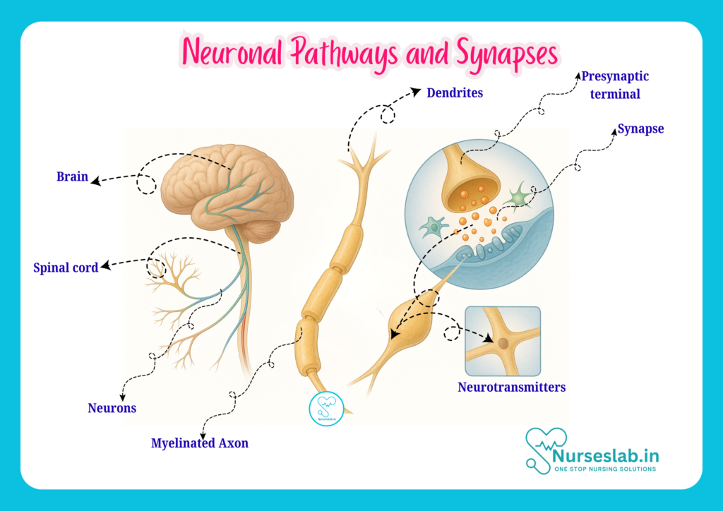
A neuronal pathway refers to a series of interconnected neurons that transmit signals from one region of the nervous system to another. These pathways are vital for relaying sensory information, executing motor commands, and integrating complex behaviours. Structurally, a neuronal pathway comprises multiple neurons connected via synapses, forming a route through which electrical and chemical signals travel.
Types of Neuronal Pathways
- Afferent (Sensory) Pathways: Carry information from sensory receptors towards the CNS. For example, touch receptors in the skin send signals to the spinal cord and brain.
- Efferent (Motor) Pathways: Transmit commands from the CNS to effectors such as muscles and glands, enabling movement and action.
- Interneurons: Located entirely within the CNS, these neurons connect sensory and motor pathways, facilitating communication and integration of signals.
Pathway Organisation
Neuronal pathways are often organised into tracts (in the CNS) and nerves (in the PNS). Tracts are bundles of axons that share a common origin, destination, and function, such as the corticospinal tract responsible for voluntary motor control. Nerves are similar bundles found in the PNS, like the sciatic nerve, which serves the lower limb. The organisation of these pathways ensures rapid, precise communication across the nervous system.
Types of Neurons
Neurons are the functional units of the nervous system, specialised for transmitting electrical and chemical signals. There are three main types:
- Sensory Neurons: Detect changes in the environment (e.g., temperature, pressure, pain) and transmit this information to the CNS. Their cell bodies are usually located in the dorsal root ganglia of the spinal cord.
- Motor Neurons: Carry signals from the CNS to muscles or glands, initiating actions such as movement or secretion. Their cell bodies are found within the CNS, and their axons extend to target tissues.
- Interneurons: Integrate and process information within the CNS, forming complex networks that enable learning, memory, and reflexes. They are the most numerous type of neuron.
Anatomy of a Neuron
Each neuron consists of distinct structural components that enable its function:
- Cell Body (Soma): Contains the nucleus and organelles, responsible for metabolic activities and maintenance of the neuron.
- Dendrites: Branched extensions that receive incoming signals from other neurons or sensory receptors.
- Axon: A long, slender projection that conducts electrical impulses away from the cell body towards other neurons, muscles, or glands.
- Myelin Sheath: A fatty insulating layer produced by Schwann cells (in the PNS) or oligodendrocytes (in the CNS), which speeds up signal transmission along the axon.
- Nodes of Ranvier: Gaps in the myelin sheath where the axon membrane is exposed, allowing rapid conduction of nerve impulses via saltatory conduction.
The precise arrangement of these structures ensures effective transmission and processing of neural signals.
Synapses
Definition and Structure
A synapse is the specialised junction where one neuron communicates with another neuron, muscle cell, or gland cell. Synapses are crucial for the transmission and modulation of information throughout the nervous system. The synapse comprises three main components:
- Presynaptic Terminal: The end of the axon of the transmitting neuron, containing synaptic vesicles filled with neurotransmitters.
- Synaptic Cleft: The small gap between the presynaptic and postsynaptic cells, typically 20–40 nanometres wide.
- Postsynaptic Membrane: The membrane of the receiving cell, equipped with receptor proteins that bind neurotransmitters.
Types of Synapses
- Chemical Synapses: The most common type, where neurotransmitters are released from the presynaptic terminal into the synaptic cleft and bind to receptors on the postsynaptic membrane. Chemical synapses allow for complex modulation and integration of signals.
- Electrical Synapses: Less common, these involve direct passage of electrical signals through gap junctions that connect the cytoplasm of adjacent cells. Electrical synapses are faster and allow for synchronised activity, such as in certain reflexes and cardiac tissue.
Chemical synapses are predominant in the human nervous system, providing flexibility and adaptability in information processing.
Synaptic Transmission
Steps in Neurotransmission
Synaptic transmission is the process by which signals are conveyed across a synapse. The key steps include:
- Action Potential Arrival: An electrical impulse (action potential) travels down the axon to the presynaptic terminal.
- Neurotransmitter Release: The action potential triggers the opening of calcium channels, causing synaptic vesicles to release neurotransmitters into the synaptic cleft.
- Neurotransmitter Binding: Neurotransmitters diffuse across the synaptic cleft and bind to specific receptors on the postsynaptic membrane.
- Generation of Postsynaptic Potential: Binding of neurotransmitters alters the postsynaptic cell’s membrane potential, potentially generating a new action potential.
- Termination: Neurotransmitters are removed from the synaptic cleft by reuptake into the presynaptic cell, enzymatic breakdown, or diffusion away from the synapse.
Neurotransmitters
Neurotransmitters are chemicals that mediate communication between neurons. Key examples include:
- Acetylcholine: Involved in muscle contraction, autonomic functions, and memory.
- Dopamine: Regulates movement, motivation, and reward.
- Serotonin: Influences mood, sleep, and appetite.
- Gamma-Aminobutyric Acid (GABA): Major inhibitory neurotransmitter, reduces neuronal excitability.
- Glutamate: Major excitatory neurotransmitter, important for learning and memory.
Receptor Types
Neurotransmitters exert their effects by binding to specific receptors on the postsynaptic membrane. There are two main receptor types:
- Ionotropic Receptors: Directly control ion channels, leading to rapid changes in membrane potential.
- Metabotropic Receptors: Activate intracellular signalling pathways, producing slower but longer-lasting effects.
Integration of Signals
Summation, Inhibition, and Facilitation
Neurons often receive input from multiple sources, requiring integration of signals. This is achieved through:
- Summation: The combined effect of multiple synaptic inputs, which can be spatial (from different locations) or temporal (over time).
- Inhibition: Inhibitory neurotransmitters (e.g., GABA) reduce the likelihood of action potential generation, balancing excitation.
- Facilitation: Repeated stimulation can enhance synaptic transmission, leading to increased responsiveness.
Effective integration of signals is essential for appropriate responses to stimuli and for complex processes like learning and memory.
Major Neuronal Pathways
Examples and Clinical Significance
- Corticospinal Tract: Originates in the cerebral cortex and descends to the spinal cord, controlling voluntary movements. Damage to this tract can result in weakness or paralysis.
- Sensory Pathways: Include the dorsal column-medial lemniscal pathway (touch, vibration, proprioception) and the spinothalamic tract (pain and temperature). Lesions in these pathways lead to sensory deficits.
- Reflex Arcs: Simple neuronal circuits that mediate automatic responses, such as the knee-jerk reflex. These pathways are important in neurological assessment.
Understanding major neuronal pathways helps nurses localise neurological injuries and interpret clinical signs.
Clinical Relevance for Nurses
Common Neurological Disorders
Neurological disorders often involve disruptions in neuronal pathways and synaptic transmission. Common conditions include:
- Stroke: Blockage or rupture of blood vessels in the brain leads to loss of neuronal function and clinical deficits such as paralysis, aphasia, and sensory loss.
- Multiple Sclerosis: Demyelination of axons impairs signal transmission, causing weakness, coordination problems, and sensory disturbances.
- Parkinson’s Disease: Degeneration of dopamine-producing neurons affects motor pathways, resulting in tremors, rigidity, and bradykinesia.
- Epilepsy: Abnormal electrical activity in neuronal networks leads to recurrent seizures.
- Peripheral Neuropathies: Damage to peripheral nerves causes pain, numbness, and muscle weakness.
Implications for Nursing Care
Nurses play a crucial role in recognising, assessing, and managing neurological disorders. Key implications include:
- Assessment: Conduct thorough neurological examinations, including evaluation of consciousness, motor and sensory functions, and reflexes.
- Monitoring: Observe for changes in neurological status, such as altered mental state, weakness, or sensory loss.
- Patient Education: Teach patients and families about disease processes, management strategies, and preventive measures.
- Rehabilitation: Support recovery through physical therapy, occupational therapy, and encouragement of mobility and independence.
- Medication Administration: Understand the pharmacology of drugs affecting neuronal pathways (e.g., antiepileptics, anticholinergics) and monitor for side effects.
Assessment Tips
Effective neurological assessment involves:
- Using standardised tools (e.g., Glasgow Coma Scale) to evaluate consciousness and responsiveness.
- Testing cranial nerve function, motor strength, coordination, and sensation.
- Observing gait, balance, and reflexes.
- Documenting findings accurately and communicating concerns promptly to the medical team.
REFERENCES
- Ross and Wilson, Anatomy and Physiology in Health and Illness, Fourteenth Edition, 1 July 2022, ISBN-13: 978-0323834612.
- Roger Watson, Anatomy and Physiology for Nurses, 14th Edition, 12-06-2018, ISBN: 9780702077418
- P.R Asha Latha, Text Book of Applied Anatomy & Physiology for Nurses, 7th Edition,3 January 2024, ISBN-13: 978-9356968622.
- Bryan H. Derikson, Tortora’s Principles of Anatomy and Physiology, 16th Edition, August 2023, ISBN: 978- 1119400066.
- Anatomy.co.uk, Reproductive System, Last updated on April 24, 2025, https://anatomy.co.uk/reproductive-system
Stories are the threads that bind us; through them, we understand each other, grow, and heal.
JOHN NOORD
Connect with “Nurses Lab Editorial Team”
I hope you found this information helpful. Do you have any questions or comments? Kindly write in comments section. Subscribe the Blog with your email so you can stay updated on upcoming events and the latest articles.


